Cholelithiasis
is a pathological condition in which stones form in the bile ducts and gallbladder. Depending on the place where the stones form and what complications they cause, several forms of gallstone disease are distinguished. In order to understand where stones form and what they can lead to, let's talk a little about bile, its functions and flow (i.e. where it flows and why). Bile is essential for digesting food, especially fats. Bile is constantly produced in the liver, then it flows through the bile ducts into the common hepatic duct, which, connecting with the gallbladder duct, flows into the duodenum and is called the common bile duct. Before the common bile duct enters the duodenum, it is joined by the pancreatic duct. Since bile is constantly produced and is released into the duodenum only during meals, it must accumulate somewhere and be stored between meals. The gallbladder serves as such a reservoir, which, at the time of eating, contracts and pushes bile into the duodenum.
The main components of bile are bile acids, cholesterol, pigments and calcium. It is from these substances that stones are formed. Depending on the predominance of one or another component, bilirubin stones, cholesterol stones and calcareous stones are distinguished.
If stones form in the gall bladder, this leads to its inflammation and is called chronic calculous (stone) cholecystitis, if a stone is strangulated in the gall bladder duct, acute calculous cholecystitis develops, if stones get into the main bile ducts, their inflammation can develop - cholangitis, and if blockage of the ducts - obstructive jaundice. If a stone becomes wedged at the point where the common bile duct enters the duodenum and the pancreatic duct joins it, the outflow of pancreatic juice is disrupted. In this condition, biliary pancreatitis develops, which is a deadly complication.
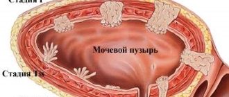
1. Gallbladder and cystic duct stones.
2. Common hepatic duct stones.
3. Common bile duct stones.
4. Wedged stone of the major duodenal papilla.
5. Stones of intrahepatic bile ducts.
Causes of the disease
There are three main reasons for the formation of stones: metabolic disorders, bile stagnation and inflammatory changes in the wall of the gallbladder. Recently, there has been an increase in the prevalence of gallstone disease, which is associated with changes in the diet and lifestyle of the modern population. Abuse of fatty and fried foods leads to changes in the composition of bile, changes in the ratio of cholesterol and bile acids. Bile becomes lithogenic, i.e. prone to stone formation. The components of bile precipitate and form a substrate for further precipitation of cholesterol, bilirubin and calcium crystals on their surface. Lithogenic bile can be compared to sticky snow during the thaw period, when snow masses easily stick to a snowball. It is known that gallstone disease often develops in patients with metabolic diseases such as diabetes, obesity and hemolytic anemia.
Damage to the gallbladder wall is also important in the formation of stones. Most patients have bacteria in their bile that leads to the destruction of the inner layer (epithelium) of the gallbladder. The desquamated particles serve as the basis for the precipitation of bile components, which were previously in dissolved form, into a crystalline state - stones are formed.
Prolonged stagnation of bile leads to its thickening and also contributes to the precipitation of bilirubin, cholesterol and calcium into crystals.
The most common site for stone formation is the gallbladder.
1.What is cholelithiasis and its causes
Gallstones
They are crystals of cholesterol or bile salts - solid particles that vary in size and shape.
The gallbladder
is an organ that is a reservoir for bile, which takes part in the digestion of fats.
The gallbladder is located on the front side of the liver and plays an important role in the functioning of the gastrointestinal tract and metabolism. That is why gallstones are cholelithiasis or cholelithiasis
- are considered a serious illness.
In order to understand the causes of gallstones, it is necessary to take a closer look at the features of its functioning. Gallbladder
- a small reservoir of bile regularly supplied from the liver. When you eat, the gallbladder releases fluid into the duodenum, improving the process of digesting food.
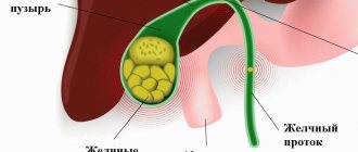
Bile has a complex chemical composition and contains quite a large amount of cholesterol and bilirubin, a pigment produced by the liver. With prolonged stagnation of bile in the gallbladder, cholesterol begins to be deposited on protein particles that are formed from the components of bile. The result of this process is the appearance of microscopic stones, which over time connect with each other, increase in size and form large stones - calculi
in the gallbladder. The formation of stones is a rather long process, taking from 5 to 20 years.
The main causes of gallstones include:
- Increased cholesterol levels in bile.
Increased cholesterol levels can be caused by eating fatty foods, obesity, metabolic disorders in the body and diseases such as diabetes. - Impaired flow of bile.
Stagnation of bile can be caused by dyskinesia - a violation of the contractile function of the gallbladder, flatulence, a sedentary lifestyle, poor diet, and pregnancy. - Infection in the gallbladder.
When an infection enters this gallbladder, it is accompanied by inflammation of its mucous membrane and the creation of favorable conditions for the deposition of bile components.
In a healthy body, bile in the gallbladder is in a liquid state and does not form solid compounds. Only under the influence of the above factors, some substances from its composition begin to precipitate, contributing to the formation of stones. Consider the following risk factors that increase the likelihood of hard compounds forming in the gallbladder:
- Genetic predisposition.
Some people have a hereditary tendency to form gallstones. Therefore, if at least one member of your family has a similar pathology, you are also at increased risk. - Obesity.
This factor is considered one of the main reasons for the development of gallstones, since obesity significantly increases cholesterol levels, which leads to disruption of the complete emptying of the gallbladder. - Ethnic origin.
Nationality also affects a person’s likelihood of developing such a pathology. Numerous studies have found that Americans, for example, are more prone to developing gallstones. - The hormone estrogen.
Estrogen in some cases contributes to the deterioration of gallbladder motility. High estrogen levels are common in pregnant women and women taking birth control pills and other hormonal medications. - Age and gender.
Most often, cholelithiasis occurs in older people and women.
A must read! Help with treatment and hospitalization!
Symptoms
Symptoms of calculous cholecystitis depend largely on the size of the gallstones. Large stones are considered to be those stones that, due to their size, cannot pass into the gallbladder duct and enter the main bile ducts, therefore, they cannot block it either. These stones are considered less dangerous than small ones. Stones of a smaller diameter, on the contrary, can pass into the gallbladder duct or into the main bile ducts and become strangulated there. It is the strangulated stone that is often the cause of biliary colic.
Pain most often occurs after an error in eating (fatty, spicy and fried foods), during physical activity, bumpy driving, or psycho-emotional stress. The pain is localized in the right hypochondrium and the solar plexus area, radiating to the lumbar region and the right shoulder blade and shoulder. An attack of biliary colic is often accompanied by nausea and repeated vomiting mixed with bile.
Outside of an attack, patients may feel completely healthy, but often they still have heaviness and dull pain in the right hypochondrium, intensifying after an error in diet, a feeling of bitterness in the mouth, diarrhea, and flatulence.
Symptoms of gallstone disease also depend on the stage and the presence of characteristic complications. In advanced forms of cholelithiasis, symptoms of peritonitis, intestinal obstruction, and obstructive jaundice may come to the fore.
Complications
Complications of cholelithiasis are often observed due to blockage of any part of the biliary tract by a stone. Let's look at the most common of them.
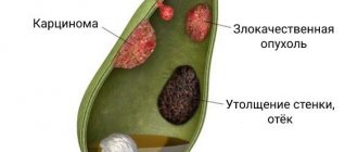
Choledocholithiasis is the presence of stones in the bile ducts. This condition may be asymptomatic if the stones do not interfere with the patency of the bile ducts. If they obstruct (clog) the bile ducts, obstructive jaundice develops. Since bile does not flow from the liver into the duodenum, it enters the blood, turning the skin yellow.
Stones located in the bile ducts not only block them, but also, by injuring them, cause inflammation - cholangitis. Stagnation of bile also contributes to inflammation. A dangerous form of cholangitis is purulent cholangitis. With the progressive rapid course of purulent cholangitis, small ulcers form not only in the bile ducts, but also in the thickness of the liver itself, which leads to the formation of multiple liver abscesses. The prognosis in such cases is unfavorable.
Acute obstructive cholecystitis develops when the cystic duct, through which bile flows from the gallbladder, is blocked by a stone. The outcome of obstructive cholecystitis can be hydrocele of the gallbladder, if the body’s strength is high enough, and blockage of the cystic duct by a stone persists. If the strength of the bacteria present in the bile exceeds the strength of the body, either empyema of the gallbladder or gangrene of the gallbladder occurs. With empyema of the gallbladder, its lumen contains pus, which can break into the abdominal cavity with all the ensuing consequences. With gangrene of the gallbladder, the death of its wall occurs, which can provoke perforation (the formation of a hole), which, as in the case of empyema, leads to diffuse peritonitis.
Large gallbladder stones can form a bedsore in the gallbladder wall. And since the wall of the gallbladder often comes into contact with the wall of the duodenum, a bedsore can form in it. Once in the intestinal lumen, the stone usually blocks the intestinal lumen, and intestinal obstruction develops, requiring emergency surgical intervention.
If a stone blocks the pancreatic duct, pancreatitis develops, i.e. inflammation of the pancreas. Pancreatitis can progress to pancreatic necrosis - death of pancreatic tissue, which often leads to death.
We tried to characterize the main complications of gallstone disease so that you understand how important it is to see a doctor in a timely manner.
3. Treatment of the disease
Treatment of gallstone disease is a systematic and multifaceted process. It includes following a diet and reducing cholesterol levels in the body. The treatment is aimed at removing stones from the gallbladder, which can be carried out in the following ways:
- Dissolving stones.
Without surgical intervention, cholelithiasis can be cured only with the help of medications: ursodeoxycholic and chenodeoxycholic acid. They reduce the level of substances in bile that contribute to the formation of stones, and increase the level of substances that dissolve solid compounds. This method is effective only in the early stages of the disease. - Crushing gallstones.
This method of treating gallstone disease is based on the effect of high pressure on gallstones through ultrasound. As a result, the stone is destroyed and crushed into small particles, which are subsequently dissolved with the help of medications. - Removal of the gallbladder.
This method of treating gallstones is used for inflammation of the walls of the gallbladder, in the presence of very large stones and other complications of gallstone disease.
The method of treatment for gallstone disease is determined by the doctor and depends on the stage of the disease, so doctors recommend that you be attentive to your health, undergo preventive examinations for gallstones if there is a tendency to develop them in your family, and immediately consult a doctor if you have symptoms of gallstones .
About our clinic Chistye Prudy metro station Medintercom page!
Diagnostics
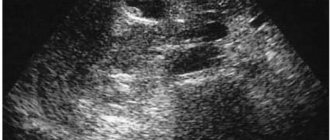
Diagnosis of cholelithiasis in the most common variant of its course does not present any particular difficulties. If stones are in the gallbladder, then with almost one hundred percent probability they are detected during an ultrasound examination of the abdominal organs. In addition to gallstones, ultrasound can evaluate the condition of the gallbladder wall, the patency of the cystic duct, and sometimes the diameter of the intrahepatic and extrahepatic bile ducts. An increase in the diameter of the bile ducts indicates increased pressure in them, which is often caused by stones located there. But to most accurately determine the location and size of stones in the bile ducts, endoscopic retrograde cholangiopancreatography, or ERCP, is necessary. The essence of this method is to take a series of x-rays of the bile ducts after a radiopaque substance has been injected into these ducts. Using an endoscope, the doctor passes through the esophagus, stomach and ends up in the duodenum, into which the common bile duct opens in the area of the major papilla. Through this papilla, the endoscopist uses a catheter to penetrate the common bile duct and pump in a radiopaque substance, which fills the lumen of the bile ducts and the pancreatic duct. Next, X-rays are taken to clearly show where the stones are and what size they are. Using the described method, you can not only carry out diagnostics, but also remove detected stones. The examination is painless and is performed without anesthesia. However, for a successful result, a highly qualified endoscopist is required.
Treatment
The main method of treating cholelithiasis is surgical. Since the main site of stone formation is the gallbladder, its removal is an integral part of surgical treatment. And although recently work has been carried out to create drugs that can dissolve small cholesterol stones, success in this matter has not been achieved. Firstly, because the composition of the stones is mixed (in addition to cholesterol, the stones contain bilirubin and calcium, which are not affected by these drugs); secondly, treatment with these drugs is long and expensive; thirdly, it is not always effective even for cholesterol stones; fourthly, when these medications are discontinued, stones form again.
So why can't these stones be crushed?
Of course, it is possible to crush stones, but with this method there are no fewer limitations than with drug therapy. Firstly, equipment for crushing stones is quite expensive and is present only in some clinics, secondly, crushing itself is associated with the danger of blocking the bile ducts with stone fragments, and, finally, the most important thing - even if the stones are successfully crushed, nothing will prevent them from forming in the gallbladder again. Because reasons such as stagnation of bile, the presence of infection and metabolic disorders are not eliminated during crushing.
Therefore, today, in fact, the only effective treatment for gallstone disease is surgical removal of the gallbladder.
Cholecystectomy - removal of the gallbladder - is currently performed in three main ways.
1. Open cholecystectomy
accompanied by a wide laparotomy incision (an incision opening the abdominal cavity) in the right hypochondrium or along the midline of the abdomen. The advantages of this method are its relative cheapness and the ability to remove the gallbladder in case of any technical difficulties. The disadvantages include high trauma, a long postoperative period and a fairly high risk of the formation of postoperative hernias.
2. Mini access cholecystectomy
−removal of the gallbladder through an incision 4-5 cm long. This method combines the advantages of open cholecystectomy and laparoscopic (removal of the gallbladder through punctures). It is performed using specialized instruments and retractors. Disadvantages of the method: an incision remains on the body, albeit a small one. Advantages of the method: the cheapest method, and there are fewer contraindications for performing cholecystectomy with this method than with laparoscopic. At the moment, this method is practically not used.
3. Laparoscopic cholecystectomy
–removal of the gallbladder using the endoscopic method, through punctures. Incisions no larger than 1 cm are made on the abdomen at four points, through which special instruments are inserted to remove the gallbladder. The surgical trauma with this method is minimal, the postoperative period is short, and the risk of postoperative hernia formation is minimized.
The only thing I would like to note is that, regardless of the method of removing the gallbladder, a competent surgeon during the operation must check all bile ducts for the presence of stones in them. Because only this approach provides a guaranteed high result in the treatment of gallstone disease.
Gallstones
Ufimtseva Irina Vladimirovna
Gastroenterologist
GSD is rightly considered the “disease of the century” and the “disease of well-being”, bearing in mind the direct connection of its development with the nature of nutrition.
In most patients, cholelithiasis develops as a result of the influence of several factors
:
- Dietary:
food excessive in carbohydrates and animal fats, poor in plant fibers and proteins, low-calorie diets with rapid reduction of body weight, violation of diet. - Constitutional:
heredity, hypersthenic type of constitution. - Medical:
diabetes mellitus, diseases of the liver, intestines, pancreas and gall bladder, hemolytic anemia, long-term parenteral nutrition, spinal cord injury. - Pharmacological:
contraceptives, fibrates, diuretics, octreotide, ceftriaxone. - Social and hygienic:
alcohol abuse, smoking, physical inactivity. - Psychological:
frequent stress, conflicts. - Pregnancy, female gender, excess body weight.
Gallstones are crystalline structures that occur in abnormal bile. Increased cholesterol content in bile promotes the growth of stones. This process is long: it can take several years until the chain “cholesterol-cholesterol flakes-cholesterol crystals-cholesterol stones” is completed.
The basis of the disease is a change in the viscosity of bile (dyscholia), associated with a violation of the physicochemical properties of bile. This involves the main pathological processes: oversaturation of bile with cholesterol, disruption of the dynamic balance between antinucleating and pronucleating factors and a decrease in the contractile function of the gallbladder.
Cholesterol stones
— this is the acquisition of the population of highly developed countries, especially those suffering from overeating. With a vegetarian diet, cholelithiasis is rare.
Treatment of cholesterol stones is currently possible, but it must be timely, at the stage of formation of a soft and small stone.
To determine the possibility of therapeutic treatment of cholecystitis with stones, it is necessary to study the gallbladder using computed tomography to determine the density of the stone and bile.
This study has many advantages over ultrasound of the abdominal cavity, because determines the density of the stone, which means its ability to dissolve. Dense, calcareous stones in Hounsfield units according to CT are more than 100 units, soft, cholesterol from 30 to 90 units. The size of the stone also matters - stones up to 10-15 mm dissolve when the gallbladder is less than 1/3 full.
At LOTOS MC we carry out this examination and treatment of patients. In 2015, a group of 112 patients was recruited to undergo litholytic therapy (therapy for dissolving stones with drugs). Over a period of 6 months, stones dissolved in 30% of patients, after 9 months in 60%, after a year in 82%. These are good indicators of litholysis, given the fact that the treatment made it possible to avoid surgery to remove the gallbladder and save the organ.
If you or your loved ones have cholelithiasis, you need to act quickly and correctly, without triggering the disease.
Correctly selected examination and treatment will help cope with the problem of gallstones. — Ufimtseva Irina Vladimirovna