Why do a plain abdominal x-ray?
This technique allows you to identify pathologies, detect their localization, the degree of damage, and life-threatening conditions.
In addition to clarifying the diagnosis, it is possible to determine the cause of the development of the pathological process. Plain radiography of the abdominal cavity reveals:
- abscess in tissues;
- hematomas;
- tumors in the abdominal organs;
- gallstones and kidney stones;
- intestinal perforation;
- organ injuries;
- abnormal development of internal organs;
- peritonitis;
- intestinal obstruction;
- diverticular disease;
- flatulence;
- presence of foreign bodies;
- hernias;
- inflammatory processes in the abdominal cavity (pancreatitis, appendicitis, etc.);
- kidney prolapse and other pathologies.
A survey radiography of the abdominal cavity is carried out to clarify the condition of the internal organs, including for preventive purposes. If there are provoking factors or predispositions, it is better to act proactively - to diagnose before the disease manifests clinical symptoms.
What does the research reveal?
Presence of free gas during perforation of a hollow organ and free liquid
Violation of the integrity of the hollow organ is the reason for the presence of free gas. This phenomenon does not occur in a healthy person. The cause is often ulcerative lesions.
Perforation, or perforation, is the occurrence of a defect in an organ, resulting in a direct communication between its contents and the environment surrounding the organ.
Perforation can appear in any hollow organ, but most often it occurs in the stomach, duodenum and intestines.
The main reasons for perforation:
- destruction of the walls by an inflammatory process, necrosis or neoplasm (most often perforated ulcers of the stomach and duodenum);
- mechanical damage to the walls due to a gunshot or knife wound;
- violation of integrity during medical manipulation (uterine curettage, biopsy during gastroscopy);
- rupture of the wall due to increasing tension (when feces accumulate, for example).
The most striking symptom of perforation is pain. Besides:
- vomiting with the development of functional intestinal obstruction;
- bloating;
- severe pain at the time of perforation of an ulcer of the duodenum or stomach, occurring in the epigastric region and right hypochondrium, etc.
Malignant tumors can lead to the accumulation of fluid: cancer of the stomach, intestines, pancreas. Sometimes the cause is pathologies such as pancreatitis (inflammation of the pancreas), peritonitis (inflammation of the peritoneum). That is why it is important to pay attention to alarming symptoms in time and undergo an examination.
Peritonitis can be a manifestation of an acute abdomen, a phenomenon that is characterized by severe pain and pathological tension of the abdominal wall. An acute abdomen is observed in gastrointestinal pathologies such as perforated gastric ulcer, appendicitis and others. This condition requires hospitalization and surgery!
Intestinal obstruction
With intestinal obstruction, food is unable to pass through the intestinal tract due to mechanical obstructions or problems with the intestines.
Main symptoms:
- bloating;
- constipation;
- bursting pain in the abdomen;
- nausea.
A test for intestinal obstruction is done if you have bloating, constipation, or other symptoms. Thanks to the study, the diagnosis is confirmed or refuted.
Obstruction may be a consequence of the appearance of polyps, tumors, or impaired intestinal motility.
Early detection of signs of intestinal obstruction allows you to start treatment on time!
With the help of an intestinal examination, a specialist can detect liquids and gases that form “bowls” and “arches”. On an x-ray, the so-called “Kloiber cups” look like gas bubbles of a semicircular shape. The cup pattern is formed when the bowel loop contains more fluid and a small amount of gas; the arches are the opposite. The bowl can be transformed into an arch, and the arch into a bowl.
With intestinal obstruction, several Kloiber cups appear at once, which are located in the center of the abdominal cavity, in the area of the loops of the small intestine. On an x-ray, it appears as swollen areas of intestine filled with fluid and gas.
Examining the patient in an upright position gives a clearer picture than when examining a patient in a lateral position.
Also, a radiologist can detect intussusception , a type of intestinal obstruction when one section of the intestine penetrates into the lumen of another. But this pathology is typical for young children.
Historical reference
The German radiologist Kloiber in 1919 first published systematic radiological observations of acute intestinal obstruction. Unlike others, he did not use contrast agents.
Modern X-ray diagnosis of acute intestinal obstruction is based mainly on the study of the accumulation of gases and liquids in the intestines. They look like light spots on the film. With such a clear picture, the use of contrast is unnecessary.
Retroperitoneal abscess
An abscess usually occurs after surgery or after injury. The disease most often occurs in people 20-40 years old.
There may be no symptoms at first. And then intoxication occurs, characterized by nausea and chills. Unpleasant sensations arise first when walking, and then at rest.
Indications for plain radiography of the abdominal cavity
Plain radiography of the abdominal cavity is performed for primary differentiation of symptoms. Many pathologies of internal organs have similar manifestations (pain, nausea, heartburn, bowel dysfunction, bloating), so it is often difficult to understand what exactly causes these symptoms.
Plain radiography of the abdominal cavity is indicated for any pain in the abdominal area, gastrointestinal disorders, abnormalities in the results of urine and blood tests, as well as after operations on internal organs and a family history.
Preparing for a plain abdominal x-ray
In 3-6 days, you need to go on a strict diet, excluding from your diet fried, too salty, peppered foods, foods that cause gas, fast food, legumes, confectionery and flour products, sausages, dumplings, and whole milk.
You can eat simple dietary dishes: low-fat cottage cheese, broths, boiled poultry, water porridge, light salads with a minimum amount of ingredients.
On the eve of the x-ray, a cleansing enema is recommended. There is an alternative to an enema - you can take laxatives. It is better to discuss this point in advance with your doctor or the doctor who referred you for the study.
12 hours before, you should completely stop eating solid food, and 6 hours before, exclude any food and water intake.
Attention: plain radiography of the abdominal cavity can be performed without preparation if the clinical case requires it and there is an urgent need to examine the internal organs.
Memo on preparing patients for x-ray examination
Preparing patients and conducting x-ray examinations of the stomach and small intestine.
How are patients prepared for x-ray examination of the stomach and small intestine?
Patients with normal bowel function do not require any special preparation for an X-ray examination of the stomach
.
In case of pathology of the stomach and intestines, 2-3 days before the study, foods that contribute to gas formation (brown bread, vegetables, fruits, legumes, milk, etc.) are excluded from the diet of the test subject. 14 hours before the examination, the patient stops eating, takes 30 ml of castor oil in the evening, and after 2-3 hours he is given a cleansing enema with 1-1.5 liters of warm water, chamomile infusion or soap solution (5 g of baby soap). 2-3 hours before the study, a repeat cleansing enema at room temperature is given. On the day of the study, the patient should not drink or smoke.
If there is a large amount of liquid, mucus, or food debris in the patient’s stomach (for example, with an organic narrowing of the gastric outlet), the stomach should be rinsed 2-3 hours before the test.
For severe flatulence and persistent constipation, a cleansing enema is recommended 1.5-2 hours before the test.
How is an X-ray examination of the stomach and duodenum performed?
As a contrast agent for x-ray examination of the stomach and duodenum
use a suspension of barium sulfate, which is prepared at the rate of 100 g of powder per 80 ml of water.
Preparing patients and conducting x-ray examinations of the gallbladder and biliary tract.
How are patients prepared for x-ray examination of the gallbladder and biliary tract?
For X-ray examination of the gallbladder and biliary tract
Two main methods are most often used:
cholecystography
(x-ray examination of the gallbladder with preliminary oral administration of a contrast agent) and
cholegraphy
(x-ray examination of the bile ducts with intravenous administration of a contrast agent). Before cholecystography and cholegraphy, the patient must follow a diet for 3 days to prevent flatulence (excluding raw cabbage, black bread, milk, etc.). Accumulations of gas in the intestines, giving rounded foci of clearing on an x-ray image, can overlap the shadow of the gallbladder, complicating the correct interpretation of the data obtained. Special mandatory cleansing enemas, as well as so-called “fat breakfasts” on the eve of the study are not required. A cleansing enema is given only in cases of severe flatulence.
How is cholecystography performed?
During cholecystography, the patient on the eve of the study takes a radiopaque iodine-containing drug (cholevid, yopagaost, etc.) at the rate of 1 g per 20 kg of the patient’s body weight, washed down with sweet tea, but 0.5 g every 5 minutes for half an hour. The contrast agent, entering the liver, is excreted in the bile and accumulates in the gallbladder. In this case, the maximum concentration of the drug in the gallbladder is observed 15-17 hours after administration; therefore, if cholecystography is scheduled for 9-10 a.m., then the drug should be taken the night before at 5-7 p.m. It is necessary to warn patients about the possibility of nausea and loose stools after taking these radiocontrast drugs.
The next day, X-rays (x-rays) of the gallbladder are taken. How is gallbladder x-ray analyzed?
When analyzing radiographs, the intensity of the shadow of the gallbladder, its shape, size, position, the presence or absence of deformation, concretions (stones), etc. are assessed.
How is gallbladder motor function tested?
To clarify the motor function of the gallbladder, the patient is given a so-called choleretic breakfast (2 raw egg yolks or 20 g of sorbitol in 100-150 ml of water), after which after 30-45 minutes (preferably serially, every 15 minutes), repeated photographs are taken and determined contractility of the gallbladder.
How is cholegraphy performed?
When conducting cholegraphy, a contrast agent (bilignost, bilitrast, etc.), which is also secreted by the liver and contrasts the bile ducts, is administered intravenously. Taking into account the possibility of allergic reactions, a test dose (1-2 ml) of a 50% solution of bilignost or biligrafin, warmed to body temperature, is first administered intravenously. If there are no allergic reactions (itching, chills) after 5-10 minutes, the main part of the drug is slowly administered. More intense filling of the ducts occurs after additional administration of 0.5 ml of a 1% morphine solution to the patient. Subsequent images are taken 20, 30-40 and 45-60 minutes after administration of the contrast agent.
How is bile duct radiographs analyzed?
On radiographs, the size, contours, lumen of the intra- and extrahepatic bile ducts, the presence or absence of stones in them are assessed, and the concentration and contractile functions of the gallbladder are clarified. To more accurately determine the condition of the common bile duct, intravenous cholegraphy is often supplemented with an X-ray examination of the duodenum ( duodenography
).
What are the contraindications for cholecystography and cholegraphy?
Cholecystography is not performed in case of severe liver damage, hypersensitivity to iodine, but cholegraphy. in addition, in acute inflammatory diseases of the bile ducts that occur with an increase in temperature (cholangitis), severe hyperfunction of the thyroid gland.
Preparing patients and conducting x-ray examination of the large intestine.
What is the purpose of an x-ray examination of the colon?
X-ray examination of the colon (irrigoscopy)
carried out using a contrast enema. The use of irrigoscopy makes it possible to determine the shape, position, condition of the mucous membrane, tone and peristalsis of certain parts of the colon and plays an important role in recognizing its various diseases - tumors, polyps, diverticula, intestinal obstruction.
How is preparation for irrigoscopy carried out?
To prepare a patient for irrigoscopy, food that promotes flatulence is excluded from his diet for 3 days, and porridge, jelly, omelettes, boiled meat and fish products are prescribed. Three times a day, chamomile infusion is given internally, a gas tube is inserted;
On the eve of the study, the patient is given 30 g of castor oil before lunch, and in the evening they give a cleansing enema, preferably twice with an interval of 1 hour. The patient does not eat dinner. In the morning, the patient is given a light breakfast and again given 2 cleansing enemas.
How is an X-ray examination of the colon (irrigoscopy) performed?
A suspension of barium sulfate is used as a contrast agent (at the rate of 400 g of powder per 1600 ml of water), which is best prepared in an electric mixer. The suspension, warmed to body temperature, is administered using an enema.
Preparing patients and conducting x-ray examination of the urinary system.
How do you prepare for an x-ray examination of the urinary system (urography)?
Before a survey of the kidneys, gas-forming foods (brown bread, potatoes, sauerkraut, legumes, sweet fruits, whole milk, etc.) are excluded from the patient’s food for 2-3 days, and saline laxatives are not prescribed. The night before, a cleansing enema of warm water with chamomile infusion is given. In the morning, 3 hours before the study, a cleansing enema is given again. On the day of the procedure, the patient should not eat or drink.
During an X-ray examination with contrast agents containing iodine, a sensitivity test is performed the day before the procedure. In case of an allergic reaction, the study is contraindicated.
urography procedure performed?
?
30 minutes before the study, the patient empties the bladder and X-rays check for the presence of gases in the intestines. If there is a large amount of gas, the enema is repeated and after 45 minutes a survey of the kidneys is taken.
In retrograde urography, a contrast agent is injected through a catheter into the bladder ( cystography
) or through special catheters into the renal pelvis. X-rays are then taken.
Preparing patients for x-ray examination of the bronchi, trachea, and chest.
How is a patient prepared for bronchography?
Bronchography
- This is an X-ray examination of the bronchi and trachea using contrast agents. During preparation, the patient’s sensitivity to iodine drugs is checked, postural drainage of the bronchi is carried out, expectorants, bronchodilators, and antibiotics are prescribed. Before the procedure, atropine is administered subcutaneously, and, if necessary, pipolfen and seduxen.
What are the features of the procedure?
Bronchography is performed under general anesthesia or local anesthesia.
After the procedure, the patient is not allowed to eat for 3 hours.
Catheters for administering contrast agents are sterilized by boiling.
How is a chest x-ray performed?
Examination of the chest (x-ray and x-ray) is carried out without special preparation of the patient. The method of photographing an X-ray image on 7x7 cm or 10x10 cm film is called fluorography.
How is a plain radiography of the abdominal cavity performed?
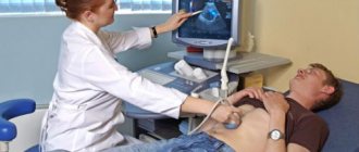
The patient is located in the X-ray room, takes off his clothes to the waist, as well as metal jewelry and accessories. After this, he stands at a given point in front of the device; The doctor fixes the position of the body and asks you to hold your breath. An X-ray scan takes literally a second.
A survey radiography of the abdominal cavity is carried out quite quickly (no more than 15 minutes in general - with preparation and conversation with a radiologist) and does not cause pain or discomfort. After the examination, you can eat and return to normal activities.
How is the examination carried out?
Immediately before starting the procedure, you will need to remove tight clothing and metal objects (jewelry, accessories). The position of the body will be determined by the radiologist depending on the preliminary diagnosis. Most often, x-rays are taken in a lying or standing position; one or more projections are used for informative images. While the camera is operating, you must hold your breath and remain still in order to get a clear picture.
Plain radiography is performed without the introduction of a contrast agent. However, for targeted diagnosis, an x-ray with contrast may be prescribed. It is performed for obstruction, narrowing or other pathologies of the gastrointestinal tract. A barium suspension is used as a contrast, and x-rays are taken in series with an interval of several minutes between them.
Based on the results of the x-ray, a conclusion is issued, which is analyzed by the attending physician to make/clarify the diagnosis.
If you are concerned about your health, do not delay visiting a doctor. We invite you to accurate diagnostics at affordable prices at the Kutuzovsky Children's Center. Pre-registration by calling the clinic.
Benefits of plain abdominal radiography
The group is an undisputed authority in the provision of medical services. We have created comfortable conditions for performing abdominal radiography and offer convenient hours for visiting the clinic.
Our advantages:
- excellent equipment (modern X-ray machines are used);
- strict adherence to study protocols;
- high diagnostic accuracy;
- a choice of clinic and doctor is provided;
- reasonable cost of radiography;
- the opportunity to consult a doctor immediately after receiving the X-ray results.
It is important to get diagnosed on time! Contact the group if you need a high-tech examination of the abdominal organs!
Contraindications to X-rays
During pregnancy, X-rays are prohibited.
As noted above, radiography of the stomach is a fairly safe type of diagnosis.
Despite this and its high information content, if certain phenomena are present, it is better to refuse the examination. In particular, X-rays are not advisable if the patient being examined:
- has continuous gastroesophageal bleeding;
- pregnant or lactating;
- susceptible to an allergic reaction to the contrast agent;
- is in serious condition.
Please note that in case of real need, solely as prescribed by a doctor, ignoring contraindications is acceptable. However, spontaneous violation of the previously noted rules is unacceptable, since in this case the diagnosis will only be harmful.