Forms of pancreatitis
Acute – characterized by acute girdling pain in the upper abdomen. Pain often appears after eating fatty foods or alcohol. Unpleasant sensations can be either barely noticeable or unbearable, radiating to the scapula or sternum. Nausea, vomiting, and stool disturbances are observed. Due to the obstructed flow of bile, the skin takes on a yellowish color.
Chronic - the main localization of pain is on the upper part of the abdominal wall with irradiation to the back, chest (left side), lower abdomen. Unpleasant sensations occur after eating heavy fatty foods, alcoholic drinks, and constant stress.
The development of chronic pancreatitis is characterized by nausea, loss of appetite, bloating, bowel dysfunction, and sometimes vomiting.
The chronic form of the pathology differs from the acute form by periods of remission and exacerbation. As the disease progresses, periods of exacerbation become more frequent; intestinal disorders, disturbances in normal digestion, and weight loss are possible.
Chronic pancreatitis often causes complications (stomach bleeding, cancer, cysts and abscesses, liver damage, diabetes mellitus, enterocolitis). That is why you need to take the disease seriously and, at the slightest suspicion of the development of inflammation, consult a doctor.
Reasons for the development of pancreatitis
The disease develops due to damage to pancreatic tissue. This happens for the following reasons:
- alcohol and tobacco abuse
- abdominal trauma, surgery
- uncontrolled and long-term use of medications: antibiotics, hormonal drugs, corticosteroids, some diuretics
- intoxication with food products, chemicals
- genetic predisposition
- improper diet with a predominance of spicy and fatty foods and long breaks between meals
Prevention
The main way to prevent pancreatitis in adults is to limit the consumption of alcoholic beverages. To prevent illness in children, proper nutrition is recommended, limiting irritating foods, fast food, and preventing injuries.
Secondary prevention, that is, prevention of relapses, exacerbations and complications, includes:
- refusal to drink alcohol;
- treatment of cholecystitis, cholelithiasis;
- constant use of enzyme preparations and diet.
If such conditions are met, pancreatitis does not lead to serious consequences, and its prognosis for life is favorable.
Important: before use, read the instructions or consult your doctor.
Symptoms of pancreatitis
Manifestations of pathology vary depending on the form - acute or chronic pancreatitis. In acute pancreatitis the following are observed:
- The pain is intense, constant, the nature of the pain is described by patients as cutting, dull.
- High body temperature, high or low blood pressure - the patient’s well-being quickly deteriorates due to the rapid development of the inflammatory process.
- Pale or yellowish complexion.
- Nausea and vomiting - dry mouth and a white coating appear, attacks of vomiting do not bring relief. The most correct step at this moment is to fast; any food intake can only worsen the situation.
- Diarrhea or constipation - stool in acute pancreatitis is most often foamy, often with a foul odor, with particles of undigested food. On the contrary, there are constipation, bloating, and hardening of the abdominal muscles, which may be the very first signal of an acute attack of pancreatitis.
- Bloating – the stomach and intestines do not contract during an attack.
- Shortness of breath - appears due to loss of electrolytes during vomiting.
Chronic pancreatitis is characterized by the following symptoms:
- Abdominal pain - may be girdling or have a clear localization radiating to the back. Appears after eating.
- Intoxication of the body - general weakness, loss of appetite, tachycardia, increased body temperature, and decreased blood pressure appear.
- Endocrine disorders - ketoacidosis, diabetes mellitus, tendency to hypoglycemia. Bright red spots may also appear in the abdomen, back, and chest, which do not disappear with pressure.
With a long course of the disease, the patient gradually develops anemia, weight loss, dry skin, brittle hair and nails, symptoms of vitamin deficiency, and increased fatigue.
brief information
The content of the article:
1.1 Definition
Acute pancreatitis (AP) is an initially aseptic inflammation of the pancreas, which may damage surrounding tissues and distant organs.
1.2 Etiology and pathogenesis
Etiological forms of pancreatitis:
- Acute alcohol-nutritional – 55%
- Acute biliary – 35%
- Acute traumatic 2 – 4%
- Other etiological forms – 6 – 8%
Factors of aggression
Primary:
- trypsin, chymotrypsin cause proteolysis of proteins;
- phospholipase A2 destroys cell membranes;
- lipase leads to lipolytic necrosis in the gland, fiber and mesentery of the intestine;
- elastase destroys the vascular wall and connective tissue structures, which leads to necrosis.
Secondary:
- activation of the kallikrein–kinin system by enzymes disrupts microcirculation.
Tertiary:
- macrophages, mononuclear cells, neutrophils produce cytokines that suppress immunity.
Quaternary:
- cytokines, enzymes, metabolites increase the permeability of the intestinal wall, which facilitates the entry of toxins into the bloodstream and lymph and damage target organs.
Factors of aggression and organ dysfunction create a syndrome of “mutual burden.”
Phases of acute pancreatitis:
- Edematous (interstitial) pancreatitis 80-85% is mild with the rare development of local complications or systemic disorders, has no phases.
- Necrotizing pancreatitis (pancreatic necrosis) in 15-20%, moderate or severe, phase course with 2 peaks of mortality - early (1 week each of phases IA and IB) and late (weeks and months).
Phase I A – up to 3 days, the formation of foci of necrosis in the parenchyma and the development of endotoxemia with multiple organ failure.
Phase I B - the body’s reaction to foci of necrosis with resorptive fever and the formation of a peripancreatic infiltrate.
Phase II – aseptic and septic sequestration.
1.3 Epidemiology
- Prevalence 32-389 per 1 million population.
- Mortality rate is 6-12 per 1 million population.
- Overall mortality 2.5%-3.5%; postoperative 20%-25%
- Since 2009, in the structure of “acute abdomen” AP has moved from 1st place to 2nd place with a share of 25%-35%, giving way to acute appendicitis.
1.4. Coding according to ICD-10
Acute pancreatitis (K85):
- K85.0 – Idiopathic acute pancreatitis;
- K85.1 – Biliary acute pancreatitis: cholelithiasis pancreatitis;
- K85.2 – Alcoholic acute pancreatitis;
- K85.3 – Drug-induced acute pancreatitis;
- K85.8 – Other types of acute pancreatitis;
- K85.9 – Acute pancreatitis, unspecified.
1.5 Classification
Mild AP - pancreatic necrosis does not form (edematous pancreatitis) and organ failure does not develop. Moderate AP - with one of the local manifestations (infiltrate, pseudocyst, abscess), or/and the development of transient - no more than 48 hours of organ failure. Severe AP - with either not limited infected pancreatic necrosis (purulent-necrotizing parapancreatitis) or/and the development of persistent organ failure.
Diagnostics
Clinical manifestations depend on the morphological form, phase of the disease, the severity of the systemic inflammatory response syndrome and organ failure.
Each phase of the disease corresponds to a clinical and morphological form, so diagnosis is carried out depending on the phase of the disease.
Scale of criteria for primary express assessment of the severity of AP (St. Petersburg Research Institute of SP, 2006):
- peritoneal syndrome;
- oliguria (less than 250 ml in 12 hours);
- skin symptoms (facial hyperemia, marbling, cyanosis);
- SBP less than 100 mmHg;
- encephalopathy;
- Hb more than 160 g/l;
- leukocytes more than 14 x109/l;
- blood glucose more than 10 mmol/l;
- urea more than 12 mmol/l;
- metabolic disorders according to ECG;
- cherry or brown-black enzymatic exudate during laparoscopy/laparocentesis;
- during laparoscopy, widespread enzymatic parapancreatitis extending beyond the boundaries of the omental bursa and spreading along the flanks;
- common steatonecrosis during laparoscopy;
- lack of effect from basic therapy.
- Scale rating:
5 signs - with 95% probability a severe form of AP. 2-4 signs – moderate AP, hospitalization in the ICU. 0 – 1 sign – mild form of AP, hospitalization in the surgical department.
The SOFA scale is used to assess organ and multiple organ dysfunctions.
If it is impossible to determine the severity of AP using scales, clinical and laboratory criteria for AP are:
- signs of systemic inflammatory response syndrome (SIRS);
- hypocalcemia <1.2 mmol/l,
- hemoconcentration: Hb> 160 g/l or Ht> 40 units, glucose> 10 mmol/l;
- CRP >120 mg/l;
- shock
- renal failure (oligo-anuria, creatinine >177 µmol/l);
- liver failure (hyperfermentemia);
- cerebral insufficiency (delirium, stupor, coma);
- gastrointestinal bleeding (more than 500 ml/day);
- coagulopathy
Urgent EPST with lithoextraction for impaction of a stone in the major duodenal papilla (MDP):
- intense pain syndrome that is not relieved by narcotic analgesics;
- rapidly progressing jaundice;
- absence of bile in the duodenum during FGDS;
- signs of biliary hypertension on ultrasound.
Indications for CT/MSCT (MRI):
- uncertainty of diagnosis and differential diagnosis;
- the need to confirm severity using clinical prognostic features;
- lack of effect from conservative treatment.
Time frame for MSCT (MRI):
- for the diagnosis of pancreatic necrosis on days 4–14 of the disease;
- as the disease progresses;
- in the absence of treatment effect;
- to clarify the localization of foci of suppuration before drainage interventions.
The Balthazar CT pancreatitis severity index is not a mandatory study, but is used to predict the severity of the disease.
First aid for an attack of pancreatitis
To reduce pain, you can use a heating pad filled with cold water. It needs to be applied to the abdominal area, namely to the epigastric region (the area under the xiphoid process, corresponding to the projection of the stomach onto the anterior abdominal wall). This allows you to reduce the intensity of pain, slightly reduce swelling and inflammation.
The patient must comply with the hospital regime. This will reduce blood flow to the organ, and therefore reduce inflammation.
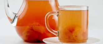
Eating is prohibited. The digestion process can cause more severe pain, nausea and vomiting. And the diet will reduce the production of enzymes that increase the inflammatory response and pain. You need to fast for 3 days. You can drink clean water without gases.
It is imperative to call a doctor for an examination, even if the patient is not exactly sure that this is an attack of acute pancreatitis. As we already know, this pathology can subside and then rapidly recur. At this time, you can take a painkiller to reduce discomfort.
Diagnosis of the disease at the private medical clinic “Medunion”
Diagnosing this disease is not difficult, since the first signs speak for themselves. However, in order to prescribe adequate treatment, it is necessary to determine the form of the disease. To do this, the doctor performs laparoscopy - a method that allows you to examine the abdominal cavity from the inside using a special instrument.
If acute pancreatitis is suspected, laboratory tests are performed:
- General blood analysis
- Blood chemistry
- Analysis of urine
- Stool analysis
- Ultrasound, MRI or radiography of the abdominal organs
- Computed tomography according to indications
In the chronic form, the same studies are carried out, but it is better to take tests during the period of exacerbation of the disease.
Surgical intervention
When the patient's body does not respond to conservative therapy, more effective treatment methods are required. Operations are performed in severe cases when mechanical restoration of the transport of pancreatic enzymes to the intestine is required. A surgical operation is performed to ensure constant drainage of secretions and resection of destroyed tissue. Foci of necrosis are removed and the ducts are expanded.
Surgical intervention is allowed only after a hardware examination and an accurate diagnosis has been established. Surgical intervention tactics are developed taking into account the assessment of the level of gland necrosis. It is important to consider possible risks:
- the occurrence of complications after surgery;
- possibility of secondary infection.
Punctures, laparoscopy, and laparotomy operations are performed. When fluid accumulates in the abdominal cavity, drainage is practiced.
Information verified by an expert
Vasiliev Vyacheslav Alekseevich
Chief physician. Urologist-andrologist, family doctor, surgeon, ultrasound specialist. Candidate of Medical Sciences Work experience - 30 years
Treatment of acute pancreatitis
If acute pancreatitis is detected, the patient should be hospitalized immediately. Treatment should take place in a hospital setting, as this condition is very dangerous.
To relieve pain, antispasmodics are taken; in difficult cases, the contents of the stomach are pumped out to relieve the load on the gland.
In case of exacerbation of pancreatitis, patients require hospitalization with daily monitoring of blood parameters, water balance, leukocyte count, and enzyme levels in the blood serum during the first week. In the first 1–3 days, fasting and taking alkaline solutions every 2 hours are recommended.
During an exacerbation of chronic pancreatitis, the patient is shown therapy similar to the acute process. The patient must follow a diet throughout his life and take drugs from the group of antispasmodics and drugs that normalize the secretory function of the organ.
The most important thing in the chronic form of the disease is to maintain a diet that involves excluding fatty and fried foods from the diet. At the slightest violation of the regimen, the patient may experience discomfort and nausea. For intense pain, the doctor prescribes antispasmodics. Antisecretory therapy can be used for a short course.
Description of the pathology
The pancreas has two main functions:
- the production of digestive enzymes, which enter the duodenum through the ducts, where they are activated and participate in the breakdown of proteins and fats;
- secretion of hormones - insulin and glucagon, which are released into the blood and regulate glucose levels.
If the patency of the ducts is disrupted for any reason, the pressure of pancreatic juice in them increases. Damage to gland cells occurs. In acute pancreatitis, the mechanism of “self-digestion” of tissues is triggered. The result of chronic inflammation is a constant deficiency of digestive enzymes, replacement of glandular cells with connective tissue, with a subsequent weakening of not only exocrine function, but also the production of hormones.
Diet for pancreatitis
For any form of the disease, the patient is prescribed a strict diet “Table No. 5p”, according to which it is forbidden to eat spicy and fried foods. All dishes are steamed, boiled or baked. Alcohol and smoking are also prohibited.
It is also necessary to limit salt intake and eat small portions 6 times a day. Dishes should always be served warm. It is necessary to exclude all products with a high content of extractives or essential oils (fish, meat broths, cocoa, coffee, etc.), fresh berries, vegetables, herbs, fruits, sour juices, carbonated drinks, marinades.
Protocol for diagnosis and monitoring of peripancreatic infiltrate in phase IB.
Peripancreatic infiltrate (PI) and resorptive fever are signs of severe or moderate pancreatitis.
Define:
- indicators of systemic inflammatory response syndrome: leukocytosis with a shift to the left, lymphopenia, increased ESR, increased fibrinogen, CRP, etc.;
- Ultrasound signs of PI (continued enlargement of the gland, blurred contours and the appearance of fluid in the tissue).
To monitor PI it is recommended:
- dynamic study of clinical and laboratory parameters;
- at least 2 ultrasound scans in the 2nd week of the disease;
- at the end of the 2nd week, CT scan of the pancreas.
Possible outcomes by the end of phase IB:
- resorption for up to 4 weeks;
- aseptic sequestration of pancreatic necrosis with the formation of a pseudocyst: stabilization of the size of the PI with normalization of health and subsidence of SIRS against the background of hyperamylasemia;
- septic sequestration (purulent complications).