Often patients who have already been treated unsuccessfully in various medical institutions come to me on the recommendation of friends. It can be seen that the examination is carried out in such a way that the picture of the disease is not identified objectively. It is necessary to send for additional examination, after which it is only possible to understand what is happening to the body, and only then can an optimal treatment plan be developed. This article is about how to check your stomach, what tests and diagnostic procedures you need to be mentally and financially prepared for if you want to take care of yourself seriously.
Capsule endoscopy
When using this technique, the study is carried out in a special capsule equipped with cameras and other sensors, which the patient must swallow. It scans the digestive tube as it naturally progresses and transmits data via wireless communication systems. This technique is one of the options for examining the intestines for diseases without the use of colonoscopy.
Main advantages:
- Simplicity and painlessness.
- Extensive data on the state of the digestive tract.
- High quality images and a lot of additional data.
Capsule endoscopy also has disadvantages, the main one being the inability to take a biopsy and perform other manipulations. The movement of the capsule cannot be controlled, so the suspicious area cannot be examined in more detail.
X-ray examination
X-ray examination of the stomach received this name due to the use of an X-ray machine. The patient is injected with contrast - a special suspension that absorbs X-rays. The specialist then takes “pictures” showing the condition of the stomach - its position in relation to other organs and various abnormalities (for example, tumors or ulcers). X-ray examination is performed less frequently than gastroscopy, since the latter is much more effective and accurate.
An X-ray of the stomach is performed if diseases of the upper gastrointestinal tract are suspected. It is used to diagnose chronic diseases as well as medical emergencies such as gastric or duodenal perforation. Preparing for an x-ray of the stomach is quite simple. The patient comes for examination on an empty stomach, having not eaten since the evening before the hospital visit. Smoking is prohibited on the day of the examination. X-rays are prohibited for pregnant women due to the risk to the fetus. The entire examination process is painless and non-invasive. After drinking the contrast, the patient turns around its axis in a lying position so that the contrast spreads well along the walls of the digestive system. In order for photographs to be taken correctly, the subject must remove outer clothing and assume a vertical position. The examination process itself takes only a few minutes. After this, the patient can safely go home.
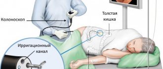
Irrigoscopy
The procedure consists of injecting a contrast agent into the large intestine (barium enema) and taking x-rays in several projections.
This allows you to accurately localize pathological areas of the intestine, detect peristalsis disorders, diverticulosis and other diseases. Irrigoscopy is safe, painless and does not take much time.
The main disadvantage of irrigoscopy is its low information content. There is also a fairly high probability of receiving false data when part of the intestinal contents is mistaken for a tumor or other pathology. As a result, a colonoscopy will still be required to clarify the diagnosis.
Progress of the procedure
The duration of the breath test for Helicobacter pylori is no more than 15 minutes. The test itself is divided into 2 stages:
The patient takes a comfortable position and fixes a special tube in the oral cavity, thereby preventing contact of the tube with the tongue.
Next, the patient is asked to take a urea solution. After entering the gastrointestinal tract, gas is formed under the influence of the urea solution. Together with the exhaled air, using a special device, the gas level is assessed and compared with the initial values.
Magnetic resonance and computed tomography
Whether an MRI can be done instead of a colonoscopy depends on the intended diagnosis and other individual characteristics of the patient. This procedure has a number of advantages:
- Simple and fast procedure.
- There is no radiation exposure to the body and no discomfort.
- Quite high accuracy.
This type of diagnosis makes it possible to detect pathological changes in the intestinal wall that are 1 cm or more in size. The main disadvantage of the method is the inability to take material for biopsy and low resolution. Typically, MRI is used to clarify data obtained by other methods, for example, to accurately localize metastases in cancer.
CT scans also provide fairly detailed images. To increase the resolution of the procedure, complete bowel emptying and the introduction of a radiocontrast agent are required. Regular air is often used for this purpose.
The main disadvantage of computed tomography is its high radiation exposure. If the patient is not properly prepared, false results can be obtained. Therefore, CT is used relatively rarely in the diagnosis of colon diseases.
Intestinal diagnostics - examination methods.
Today, medicine has a wide range of diagnostic methods for conducting high-precision examination of a number of problems with the large intestine, anal canal and perineum, as well as ample opportunities for treating a number of diseases. The range of diagnostics includes instrumental and non-instrumental rectal examination methods, general tests, as well as a number of studies.
Conducting a general examination by a doctor
The coloproctologist begins the examination by identifying complaints, conducting a general examination and collecting information. Examination is important and helps to establish a diagnosis. With the help of an initial examination, you can notice a number of problems: bloating of the entire abdomen, some of its parts, peristalsis, the presence of a tumor, protrusion of the abdominal wall forward, etc.
Methods for examining the intestines?
Tapping or percussion allows you to determine the presence of accumulated fluid in the abdominal cavity, the presence of gases in it, flatulence, and also calculate the approximate limits of a swollen intestinal loop.
Listening to noises or auscultation allows you to hear and evaluate peristalsis, determine signs of problems with patency, as well as a number of other problems.
Palpation or so-called palpation of the abdomen is considered one of the most useful primary methods of determining:
- excessive tension in the abdominal muscles;
- location, size, type, mobility of the tumor, as well as contraction of spastic type loops, ascites and many other problems and pathologies;
When the examination of the perineum is completed, the doctor begins a digital examination of the rectum, and then anoscopy or sigmoidoscopy, depending on the specific situation. During the examination of the perianal area, reflexes are checked in the form of contraction of the sphincter muscles with slight irritation of the skin around it.
Examination of the rectum using a finger
It is carried out if there are complaints of discomfort in the abdominal area and problems with the pelvic organs and gastrointestinal tract. The study is carried out using the index finger. Wearing a glove, the specialist carefully conducts the inspection. To relax the muscles, the patient needs to push a little.
Sigmoidoscopy (rectoscopy) method
This method involves inserting a sigmoidoscope through the anus to a depth of twenty to thirty centimeters. Thanks to the magnifying optics that are used during the procedure, even the smallest disorders of the mucous membrane can be examined.
If there are suspicions that the neoplasms are of poor quality, the doctor may select material for tissue analysis. Thanks to a biopsy, you can get an accurate answer about the type of tumor the patient has.
If you are over forty years old, then it is best to perform rectoscopy at least once a year - this will prevent the development of low-quality intestinal tumors.
The procedure is virtually painless, but in some cases local anesthesia may be used. In order to properly perform sigmoidoscopy, the day before the procedure, you should thoroughly cleanse the large intestine of feces. This can be done with bowel relaxants or an enema.
Patients should follow a special diet, drink only tea in the evening, and conduct the study itself without taking any food beforehand. Proper preparation for this type of procedure allows you to get a guarantee of an accurate result, so you should not neglect it.
Anoscopy
Anoscopy is a method of examining the intestines by examining it from the inside using a special anoscope device, which is inserted through the anus up to 14 centimeters in depth. Anoscopy is performed if there are complaints of pain in the anal area, bleeding, stool disturbances, as well as suspicion of diseases of the rectum. Anoscopy complements the usual rectal examination. Preparation for anoscopy is no different from preparation for sigmoidoscopy, and is no less important and responsible.
Biopsy
Tissues to study the causes of a tumor or polyp are taken using special endoscopic instruments. There are two types of biopsy: “Targeted” or “Blind”. The first is carried out when there is a suspicion of a tumor or Crohn's disease, and the second when there is a high risk of diffuse intestinal damage.
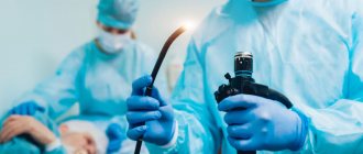
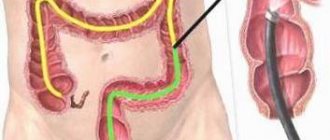
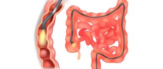
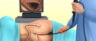
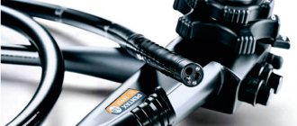
Colonoscopy
Colonoscopy is a method of endoscopic diagnosis of diseases of the large intestine, which involves the use of a colonoscope.
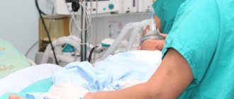
Often, no anesthesia is required for a colonoscopy examination. Patients with severe pain in the anus require local anesthesia, and if there is significant tissue destruction, general anesthesia is also used.
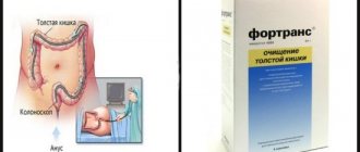
Colonoscopy is a fairly informative method of examination only when the patient strictly follows the doctor’s instructions regarding bowel cleansing. Vegetables, fruits, herbs, mushrooms, potatoes and beans should be completely avoided.
The day before a colonoscopy, you can only eat liquid and easily digestible foods. Enema is carried out until the water comes out in its pure form. Don't underestimate the importance of preparing for bowel testing procedures. Proper preparation guarantees an accurate result.
During a colonoscopy examination, patients feel fullness of the intestines with gases and the urge to defecate. After the study is completed, the air introduced into the intestine is sucked out through a special channel of the endoscope. The patient may experience short-term minor discomfort. If the feeling of fullness of the intestines with gases has not passed, take a dozen tablets of activated carbon, washed down with boiled water at room temperature.
Laboratory research
A general stool test can detect blood, bile pigments and parasites. Such an analysis makes it possible to detect the inflammatory process, invasion, composition and condition of the flora, beginning ulcers, etc. Testing stool for occult blood can help identify bleeding in parts above the colon.
As a result of the analysis, it is possible to diagnose the presence of gastrointestinal tract veins enlarged by varicose veins, peptic ulcers, dysentery and many similar pathological conditions. It is worth noting that early diagnosis of intestinal diseases, just like any other, significantly increases the chances of a speedy recovery and minimizes the consequences of the illness.
Diagnosis of intestinal cancer
Full manifestations of colon cancer do not appear immediately: patients often complain of discomfort in the abdominal area, severe symptoms of intestinal upset, constant constipation or diarrhea, significant and rapid loss of body weight, as well as very high fatigue.
In the case of an actively developing disease, there may be a deterioration in intestinal patency, and if the tumor has grown greatly, the patient may feel it in the abdomen. These symptoms are often accompanied by a significant decrease in appetite, weight loss, disability and dehydration, however, they are not obligatory.
Oncological diseases of the colon can be diagnosed by a whole range of studies and tests listed earlier. One of the most highly informative methods for establishing a predisposition to oncology can be embryonic cancer antigen, which is considered an indicator of the presence of tumor formation processes in the intestine.
CA 19.9 is a tumor marker for malignant diseases of the gastrointestinal tract and pelvic organs.
Its highest sensitivity can be observed in the case of pancreatic cancer, low-quality neoplasm in the biliary tract, as well as in the liver. In the case of gastrointestinal and intestinal oncology, the content of tumor markers increases significantly. A slight increase in the levels of this tumor marker can be observed in such unpleasant diseases as cholecystitis, as well as acute pancreatitis.
The intestine is a very important organ of the human body. The health of not only the gastrointestinal tract, but also the entire human body depends on its condition, stable and uninterrupted operation, as well as on the state of the microflora. Therefore, if you begin to notice various symptoms of a pathological nature, be it constipation, diarrhea, excessive formation of gases in the intestines, bloating, skin rashes, high fatigue, deterioration in the quality of sleep, pain in the abdominal area and some others - this is a reason to pay attention on your intestinal health. In this case, you may be recommended to study a comprehensive gastrointestinal tract plan.
Intestinal diagnostics at the Miracle Doctor clinic
Do not be afraid of various methods of medical examination of the intestines. Now you have information regarding different diagnostic methods, their implementation and the necessary preparation. Reliable and high-quality equipment, as well as the tact and patience of our employees will help to carry out the fastest, most painless and effective examination. All that is required from the patient is to carefully and accurately follow all the doctor’s advice and recommendations. Together we can restore your gut health!
Author
Zagirov Fizuli Abumuslimovich
phlebologist, surgeon, proctologist
18 years of experience
+7
Stool tests
How to check in another way is a question that is relevant for most people over 40 years old. Regularly performing this endoscopic procedure is associated with quite a lot of inconvenience for the patient and requires careful preparation and diet. An alternative may be DNA testing of stool. They are aimed at identifying specific DNA molecules belonging to tumor cells.
Stool can also be analyzed for the presence of occult blood and a fecal immunochemical test can be performed. All these laboratory techniques make it possible to accurately detect cancer, even at the earliest stages of development.
Important
Please note that any intestinal examination cannot serve as a complete replacement for colonoscopy. In most cases, they are used as screening, that is, to detect signs of pathology in the early stages of development.
Serological study
To carry out a serological test, it is enough to donate blood. The collected sample is analyzed for the presence of antibodies or antigens. In the case of the stomach, specialists look for antibodies in the blood against the bacterium Helicobacter pylori, which causes numerous gastric diseases. It also provokes stomach ulcers. In very advanced cases, lack of treatment can lead to cancer.
There is no need to prepare for a serological test. There are also no contraindications to it. If treatment for the gastrointestinal tract has already been completed, and the text shows a positive result, do not be alarmed. Antibodies can remain in the body for a long time. That is why serological testing of the stomach cannot be the only method for diagnosing gastrointestinal diseases.