Diagnosis of diseases of the digestive tract often requires the use of instrumental methods. Carrying out some research methods can be accompanied by significant discomfort and even pain.
Gastroscopy under anesthesia is an excellent way to make an informative examination of the gastrointestinal tract painless for the patient. This procedure has contraindications that should be discussed with your doctor.
What is gastroscopy?
Gastroscopy is performed both under anesthesia and under local anesthesia
Gastroscopy is also called esophagogastroduodenoscopy. This is a diagnostic procedure during which the organs of the digestive system are examined.
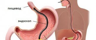
Directly during gastroscopy, organs such as the esophagus, stomach and duodenum are examined.
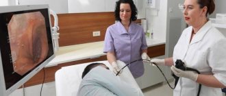
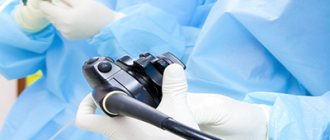
To conduct the study, the diagnostician inserts a small flexible endoscope into the patient’s mouth and sequentially moves it along the digestive tract.
Gastroscopy is used for both diagnostic and therapeutic procedures. During the examination, you can examine the digestive organs, collect material for laboratory testing, and also stop internal bleeding or remove an object stuck in the digestive tract.
New endoscopes have video chips that allow the doctor to view the image on a monitor. In different countries there are separate nuances of gastroscopy. In the United States, the procedure is usually performed using general anesthesia, since insertion of the tube is accompanied by quite severe discomfort.
In Russia, the procedure is more often performed under local anesthesia, although there is often the option of choosing general anesthesia. Gastroscopy is usually performed in a specialized endoscopy unit in a hospital or in an outpatient department. The procedure is also often performed in hospital emergency departments.
Our fears depend on us
This procedure cannot be called pleasant, especially remembering the methods used ten to twenty years ago: nausea, gag reflex, and drooling were almost impossible to avoid. Now the situation has changed for the better: very thin endoscopes have appeared, which in our clinic are inserted through the nose - nasal gastroscopy. Is it worth using anesthesia, because it “does not hurt”, “is not scary”, “is calm”? It’s not for nothing that all the epithets are put in quotation marks, because they are most often caused by our attitude towards examination - what we inspired ourselves to do is what we got. To paraphrase the famous phrase of Faina Ranevskaya: “all fears and illnesses come from nervous experiences and our attitude towards them.” If you take the examination calmly, and do not conduct surveys of your friends about “was it scary, did you feel very sick,” then the procedure will turn out to be the most ordinary, if the doctor’s hands turn out to be skillful, and the specialist is experienced - like our doctors.
Indications for gastroscopy
Gastroscopy itself is an unpleasant procedure.
In most cases, gastroscopy is prescribed to diagnose diseases of the gastrointestinal tract.
An endoscope equipped with a cold light source and a video camera allows you to quickly “walk” through the entire upper part of the digestive tract and detect any possible abnormalities.
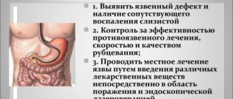
Possible deviations include ulcerative lesions, erosions, inflammatory processes, wounds and other defects of organ walls. Also, special tools allow you to collect tissue for further laboratory study.
Main indications for the procedure:
- Severe chronic heartburn.
- Vomiting blood.
- The appearance of black tarry stools.
- Pain in the upper abdomen.
- Anemia without obvious clinical reasons.
- Constant feeling of nausea.
- Unexplained weight loss.
- Feeling of fullness in the stomach even after eating a small amount of food.
- Frequent bloating.
- Pain or discomfort when swallowing.
The procedure is also prescribed to confirm a particular diagnosis. The following diseases can be diagnosed using gastroscopy:
- Gastroesophageal reflux disease (GERD).
- Varicose veins of the esophagus.
- Ulcerative and inflammatory diseases of the stomach and duodenum.
- Malignant and benign neoplasms.
- Hiatal hernia.
- Mechanical damage to the walls of the digestive organs.
- Celiac disease.
- Crohn's disease.
- Infection of the upper digestive tract.
Endoscopic examination does not allow making a final diagnosis in all of the above cases, but it makes it possible to see an objective picture of the condition of the abdominal organs.
Possible complications
- Pain and cramping in the stomach and esophagus caused by the insertion of the tube. The device used in the “Open Clinic” has a very thin tube, so it practically does not cause irritation of the mucous membrane and spasms. However, if discomfort is severe, you can take an antispasmodic drug.
- Pain in the gastric area may occur during examinations involving the removal of polyps, tissue sampling, and other types of procedures. In this case, discomfort can be relieved by taking painkillers.
Unpleasant phenomena go away quite quickly on their own and do not require treatment.
Examination of the stomach, FGDS and endoscopy, which are carried out during sleep, are convenient for the patient and the doctor. These diagnostic methods do not cause discomfort or complications and allow the doctor to carefully examine the organs in a relaxed atmosphere.
Preparing for the study
Before carrying out the procedure, it is necessary to inform the diagnostician or attending physician about conditions such as pregnancy, diseases of the cardiovascular system, respiratory diseases and allergic reactions to certain medications.
You should also tell your doctor about the following conditions:
- Previous surgery on the esophagus, stomach or intestines.
- Endocarditis.
- Presence of an artificial heart valve.
- Rheumatic carditis.
If the patient has one of these conditions, antibiotics may be required before gastroscopy.
Eight hours before the procedure you should stop eating. Immediately before the test you should also refrain from drinking any liquid. The optimal solution can be considered a small dietary dinner before six to eight o'clock in the evening on the eve of gastroscopy.
This is necessary to ensure that the digestive tract is empty and the patient does not have pronounced gag reflexes.
Before the procedure, if necessary, you can take medications to treat high blood pressure, heart disease, or thyroid disease. If the patient is diagnosed with diabetes mellitus, it is necessary to adjust the insulin dosage on the day of the test.
Gastroscopy under anesthesia requires the presence of a person who can help the patient get home. After such a procedure, the patient will experience drowsiness, dizziness and poor attention. In this condition, it is prohibited to drive a vehicle for eight hours after the procedure.
Gastroscopy in a dream
Gastroscopy while sleeping allows you to carry out the procedure as comfortably as possible. This diagnostic option is a priority in countries with a high level of medical development (Israel, Germany, USA).
To put a patient into medicated sleep, there must be no cardiac contraindications, so the diagnostic standard includes electrocardiography. The anesthesiologist examines the patient, after which he administers a drug intravenously that blocks nerve impulses and relieves psycho-emotional stress. The person falls asleep, and the doctor begins to perform an endoscopy. Awakening occurs almost immediately after the end of the procedure. It is recommended to get behind the wheel 5-6 hours after fully waking up.
The duration of the study is 10-20 minutes.
Features of gastroscopy under anesthesia
Gastroscopy under anesthesia
Sedation during gastroscopy can vary from minimal sedation to general anesthesia. Anti-anxiety medications are needed to relieve anxiety, stress, discomfort and pain.
The procedure itself can be successfully performed without any drug preparation for the patient, but it is preferable to use some form of anesthesia.
In a conscious sedative state, patients can perform purposeful actions and respond to auditory and visual stimuli. In this state, patients maintain certain vascular and respiratory parameters.
What is sedation and why is it needed?
In order for a probe with a built-in camera to examine internal organs to move freely inside the organ and “examine” in detail all hard-to-reach areas, the patient needs to relax. It is extremely difficult to do this when a foreign object is introduced inside: vomiting, nausea begin, and discomfort is felt in the intestines. To reduce this sensitivity, sedatives are used. They help the patient to relax and the doctor to do his job quickly and efficiently.
Sedation is used during gastroscopy or esophagogastroduodenoscopy - a visual examination of the esophagus, stomach and duodenum.
Sedation is used:
- to take a piece of tissue for research - biopsy;
- removal of polyps;
- injections and irrigation of mucous membranes;
- stopping internal bleeding;
- expansion of the lumen of the esophagus;
- insertion of a feeding tube through a tube in the stomach.
Differences between gastroscopy under local anesthesia and light sedation
If local anesthesia is chosen for anesthesia, the patient is given an anesthetic solution, and the mouth and throat are treated with an anesthetic spray. The person remains fully conscious and understands everything that is happening. However, at the same time, he slightly feels the impact of the endoscope in the esophagus and may experience unpleasant feelings.
Light sedation is the pain management option preferred in many developed countries. The technique occupies an intermediate place between general and local anesthesia. The patient is injected with anesthetics to relax, calm and immerse himself in a feeling of drowsiness. On the one hand, he feels much less than with local exposure, but on the other hand, he still remains an active participant in the process, which greatly simplifies medical manipulations.
The choice of pain relief option is up to the doctor, but the most extreme option - sleep - is chosen extremely rarely, and not at the request of the patient, but solely for medical reasons.
Traditional approach
Gastroscopy is a traditional way to assess the condition of the gastrointestinal tract. The method allows you to analyze all changes in the esophagus and stomach, identify inflammatory processes, tumors, detect precancerous conditions at an early stage, and also take samples of the affected areas for biopsy. The procedure is carried out under local anesthesia: before this, the patient is tested for allergies, which are possible to the selected anesthetic.
The effect of the medication ends a few hours after administration, when you can return to everyday activities. Some medications aimed at mild sedation put the patient into a state of lethargy, so doctors recommend stopping driving for at least a day.
General anesthesia is prescribed extremely rarely. In this condition, it is difficult for the doctor to assess the patient’s condition and understand whether the medical actions are correct. It is always more difficult for a patient to survive his new state after such anesthesia than if he were a full participant in the manipulation. Therefore, doctors, as a last resort, if local anesthesia is not possible, prefer to put the person into a state of light sedation or drowsiness instead of deep sleep.
Video gastroscopy
In IMMA clinics, endoscopic examination of the stomach is performed using the digital video endoscopic system PENTAX EPK-1000 (Japan).
For video gastroscopy, the PENTAX EG-2490K device is used. It has an ultra-thin tube, so the patient experiences minimal discomfort. Another advantage of using a video gastroscope is that the doctor sees the endoscopic image on the monitor already during the procedure. On the distal part of the device there is a CCD matrix that converts the optical image into electronic signals and transmits them to the processor for processing. After this, the resulting image is displayed on the screen. Diagnostics takes a short time with maximum information content. In our clinics, we pay great attention to patient safety. To clean the device after video gastroscopy, we use the BANDEQ CYW-100N sterilization machine (South Korea). It provides a full cycle of leak testing, disinfection, washing, sterilization and drying of gastroscopic equipment. This is the total destruction of infections and viruses!
Preparation
In preparation for gastroscopy, it is necessary to stop (2-3 days in advance) from drinking alcoholic beverages and foods that irritate the mucous membrane of the digestive organs. Diagnosis is carried out on an empty stomach. Dinner on the eve of the examination should be easily digestible. If the procedure is not performed in the morning, it is recommended not to eat a few hours before it. You are allowed to drink some still water 2–3 hours before. Your doctor will tell you about all the nuances of preparing for gastroscopy during your consultation.
More detailed information about preparing for the procedure...
Local anesthesia
The simplest and most common way to make an unpleasant diagnosis a little easier is local anesthesia. Typically, a lidocaine spray is used for this. The medicine is applied to the root of the tongue, which reduces its sensitivity and completely blocks the gag reflex. Thus, the doctor easily inserts a flexible hose with a camera into the esophagus. The action of the anesthetic gives the doctor enough time to examine the mucous membrane of the stomach, esophagus, and, if necessary, parts of the duodenum.
Since local anesthesia has virtually no contraindications, it is used very widely. Usually the price of gastroscopy in clinics is indicated taking into account this type of anesthesia.