08.05.2019
Quantity
Stomach ulcer
– a chronic polyetiological pathology that occurs with the formation of ulcerative lesions in the stomach, a tendency to progression and the formation of complications. The main clinical signs of peptic ulcer disease include pain in the stomach and dyspeptic symptoms. The diagnostic standard is an endoscopic examination with a biopsy of pathological areas, radiography of the stomach, and detection of H. pylori. Treatment is complex: diet and physiotherapy, eradication of Helicobacter pylori infection, surgical correction of complications of the disease.
General information
Gastric ulcer (GUD) is a cyclically relapsing chronic disease, the characteristic feature of which is ulceration of the stomach wall. PUD is the most common pathology of the gastrointestinal tract: according to various sources, from 5 to 15% of the world's population suffer from this disease, and among urban residents the pathology is five times more common. Many specialists in the field of gastroenterology combine the concepts of gastric ulcer and duodenal ulcer, which is not entirely correct - ulcerations in the duodenum are diagnosed 10-15 times more often than ulcers in the stomach. However, PU requires careful study and development of modern diagnostic and treatment methods, since this disease can lead to the development of fatal complications.
About 80% of cases of primary detection of gastric ulcers occur in working age (up to 40 years). In children and adolescents, gastric ulcers are diagnosed extremely rarely. Among the adult population, there is a predominance of men (women suffer from peptic ulcers 3-10 times less often); But in old age, sex differences in incidence are smoothed out. In women, the disease is milder, in most cases asymptomatic, and is rarely complicated by bleeding and perforation.
Gastric ulcer ranks second among the causes of disability in the population (after cardiovascular pathology). Despite the long period of study of this nosology (more than a century), therapeutic methods of influence that can stop the progression of the disease and completely cure the patient have not yet been found. The incidence of gastrointestinal ulcers throughout the world is continuously growing, requiring the attention of therapists, gastroenterologists, and surgeons.
Causes
Gastric and duodenal ulcers develop in conditions of an imbalance of factors of aggression and protection of the mucous membrane due to improper neurohumoral regulation, disorders of blood supply and nutrition of organ tissues, drug or toxic damage.
The following factors may contribute to this:
- smoking;
- frequent consumption of alcoholic and energy drinks, coffee;
- chronic overwork, acute or prolonged stress;
- serious illnesses and injuries;
- unhealthy diet with a lot of refined foods, fast food, spicy, sour, salty or pickled, excessively cold or hot foods;
- lack of proper sleep;
- taking drugs based on digitalis, from the group of NSAIDs, steroid hormones, sulfonamides, potassium chloride, anticoagulants, nitrofurans;
- genetic predisposition;
- presence of H.pilori in the stomach;
- hyperparathyroidism;
- hormonal age-related changes;
- chronic gastritis;
- diabetes.
Classification
Until today, scientists and clinicians around the world have not been able to reach agreement on the classification of gastric ulcers. Domestic experts systematize this pathology according to the following criteria:
- causative factor
– ulcers associated or not associated with H. pylori, symptomatic ulcers; - localization
- ulcer of the cardia, antrum or body of the stomach, pylorus; greater or lesser curvature, anterior, posterior wall of the stomach; - number of defects
- single ulcer or multiple ulcers; - size of the defect
– small ulcer (up to 5 mm), medium (up to 20 mm), large (up to 30 mm), giant (more than 30 mm); - stage of the disease
- exacerbation, remission, scarring (red or white scar), cicatricial deformation of the stomach; - course of the disease
- acute (the diagnosis of gastric ulcer is established for the first time), chronic (periodic exacerbations and remissions are noted); - complications
- gastric bleeding, perforated gastric ulcer, penetration, cicatricial ulcerative gastric stenosis.
Predisposing factors of the disease
Indirectly, the development of the pathological process can be facilitated by:
- stressful situations, injuries, hormonal imbalance. These processes themselves do not have a negative effect on the mucous membrane. However, they provoke the release of hydrochloric acid, as a result of which the acidity of gastric juice increases;
- inflammation of the gastric mucosa. This condition precedes peptic ulcer disease. As a result of the inflammatory process, the integrity of the walls of the stomach is disrupted, which subsequently contributes to the appearance of ulcerative defects;
- eating disorder. The abundance of spicy, sour, salty foods in the daily diet negatively affects the condition of the mucous membrane. As a result of drinking alcoholic beverages and strong coffee, the situation worsens significantly;
- frequent respiratory diseases. As a rule, exacerbation of peptic ulcer disease occurs during the off-season - autumn and spring. Frequent colds at this time help reduce the body's defenses. The situation is also worsened by taking anti-inflammatory drugs.
Causes and pathogenesis of gastric ulcer
The main etiological factor in the formation of gastric ulcer is infection with H. pylori - more than 80% of patients have positive tests for Helicobacter pylori infection. In 40% of patients with gastric ulcer infected with the Helicobacter bacterium, anamnestic data indicate a family predisposition to this disease. The second most important cause of gastric ulcer formation is considered to be the use of non-steroidal anti-inflammatory drugs. Rarer etiological factors of this pathology include Zollinger-Ellison syndrome, HIV infection, connective tissue diseases, liver cirrhosis, heart and lung disease, kidney damage, exposure to stress factors that lead to the formation of symptomatic ulcers.
The main significance for the formation of gastric ulcer is an imbalance between the protective mechanisms of the mucous membrane and the influence of aggressive endogenous factors (concentrated hydrochloric acid, pepsin, bile acids) against the background of a disorder of the evacuation function of the gastrointestinal tract (gastric inactivity, duodenogastric reflux, etc.) . Inhibition of protection and slower recovery of the mucous membrane is possible against the background of atrophic gastritis, with chronic Helicobacter pylori infection, ischemia of stomach tissue against the background of collagenosis, long-term use of NSAIDs (the synthesis of prostaglandins slows down, which leads to a decrease in mucus production).
The morphological picture of gastric ulcer undergoes a number of changes. The primary substrate for the occurrence of ulcers is erosion - superficial damage to the gastric epithelium, which forms against the background of necrosis of the mucous membrane. Erosions are usually detected on the lesser curvature and in the pylorus of the stomach; these defects are rarely isolated. The size of erosions can range from 2 millimeters to several centimeters. Visually, erosion is a mucosal defect that does not differ in appearance from the surrounding tissues, the bottom of which is covered with fibrin. Complete epithelization of erosion with a favorable course of erosive gastritis occurs within 3 days without the formation of scar tissue. If the outcome is unfavorable, erosions transform into acute gastric ulcers.
An acute ulcer is formed when the pathological process spreads deep into the mucous membrane (beyond its muscular plate). The ulcers are usually single, take on a rounded shape, and look like a pyramid when cut. In appearance, the edges of the ulcer also do not differ from the surrounding tissues; the bottom is covered with fibrin deposits. Black coloration of the bottom of the ulcer is possible when the vessel is damaged and hematin is formed (a chemical substance formed during the oxidation of hemoglobin from destroyed red blood cells). A favorable outcome of an acute ulcer consists of scarring within two weeks; an unfavorable outcome is marked by the transition of the process to a chronic form.
The progression and intensification of inflammatory processes in the area of the ulcer leads to increased formation of scar tissue. Because of this, the bottom and edges of a chronic ulcer become dense and differ in color from the surrounding healthy tissue. A chronic ulcer tends to enlarge and deepen during an exacerbation; during remission it decreases in size.
Peptic ulcer: modern approaches to diagnosis and therapy
In 1586, Marcellus Donatus of Mantua, during an autopsy, first described a gastric ulcer, and almost a century later, in 1688, Johannes von Marault described a duodenal ulcer [1]. However, despite the improvement of preventive, therapeutic and diagnostic methods, among diseases of the digestive system, peptic ulcer disease (PU) continues to be one of the most common reasons for patients seeking medical help, and today it is difficult to imagine a practicing doctor who would not be familiar with this pathology .
In the International Classification of Diseases, X Revision, the term Peptic Ulcer is absent, which reflects two modern approaches to understanding the essence of the disease: on the one hand, consideration of peptic ulcer as a heterogeneous group of diseases united by the presence of a chronic ulcerative defect in the gastroduodenal zone, and on the other hand, as a nosological unit (disease ) with some generalized view of pathogenesis, clinical manifestations and standardized therapy. Both of these approaches are reflected in the definition of the disease: “Peptic ulcer (synonyms: duodenal ulcer, gastric ulcer) is the formation of an ulcer in the stomach or duodenum, resulting from an imbalance between the protective factors of the mucous membrane and various factors that damage the mucous membrane, understood as “etiology.” "[2].
Among the etiological factors of ulcerogenesis of the gastroduodenal zone (.), infection with Helicobacter pylori and the use of non-steroidal anti-inflammatory drugs (NSAIDs) dominate. Actually, the bacterium itself causes relatively modest damage to the epithelium - flattening and sometimes disappearance of microvilli in places of contact with H. pylori, a decrease in the number and volume of secretory granules with a corresponding decrease in mucus secretion. However, the main thing in ulcerogenesis is changes in signaling systems caused by the infection. In response to the action of signaling molecules (primarily pro-inflammatory cytokines), neutrophilic leukocytes penetrate into the mucous membrane, which in turn destroy intercellular contacts with their enzymes and free oxygen radicals, disrupting the integrity of the mucous membrane and forming a surface defect. However, it is well known that such mucosal defects can heal very quickly, leaving not only consequences, but also no traces.
With superficial damage, emergency closure occurs due to migration of the epithelium from the edges, even without increased proliferation. However, persistent infection delays the healing of ulcers, being the main cause of relapses. H. pylori inhibits the first - emergency reaction of the mucous membrane to damage - migration of the epithelium, necessary for the speedy closure of the defect; stimulates apoptosis, thereby increasing cell death in the edges of ulcers and complicating healing, and also suppresses the synthesis of epidermal growth factor with blockade of receptors for it; causes disruption of microcirculation and tissue trophism.
The main mechanism for the development of gastric and duodenal ulcers associated with NSAID use is associated with blocking the synthesis of prostaglandins. A decrease in the synthesis of prostaglandins leads to a decrease in the synthesis of mucus and bicarbonates, which are the main protective barrier of the gastric mucosa from aggressive factors of gastric juice. In turn, a decrease in the synthesis of prostacyclin and nitric oxide adversely affects microcirculation and creates an additional risk of damage to the mucous membrane of the stomach and duodenum. Changing the balance of protective and aggressive gastric environments leads to the formation of ulcers and the development of complications: bleeding, perforation, penetration.
The level of prevalence of etiological factors of ulcer in the population also determines the current epidemiological trends of the disease. Thus, if back in the 70–80s of the last century it was generally accepted that every tenth person may develop ulcer in his life, now the prevalence of ulcer has decreased several times and is, for example, 2.5% in the USA [3]. At the same time, while the frequency of ulcer perforation remained at the same level, the frequency of ulcer bleeding increased significantly (due to gastric ulcers), which is caused by the increasing intake of NSAIDs and Aspirin (Fig.). However, H. pylori infection and NSAID use alone do not exhaust all etiological factors of ulcer. About 5% of ulcerative defects of the gastroduodenal zone in the eastern population and about 30% in the western population are not associated with H. pylori/NSAIDs [4].
Thousands of research papers and reviews have been devoted to the consideration of the causative factors of ulcer formation in the gastroduodenal zone, and only a cursory description of the features of ulcerogenesis would require a separate publication. In each clinical case, the attending physician should strive for the most complete elucidation of etiological factors, their influence or on increasing the factors of aggression (increased impact of the acid-peptic factor associated with an increase in the production of hydrochloric acid and pepsin; impaired motor-evacuation function of the stomach and duodenum: delay or acceleration of the evacuation of acidic contents from the stomach, duodenogastric reflux), or on the inhibition of protective factors (resistance of the mucous membrane to the action of aggressive factors; mucus formation; adequate production of bicarbonates; active regeneration of the surface epithelium of the mucous membrane; sufficient blood supply to the mucous membrane; normal content of prostaglandins in the wall of the mucous membrane ; immune defense).
It is important to note that many of these factors of aggression and defense are genetically determined, and the balance between them is maintained by the coordinated interaction of the neuroendocrine system, including the cerebral cortex, hypothalamus, peripheral endocrine glands and gastrointestinal hormones and polypeptides.
Today, a number of genetic factors have been identified, the presence of which contributes to the occurrence of peptic ulcer disease:
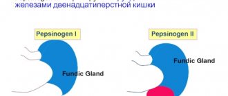
- hereditarily caused increase in the mass of parietal cells, their hypersensitivity to gastrin, increased formation of pepsinogen-1 and gastroduodenal motility disorder can lead to damage to the mucous membrane of the stomach and duodenum;
- congenital deficiency of mucus fucomucoproteins, insufficiency in the production of secreted IgA and prostaglandins reduce the resistance of the mucous membrane;
- blood group 0 (1), positive Rh factor, the presence of HLA antigens B5, B15, B35, etc. increase the likelihood of ulcer disease;
- polymorphism of the TNF-alpha cytokine gene (TNF-alpha promoter single nucleotide polymorphism). Carriers of TNF-alpha-1031C and 863A have a high risk of developing duodenal ulcer and gastric ulcer in the presence of H. pylori infection: if carriers had either 1031C or 863A alleles, then the risk of developing ulcer in the presence of H. pylori is increased by 2.46 times, and in the presence of both alleles, the risk of developing ulcer in the presence of H. pylori is increased by 6.06 times. The 863CC genotype is associated with a high risk of developing intestinal metaplasia in patients with gastric ulcer.
The diagnostic process of ulcer disease includes analysis of the clinical manifestations of the disease, collection of anamnesis, examination of the patient, general clinical laboratory tests, esophagogastroduodenoscopy with biopsy, diagnosis of H. pylori infection, and (if necessary) other tests. In the Russian population, where the infection rate of the adult population in some regions reaches 90%, it is necessary to ensure the absence of bacteria in a given clinical situation, critically evaluate both the diagnostic methods used, and assess the likelihood of a false negative result in a given clinical case. If, however, the absence of infection is proven, the doctor receives an additional incentive to search for other etiological factors of ulceration in the gastroduodenal zone.
Despite the apparent simplicity of the issue, the clinical symptoms of ulcer require special attention. Which of the doctors who have undergone internship is not familiar with the description of the classic stigmas of gastroduodenal ulcers? The traditional description of the pain syndrome is nighttime, hungry, late pain in the localization of the ulcer in the duodenum and early pain in the gastric localization. However, in recent decades, the symptoms and course of ulcerative disease have become polymorphic, masked by dyspeptic symptoms of an uncertain nature, and in a certain percentage of cases (about 10%) the disease has no clinical manifestations [6]. Gastroduodenal ulcers in most elderly patients occur with a mild clinical picture and often manifest with complications, the frequency of which increases from 31% at the age of 60–65 years to 76% at the age of 75–80 years. More than 60% of bleeding from ulcerative defects of the gastroduodenal zone occurs in people over 60 years of age, who more often suffer from chronic diseases and take NSAIDs/Aspirin more often than younger people.
Once a diagnosis of ulcer is made, a decision must be made about where to treat the patient. Compliance with treatment standards using highly effective and safe agents allows one to achieve similar treatment results both in outpatient and inpatient treatment [7, 8]. Indications for hospitalization are: newly diagnosed ulcers (exclusion of symptomatic ulcers) and/or deep ulcers; persistent and severe pain lasting more than 7 days; carrying out a differential diagnosis with a tumor process); large (more than 2 cm) and long-term (more than 4 weeks) non-scarring ulcers (the need for additional examination, individual selection of medicinal and non-medicinal treatments); weakened patients or ulcers due to severe concomitant diseases. If therapy during an exacerbation of the disease is carried out on an outpatient basis, the patient should limit physical activity and follow dietary and lifestyle recommendations.
What goals do we strive to achieve when treating patients with ulcer? After all, it is important not only to relieve symptoms (if any), but also to prevent complications and relapses of the disease, and improve the quality of life of patients.
Depending on the mechanism of action, the following groups of drugs used in the treatment of patients with ulcer are distinguished:
- antacids;
- antisecretory agents (anticholinergics, H2 blockers, proton pump inhibitors (PPIs));
- drugs with local protective action (cytoprotectors, reparants);
- drugs that affect neurohumoral regulation (psychotropic drugs, regulators of motor-evacuation function - prokinetics, gastrointestinal hormones);
- anti-Helicobacter therapy.
The first component of therapy is eradication when H. pylori is detected. The feasibility of eradicating H. pylori in patients with ulcer disease today is beyond doubt. Eradication of the bacterium leads to faster and better scarring of ulcerative defects, and also reduces the risk of relapse of the disease within a year from 70% to 4–5% and the likelihood of complications. These facts, obtained from studies that meet the requirements of evidence-based medicine, served as the basis for inclusion in the final document of both the 2000 and 2006 Maastricht Consensus as a strongly recommended indication for eradication therapy with the highest degree of scientific evidence, H. pylori-associated ulcer both in the acute stage and in remission, including complicated forms. The same agreement also established very stringent requirements for combination eradication therapy, capable of destroying the bacterium in controlled studies in at least 80% of cases and not causing forced withdrawal of therapy by the doctor due to side effects (acceptable in less than 5% of cases) or discontinuation the patient takes medications according to the regimen recommended by the doctor. According to consensus, therapy is carried out according to standard regimens in which PPIs are the basic drugs.
First line therapy (duration 10–14 days):
- PPI in a standard dose 2 times a day;
- clarithromycin 500 mg 2 times a day;
- amoxicillin 1000 mg 2 times a day.
- Second line therapy (duration 10–14 days):
- PPI in a standard dose 2 times a day;
- colloidal bismuth subcitrate (De-nol) 120 mg ´ 4 times;
- Tetracycline 500 mg ´ 4 times a day;
- Metronidazole 500 mg ´ 3 times a day.
When starting treatment for ulcer associated with H. pylori infection, control of infection eradication should be planned, and diagnostic tests for Helicobacter should be performed no earlier than 4–6 weeks from the end of taking the drugs.
The second component of therapy is suppression of the acid-peptic factor [9]. During an exacerbation of the disease, it is important to maintain a certain pH level, since healing of a mucosal defect in a duodenal ulcer occurs on average in 3–4 weeks, and in a stomach ulcer in 4–6 weeks, provided that it is possible to maintain a pH in the stomach > 3 less than 18 hours [10]. Compliance with this rule is impossible without the use of IPP. According to the chemical structure, PPIs belong to the class of substituted pyridine-methyl-sulfonyl-benzimidazoles, differing in the radicals in the pyridine and benzimidazole rings. Differences in chemical structure determine differences in pharmacokinetic properties. Are these differences clinically significant? Yes, they do. For example, pantoprazole (Controloc) causes the longest suppression of acid secretion compared to other PPIs, which is due to its specific binding to the 822-position cysteine, which is immersed in the transport domain of the gastric acid pump.
This is an important factor since restoration of acid production is entirely dependent on the self-renewal of proton pump proteins. The same pharmacokinetic features also determine the low level of drug interactions, which is especially important when carrying out preventive antisecretory therapy in individuals taking NSAIDs, Aspirin, and anticoagulants [11]. Thus, in recent years it has been found that long-term use of PPIs, with the exception of pantoprazole, is associated with a decrease in the effectiveness of clopidogrel and an increase in coronary mortality by 40% [12].
The third component of therapy is cytoprotection, which is achieved by prescribing agents that stimulate the synthesis of prostaglandins and bicarbonates, normalizing motility and microcirculation. The choice of drug (bismuth drugs, antacids, antispasmodics, prokinetics, etc.) is determined by the specific clinical situation. For example, in the absence of H. pylori infection, the basis of therapy is the choice of drugs that maximally suppress aggression factors with the obligatory prescription of cytoprotective agents [13].
For uncomplicated peptic ulcers of small and medium size, endoscopy monitoring with assessment of scarring of the ulcerative defect is performed after 14 days, and for larger ulcerative defects - after 21 days from the start of therapy.
If scarring of the ulcerative defect is not achieved, it is important to exclude the most common causes of failure (slow scarring of the ulcerative defect):
- persistence of H. pylori infection;
- inadequate cooperation between doctor and patient (low compliance);
- taking NSAIDs (including hidden ones);
- dense fibrosis, heavy smoking;
- inadequate inhibition of hydrochloric acid secretion;
- rare causes of ulcers.
If scarring of the ulcer has taken place, and the symptoms persist, it is necessary to reconsider the diagnostic strategy for the patient with an active search for other diseases (irritable bowel syndrome, pathology of the biliary tract, pancreas, etc.).
After achieving scarring of the ulcer and relief of symptoms, the next stage of patient supervision is making a decision on the need for maintenance therapy. Who needs it? We are talking about those patients who retain the etiological factors of ulcerogenesis of the gastroduodenal zone. These are people who are forced to continue taking NSAIDs/Aspirin, cytostatics, glucocorticosteroids, with Zollinger-Ellison syndrome, etc. Prevention is carried out by taking PPIs at half the dosage (for example, Controloc 20 mg).
Clinical observation of patients with an uncomplicated course of ulcerative disease and achieved eradication is carried out for 5 years with an annual endoscopic examination of the upper digestive tract and assessment of H. pylori infection.
A modern internist has impressive diagnostic and treatment capabilities and standards for managing a patient with ulcerative disease. However, their implementation is possible only with a meaningful analysis of the clinical picture and the nature of the course of the disease, an in-depth study of the causative factors of ulcerogenesis, and the appointment of treatment regimens with the maximum degree of evidence.
Literature
- Unge Peter. Helicobacter pylori treatment in the past and in the 21st century.” in Barry Marshall. Helicobacter Pioneers: Firsthand Accounts from the Scientists Who Discovered Helicobacters. 2002 Victoria, Australia: Blackwell Science Asia. pp. 203–213. ISBN 0–86793–035–7.
- Ferri's Clinical Advisor: Instant Diagnosis and Treatment, 2003 ed., Copyright © 2003 Mosby, Inc. www.mdconsult.co.
- Vakil Nimish Dyspepsia, Peptic Ulcer and H. pylori: A Remembrance of Things Past //Am J Gastroenterol. 2010; 105:572–574; doi: 10.1038/ajg.2009.709.
- Shiu Kum Lam. Differences in peptic ulcer between East and West Clinical Gastroenterology. Vol. 14, no. 1, pp. 41–52, 2000 doi:10.1053/bega.1999.0058.
- Weil J., Colin-Jones D., Langman M., Lawson D., Logan R., Murphy M., Rawlins M., Vessey M., Wainwright P. Prophylactic aspirin and risk of peptic ulcer bleeding // BMJ. 1995, Apr 1; 310(6983):827–830.
- Lee SW, Chang CS, Lee TY, Yeh HZ, Tung CF, Peng YC Risk factors and therapeutic response in Chinese patients with peptic ulcer disease // World J Gastroenterol. 2010; 16 (16): 2017–2022.
- Order of the Ministry of Health and Social Development of Russia No. 612 of September 17, 2007 “On approval of the standard of medical care for patients with stomach ulcers (when providing specialized care).”
- Order of the Ministry of Health and Social Development of Russia No. 611 of September 17, 2007 “On approval of the standard of medical care for patients with duodenal ulcers (when providing specialized care).”
- Karen van Rensburg. Acid suppressants and peptic ulcer disease // SAPJ. April2010, pp. 33–37, indd 33.
- Could ML, Enas N., Humphries TJ, Bassion S. Results of three placebo-controlled dose-response clinical trials in duodenal ulcer, gastric ulcer and gastroesophageal reflux disease (GERD) // Dig. Dis. Sci. 1998. Vol. 43. P. 993–1000.
- Thomson ABR, Sauve MD, Kassam N., Kamitakahara H. Safety of the long-term use of proton pump inhibitors // World J Gastroenterol. 2010; 16(19):2323–2330.
- Juurlink David N. A population-based study of the drug interaction between proton pump inhibitors and clopidogrel // CMAJ. 2009;180(7):713–718.
- Kenneth EL McColl, How I. Manage H. ylori Negative, NSAID/Aspirin-Negative Peptic Ulcers // Am J Gastroenterol. 2009; 104: 190–193; doi: 10.1038/ajg.2008.11.
M. A. Livzan, Doctor of Medical Sciences, Professor M. B. Kostenko
PDO GOU VPO OmSMA Roszdrav , Omsk
Contact information about the author for correspondence
peptic ulcer factors
Symptoms of stomach ulcer
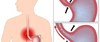
The clinical course of gastric ulcer is characterized by periods of remission and exacerbation. Exacerbation of peptic ulcer is characterized by the appearance and increase of pain in the epigastric region and under the xiphoid process of the sternum. With an ulcer of the body of the stomach, the pain is localized to the left of the center line of the body; in the presence of ulceration of the pyloric region - on the right. Pain may radiate to the left half of the chest, shoulder blade, lower back, and spine. Gastric ulcer is characterized by the onset of pain immediately after eating with increasing intensity within 30-60 minutes after eating; pylorus ulcer can lead to the development of night, hunger and late pain (3-4 hours after eating). The pain syndrome is relieved by applying a heating pad to the stomach area, taking antacids, antispasmodics, proton pump inhibitors, and H2-histamine receptor blockers.
In addition to the pain syndrome, ulcerative gastrointestinal tract is characterized by a coated tongue, bad breath, and dyspeptic symptoms - nausea, vomiting, heartburn, increased flatulence, and stool instability. Vomiting mainly occurs at the height of stomach pain and brings relief. Some patients tend to induce vomiting to improve their condition, which leads to progression of the disease and complications.
Atypical forms of gastric ulcer can manifest as pain in the right iliac region (appendicular type), in the heart (cardiac type), and in the lower back (radiculitis pain). In exceptional cases, pain syndrome with gastric ulcer may be completely absent, then the first sign of the disease is bleeding, perforation or cicatricial stenosis of the stomach, for which reason the patient seeks medical help.
Types of ulcers
The following types of pathology are distinguished:
- Perforated ulcer. This pathological condition is characterized by the appearance of a hole in the wall of the organ. As a result, food debris mixed with acid enters the abdominal cavity. As a result, an inflammatory process begins in the tissues.
- Stress ulcer. As a result of emotional shocks, the body begins to produce adrenocorticotropic hormone. It suppresses the secretion of natural mucus inside the stomach. As a result, the mucous membrane does not have time to recover, its blood supply deteriorates, and the acid-peptic balance is disrupted.
- Antral ulcer. Most often this disease is diagnosed in young people. The pathological process develops in the stomach, at the border with the duodenum. The disease is accompanied by a gag reflex, heartburn, and a constant feeling of heaviness in the stomach.
- Cardiac ulcer. The pathology is characterized by severe pain after eating. Patients note the appearance of a bitter taste in the mouth and frequent belching. A coating forms on the tongue, and pleurisy may develop. In an advanced stage, this pathology requires surgical treatment.
- Drug ulcer. The development of the pathological process in this case is facilitated by the use of corticosteroids, salicylates, etc. With prolonged treatment with these drugs, gastric and duodenal ulcers form. This pathology can cause bleeding.
- Chronic ulcer. The disease is characterized by a slow course and in most cases worsens during the off-season. At this time, severe heartburn, dyspepsia, severe pain, nausea and vomiting after eating, etc. are observed.
- Callous ulcer. Most often, the disease develops against the background of chronic pathology. If left untreated, callous ulcers of the stomach and duodenum can lead to cancer.
- Peptic ulcer. The pathological process develops in the lower part of the stomach. The disease appears as a result of infection. Pathology is indicated by severe frequent vomiting with blood clots, diarrhea, a sharp deterioration in appetite and other symptoms.
- Mirror ulcer. Pathology develops in the area of inflammation and, as a rule, affects the lower parts of the stomach. The ulcer involves several layers of the organ walls. The disease is accompanied by severe, prolonged pain.
- Endocrine ulcer. The disease occurs as a result of a sharp increase in the secretion of gastric juice. The defect develops quickly and has all the symptoms inherent in an ulcer.
Often the pathological condition appears against the background of other diseases: cirrhosis, kidney failure, hepatitis, diseases of the pancreas, cardiovascular system, etc.
Diagnostics
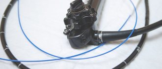
The gold standard for diagnosing gastric ulcers is esophagogastroduodenoscopy. Endoscopy allows you to visualize the ulcerative defect in 95% of patients and determine the stage of the disease (acute or chronic ulcer). Endoscopic examination makes it possible to promptly identify complications of gastric ulcer (bleeding, cicatricial stenosis), conduct endoscopic biopsy, and surgical hemostasis.
X-ray of the stomach (gastrography) is of paramount importance in the diagnosis of cicatricial complications and penetration of ulcers into nearby organs and tissues. If endoscopic visualization is not possible, radiography can verify a gastric ulcer in 70% of cases. For a more accurate result, it is recommended to use double contrasting - in this case, the defect is visible in the form of a niche or a persistent contrasting spot on the wall of the stomach, to which the folds of the mucous membrane converge.
Considering the huge role of Helicobacter pylori infection in the development of peptic ulcer, all patients with this pathology undergo mandatory tests to detect H. pylori (ELISA, PCR diagnostics, breath test, examination of biopsy specimens, etc.).
Of auxiliary importance for gastric ulcers are ultrasound of the gastrointestinal tract (detects concomitant pathology of the liver, pancreas), electrogastrography and antroduodenal manometry (makes it possible to assess the motor activity of the stomach and its evacuation ability), intragastric pH-metry (detects aggressive factors of damage), stool analysis for hidden blood (done if gastric bleeding is suspected). If a patient is admitted to the hospital with a clinical picture of an “acute abdomen,” diagnostic laparoscopy may be required to exclude gastric perforation. Gastric ulcers must be differentiated from symptomatic ulcers (especially medicinal ones), Zollinger-Ellison syndrome, hyperparathyroidism, and gastric cancer.
Treatment of gastric ulcer
The main goals of therapy for peptic ulcer include repair of the ulcer, prevention of disease complications, and achievement of long-term remission. Treatment of gastric ulcer includes non-drug and medicinal treatments, and surgical methods. Non-drug treatment of peptic ulcer involves following a diet, prescribing physiotherapeutic procedures (heat, paraffin therapy, ozokerite, electrophoresis and microwave exposure), it is also recommended to avoid stress and lead a healthy lifestyle.
Drug treatment should be comprehensive and affect all parts of the pathogenesis of ulcerative gastrointestinal tract. Anti-Helicobacter therapy requires the use of several drugs to eradicate H. pylori, since the use of monoschemes has shown to be ineffective. The attending physician individually selects a combination of the following drugs: proton pump inhibitors, antibiotics (clarithromycin, metronidazole, amoxicillin, tetracycline, furazolidone, levofloxacin, etc.), bismuth preparations.
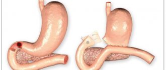
If you seek medical help in a timely manner and carry out a complete anti-Helicobacter treatment regimen, the risk of complications of gastric ulcer is minimized. Emergency surgical treatment of gastric ulcer (hemostasis by clipping or suturing a bleeding vessel, suturing the ulcer) is usually required only for patients with a complicated pathology: perforation or penetration of the ulcer, bleeding from the ulcer, malignancy, and the formation of scar changes in the stomach. In elderly patients, if there is a history of indications of complications of gastric ulcer in the past, experts recommend reducing the duration of conservative treatment to one to one and a half months.
Absolute indications for surgical intervention: perforation and malignancy of the ulcer, massive bleeding, cicatricial changes in the stomach with disruption of its function, ulcer of the gastroenteroanastomosis. Conditionally absolute indications include penetration of ulcers, giant callous ulcers, recurrent gastric bleeding during conservative therapy, and lack of ulcer repair after suturing. A relative indication is the absence of a clear effect from drug therapy for 2-3 years.
For decades, surgeons have been discussing the effectiveness and safety of various types of surgical interventions for gastric ulcers. Today, gastrectomy, gastroenterostomy, and various types of vagotomies are recognized as the most effective. Excision and suturing of a gastric ulcer is used only in extreme cases.
Complications of the disease
In the absence of timely treatment for gastric and duodenal ulcers, the following consequences may occur:
- bleeding. The complication is associated with damage to the blood vessels inside the ulcer. The stomach gradually fills with blood. The main danger is that the patient does not feel pain. Vomiting with blood clots indicates internal bleeding. Blood pressure gradually decreases, the skin becomes pale, weakness and shortness of breath appear. Bleeding from a duodenal ulcer is accompanied by dark stools;
- perforation. The complication occurs as a result of a violation of the integrity of the layers of the gastric wall. Perforation of an ulcer requires mandatory surgical intervention;
- penetration. With this pathology, the ulcer passes through the wall of the stomach to nearby organs. The pancreas is most often damaged, sometimes the liver is affected. In this case, signs of pancreatitis are added to the characteristic symptoms of peptic ulcer;
- stenosis of the gastric outlet. Complications arise when pathology is localized in this area. As a result of the development of a defect, food cannot pass from the stomach to the intestines. To restore normal functioning of the gastrointestinal tract, an operation is performed;
- perigastritis. Pathology involves the development of an inflammatory process around the ulcer. In the absence of proper therapy, adhesions form on the outside of the stomach. As a result, deformation of the stomach is possible;
- malignancy. Pathology suggests the appearance of a malignant neoplasm at the site of the ulcer.
Prognosis and prevention
The prognosis for gastric ulcer largely depends on the timeliness of seeking medical help and the effectiveness of anti-Helicobacter therapy. Peptic ulcer is complicated by gastric bleeding in every fifth patient, from 5 to 15% of patients suffer perforation or penetration of the ulcer, and 2% develop cicatricial stenosis of the stomach. In children, the incidence of complications of gastric ulcer is lower - no more than 4%. The likelihood of developing stomach cancer in patients with peptic ulcer is 3-6 times higher than among people who do not suffer from this pathology.
Primary prevention of gastric ulcer includes preventing infection with Helicobacter pylori infection, eliminating risk factors for the development of this pathology (smoking, cramped living conditions, low standard of living). Secondary prevention is aimed at preventing relapses and includes following a diet, avoiding stress, and prescribing an anti-Helicobacter drug regimen when the first symptoms of peptic ulcer appear. Patients with gastric ulcer require lifelong monitoring, endoscopic examination with mandatory testing for H. pylori once every six months. Source: https://www.krasotaimedicina.ru/diseases/zabolevanija_gastroenterologia/stomach-ulcer
Nutrition for stomach and duodenal ulcers
A person facing this problem needs to plan and establish a diet. The recommended number of meals is at least five. Portions should be small.
During an exacerbation of the pathological process, the patient should exclude the following foods and drinks from the diet:
- alcohol;
- meat, fish, mushroom, vegetable soups;
- black bread;
- fatty and fried foods;
- pickled vegetables;
- smoked meat;
- coffee and strong tea;
- onion and garlic;
- radish and horseradish;
- sparkling water.
The daily diet should contain foods that do not provoke increased secretion of gastric juice. These include:
- milk and dairy products (cream, cottage cheese);
- chicken egg white;
- lean beef and chicken meat, previously cleaned of veins, skin, etc.;
- boiled and stewed vegetables (potatoes, carrots, zucchini, cauliflower);
- low-fat soups cooked in a second broth;
- pate made from beef, poultry, fish, etc.