Gastric leiomyoma is absolutely benign in morphological essence, grows very slowly and in most cases does not manifest clinical symptoms. Large nodes can disrupt the normal functioning of the gastrointestinal tract and lead to dangerous complications.
- General information
- Kinds
- Reasons for appearance
- Symptoms
- Diagnostics
- Treatment
- Possible complications
- Prevention
- Forecast
Externally, leiomyoma cannot be distinguished from malignant neoplasms, so it is removed for microscopic and IHC studies, which prove the absence of signs of malignancy.
General information
Leiomyomas develop from cells of the muscular layer of the stomach, which is why their second name is “fibroids.” The tumor develops mainly in older people, but there are known cases of diagnosis of formation in childhood. In the scientific literature of the last century, a more frequent incidence of leiomyoma in women was noted; today, pathology is more often found in men from 40 to 70 years old.
Leiomyoma is a rare pathology and accounts for 3% of all organ neoplasms, but it is also the most common benign gastric tumor - it accounts for almost 45%. Since the process is asymptomatic, in the vast majority of cases, fibroids in the stomach are discovered accidentally during an examination for another reason or at an autopsy. In the last decade, the frequency of pathology has increased; it is believed that the reason for this is the greater availability of medical examinations and mandatory medical examinations.
Differences between uterine leiomyoma and fibroids
Many women are interested in the differences between fibroids, fibroids, fibromyomas and uterine leiomyomas. The medical history of patients often contains both one and the other diagnosis. Essentially, uterine fibroids are the common name for fibroids, fibroids, and leiomyomas. In turn, some differences between leiomyoma, fibroma and fibromyoma still exist and they relate to the structure of the tumors. Leiomyomas contain predominantly smooth muscle cells, whereas fibroids and fibroids are composed of fibrous tissue cells.
Kinds
A generally accepted classification of gastric leiomyoma has not been developed.
Gastric fibroids are usually divided according to the location of the node:
- submucosal or submucosal;
- inside the muscular layer of the stomach - intramural;
- subserous, growing predominantly outward, that is, into the abdominal cavity.
Some experts classify fibroids as:
- internal, growing mainly inside the stomach - endogastric;
- external - with growth directed into the abdominal cavity - exogastric.
By localization in the stomach it is designated as follows: located in the upper third or antrum; in the middle and lower third of the organ.
For gastric fibroids, a typical localization has not been established: approximately a fifth is located in the upper parts, about a third - in the middle part.
Reasons for appearance
The list of reasons for the development of a tumor in the muscular layer of the stomach includes a dozen “general” assumptions, that is, all known risk factors for tumors of any nature are mentioned.
First of all, heredity is suspected - a family history of gastrointestinal neoplasms is assumed without indicating the specific genes “responsible” for initiating the process or hereditary syndromes associated with leiomyoma. The negative impact of radiation, from electromagnetic to radioactive and solar, cannot be ruled out.
Possible factors favoring tumor pathology are immunodeficiency in HIV, as well as hormonal imbalance.
The harmful effects of the environment, unhealthy lifestyle with irregular and unhealthy diet, alcohol abuse and smoking cannot be excluded. And, of course, it was not without the complicity of nervous breakdowns in the initiation of tumor growth.
In fact, none of these reasons has reliable evidence and it would be correct to state the following - the reasons for the development of leiomyomas in the stomach are unknown to science.
Why does it occur
Causes of neoplasm:
- Unhealthy Lifestyle. The presence of bad habits in a person’s daily life: using tobacco products, drinking alcohol, unhealthy diet. The systematic consumption of junk food, a large amount of spicy, salty, fatty foods is to blame. Fast food and fried foods in recycled oil are dangerous.
- Presence of harmful and dangerous production factors.
- A disease acquired by inheritance due to genetic predisposition. The chance of acquiring the disease increases if a close relative had cancer of the specified organ.
- Environmental conditions harmful to health.
- Reduced immunity.
Many of the listed causes that precede the occurrence of esophageal leiomyoma are similar to the causes of cecal disease and other intestinal diseases.
Symptoms
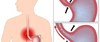
The slow growth and benign course of the myomatous node with high extensibility of the gastric wall in most cases contributes to the absence of complaints of discomfort. It is clear that the severity and range of clinical manifestations are determined by the size and location of the tumor:
- Small or “miniature” leiomyomas do not manifest themselves in any way.
- Large nodes with intracavitary growth can disrupt the evacuation function of the stomach, interfering with the movement of food masses, clinically manifested by belching, vomiting and a feeling of fullness of the stomach after eating.
- Externally located large fibroids, especially on a narrow base - a pedicle, can put pressure on neighboring organs and disrupt their function, as well as twist in the pedicle and simulate an “acute abdomen” clinic with severe pain, vomiting and intoxication syndrome.
The main danger of gastric fibroids is the complicated course of very large nodes, which we will consider further.
Since there is no generally accepted classification of leiomyomas, nodules less than 2 centimeters are considered small, large ones - more than 5 centimeters; the literature contains cases of surgical removal of huge leiomyomas that were masquerading as tumors of the abdominal cavity or ovaries.
Reviews, prognosis and prevention
According to doctors, if detected early and treated correctly, leiomyoma has a favorable prognosis. Organ-conserving surgeries performed on young women allow them to preserve their reproductive function. Radical surgery may be required only if the fibroids grow rapidly and some other factors, when removal of the uterus becomes inevitable.
In order to prevent the development of this pathology, it is imperative for every woman to regularly visit a gynecologist and perform ultrasound diagnostics, which will allow timely detection of a tumor in the uterus.
The Yusupov Hospital offers a comprehensive examination using modern high-tech equipment. Thanks to the modern equipment of the hospital, the experience and high professionalism of our specialists, we guarantee the most accurate results in the shortest possible time.
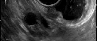
Diagnostics
Mostly the tumor looks like a nodular formation with a smooth surface, but it can also consist of several nodules. An experienced endoscopist will detect it by bulging of the wall, if growth is directed into the gastric lumen, and a change in the peristaltic “wave”.
A biopsy of the submucosal node is often inconclusive due to its deep location, so in hardly a third of cases it is possible to judge the true nature of the neoplasm.
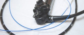
High-tech diagnostics - CT and MRI are practically powerless for pathology of the gastric wall; the most effective detection method is endoscopy or endoscopic ultrasonography combined with ultrasound, which allows you to examine the formation from the stomach cavity, determine its boundaries inside the gastric wall, and see the structure and structure of the node.
Treatment
There is no need to remove small tumors that do not bother the patient if there is 100% confidence in their benign nature, but with submucosal growth such confidence is excluded. Benign fibroids and malignant GISTs are visually and morphologically indistinguishable and are differentiated only by immunohistochemical analysis of tumor sections obtained surgically.
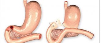
The final diagnosis and simultaneous treatment with a diagnosis of gastric leiomyoma consists of their surgical removal. The extent of intervention and access to the organ is determined by the size and location of the tumor, the presence of complications and the patient’s initial health.
In most cases, organ-preserving endoscopic interventions are resorted to, of course, with urgent histological examination during surgery. Laparoscopic resections are quite sufficient and adequate, allowing for rapid recovery of the patient.
In case of a large complicated neoplasm that has caused ulceration of the mucous membrane, perforation of the gastric wall or bleeding, classical operations with dissection of the abdominal wall and gastrectomy are resorted to.
STOMACH POLYPS
Gastric polyps are benign tumors of various origins that protrude into the lumen.
There are adenomatous polyps, which are considered true neoplasms, and hyperplastic (regenerative) polyps. Adenomatous polyps (adenomas) are benign tumors of the glandular epithelium, forming papillary and/or tubular structures with varying degrees of cellular dysplasia and atypia. These polyps in the stomach are rare and have a high potential for malignancy, just like colon polyps. Their endoscopic removal or gastrectomy (removal of part of the stomach) is indicated. The frequent combination of adenomatous polyps of the stomach and colon requires a diagnostic colonoscopy.
Hyperplastic polyps are not true benign tumors. They are a consequence of disregenerative processes that usually occur against the background of chronic active HP gastritis. Gastric polyps are often detected during endoscopic examination. Their biopsy and removal are mandatory to detect early gastric cancer. All patients after removal of gastric polyps in the long term are subject to dispensary observation with repeated endoscopic examinations 1-2 times a year.
Possible complications
Large leiomyomas growing into the stomach cavity disrupt the structural integrity of the mucous membrane, causing its ruptures - erosion and forming ulcers. Ulcerations in clinical manifestations are indistinguishable from a banal ulcer - pain, discomfort, episodes of vomiting, changes in appetite.
And just like ordinary ulcers, they can be complicated by acute or chronic bleeding. Acute gastric bleeding is manifested by vomiting coffee grounds and black stools, decreased vascular pressure up to shock. Chronic blood loss is manifested by anemia syndrome, changes in stool, and weight loss. Insufficient nutrition of leiomyoma leads to the disintegration of its tissues with possible rupture of the gastric wall - perforation and the development of peritonitis.
The complicated course of a benign stomach tumor requires active therapy followed by long-term rehabilitation.
Forecast
Leiomyoma after proper removal does not recur and does not affect the patient’s life expectancy. The literature mentions the possibility of malignancy, which from an oncological point of view is an initially undiagnosed malignant slow-growing GIST; it is because of such cases that gastric leiomyoma must be removed.
A patient with suspected gastric leiomyoma should be operated on by oncological surgeons in a clinic with a powerful diagnostic service and a morphological laboratory focused on oncopathology. During surgery, it is necessary to differentiate a benign process from a malignant one in order to determine the optimal size of removal. Euroonko is one of the few medical institutions that meet these important requirements for the patient.
Book a consultation 24 hours a day
+7+7+78
Bibliography:
- Gankov V.A., Maslikova S.A., Lazarev A.F., Oskretkov V.I./Surgical tactics for submucosal formations of the esophagus, stomach and duodenum//Bulletin of Med. Sciences No. 2 (10) 2018
- Starkov Yu.G., Solodinina E.N., Novozhilova A.V. /Submucosal neoplasms of the gastrointestinal tract in endoscopic practice// Surgery, 2010; 2.
- Firsov E.I., Vorotyntseva N.S., Novikov M.V. /Comprehensive clinical and radiological diagnosis of benign non-epithelial tumors of the stomach// Kursk scientific-practical. Bulletin "Man and his health", 2009, No. 2.
- Bucber P., Villiger P., Egger JF., et al. /Management of gastrointestinal stromal tumors: from diagnosis to treatment// Swiss. Med. wkly., 2004; 134.
- Greenson JK /Gastrointestinal stromal tumors and other mesenchymal lesions of the gut// Mod. Pathol., 2003; 16(4).
- Nishida T., Blay JY, Hirota S., et al. /The standard diagnosis, treatment, and follow-up of gastrointestinal stromal tumors based on guidelines // Gastric Cancer., 2016 Jan., 19 (1).