One of the causes or consequences of the development of acute erosive and ulcerative lesions is a violation of microcirculation [1] of the parasympathetic and sympathetic nervous system in the gastric mucosa in patients with acute ulcerations of various etiologies [2]. It is not clear what is primary: 1) acute ulcerations of the mucous membrane of the stomach and duodenum (DPC), which lead to a violation of the density of cholinergic and adrenergic fibers, or 2) disorders and features of the autonomic innervation of the mucous membrane of the stomach and duodenum, which cause the development of acute ulcers and erosions under the influence of various factors of acute ulceration. One thing is certain - not everyone experiences acute ulcerations of the gastric and duodenal mucosa, even when exposed to multiple ulcer-forming factors.
The purpose of this work is a morphological and histochemical study of the pathogenesis of the development of acute ulcerations of the stomach and duodenum with various etiological factors of acute gastropathy.
What is a gastric biopsy
What is a biopsy - this is the process of obtaining a sample of altered tissues with their subsequent morphological (histological) examination. The material is collected during gastroscopy (EGD, FGDS). Why is it carried out - to establish an accurate diagnosis if a malignant process is suspected.
To obtain a piece of tissue from the stomach, the doctor uses a special device - a flexible esophagogastroscope equipped with fiber optics. The esophagogastroscope is characterized by elasticity, the ability to clearly visualize and record images, and a low risk of trauma to the digestive tract.
Before the procedure, the patient is given premedication and the oropharynx area is treated with lidocaine using a special spray. According to indications, medicated sleep is used, but usually the patient remains fully conscious. The patient takes a position on his left side and clamps an endoscopy mouthpiece in his mouth - a special device to protect the endoscope from accidental biting. Through the mouthpiece, the doctor inserts the endoscope into the pharynx and esophagus. When passing the mouth of the esophagus, a gag reflex occurs. To overcome it, the patient makes a swallowing movement.
Next, air is supplied inside in small portions, mucus and gastric juice are sucked out. The doctor examines the inner surfaces of the walls and plucks off several pieces with a diameter of 2–3 mm from the required areas. It is important to obtain at least 5 samples. Pinching is performed using biopsy forceps. Biopsy forceps are adapted to the curves of the gastrointestinal tract and allow obtaining good tissue samples. If it is necessary to remove a large fragment, an electrosurgical loop is used.
Patients are concerned about whether it hurts to do a biopsy. The most uncomfortable moment in the entire procedure is the passage of the endoscope through the oropharynx area. No pain is felt during tumor tissue collection.
Indications and contraindications
Stomach cancer is not the only gastrointestinal disease for which a biopsy is indicated. This is an informative study that can tell a lot about the state of the digestive tract. In what cases does the doctor prescribe a gastric biopsy:
- the patient has clinical symptoms suspicious for cancer: epigastric pain combined with nausea, vomiting and weight loss;
- other studies have shown the presence of a neoplasm;
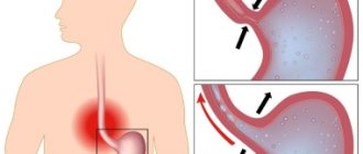
- the patient has gastroesophageal reflux disease - the throwing of part of the gastric juice and stomach contents back into the esophagus;
- an ulcer or atrophic gastritis was previously diagnosed.
In the presence of gastric and duodenal ulcers, endoscopic examination is recommended once a year. Preventive FGDS allows timely tracking of the development of a malignant process, since ulcerative lesions of the mucous membrane may have a tendency to malignancy (malignancy). If during a preventive FGDS the doctor notices suspicious areas on the mucous membrane, he will immediately take tissue samples. Early diagnosis of stomach cancer is good luck, since most patients see a doctor at the third stage of the tumor, when the chances of remission drop due to metastases.
The analysis is taken for an ulcer that does not heal for a long time, for chronic gastritis to clarify the stage of the process, to diagnose a polyp, and to monitor postoperative complications.
A biopsy is contraindicated in the presence of the following problems:
- stroke, heart attack and other acute vascular pathologies;
- esophageal stenosis;
- bronchial asthma in the acute stage;
- infectious process in the body;
- epilepsy.
It is necessary to stabilize the patient's condition as much as possible before examination, or find another way to study the tumor.
Indications for histological examination
- hypoacid gastritis (this stomach disease is considered precancerous);
— diagnosis of special forms of gastritis (granulomatous, eosinophilic, lymphocytic gastritis);
- chronic peptic ulcer disease;
- dysphagia;
- Barrett's esophagus;
- weight loss, lack of appetite, anemia;
- stable discomfort in the stomach, aversion to meat food and others.
How to prepare for an endoscopic examination
— The best time to perform a biopsy followed by histological analysis is morning. The study should be carried out on an empty stomach; the night before, only a light dinner is allowed; the consumption of fried and fatty foods is excluded.
— It is necessary to stop taking medications that contain activated carbon and iron.
— On the eve of histological examination, chewing gum and smoking are excluded. Suspicious patients are advised to take sedatives the day before the procedure.
Biopsy collection procedure
The biopsy is taken during an endoscopic examination. In this case, the doctor inserts an endoscope, which is equipped with a camera and small forceps, through the mouth and esophagus into the stomach. Using a camera, the doctor identifies suspicious areas on the gastric mucosa from which a biopsy must be taken. Then the endoscope is brought to these defects and, using forceps, bites off small pieces. Because small pieces are removed during the biopsy, the procedure is virtually painless. After collecting all the necessary pieces, the endoscope is brought out. The pieces are processed like histological preparations, and then a pathologist examines them under a microscope, identifying the presence or absence of atypical (cancerous) cells. Sometimes even experienced specialists are not able to take pieces from all parts of the ulcer. In this case, it is necessary to perform a biopsy again after some time. A proper biopsy is multiple, that is, the doctor takes several pieces for examination from the edges and bottom of each ulcerative defect. In addition, several pieces are taken from each scar visible at the site of the healed ulcer. It is also recommended to take several pieces of biopsy from each suspicious location on the gastric mucosa. It has now been proven that 100% accuracy in diagnosing cancer is achieved by taking at least 6 pieces from different parts of each ulcerative and scar defect. Examination of only 1–2 pieces is uninformative, since it allows detecting cancer in the early stages only in half of the cases.
Preparation for the procedure
Carrying out an FGDS and gastric biopsy to diagnose cancer is usually a planned event. Emergency gastroscopy is indicated for internal bleeding when the patient needs emergency care. In other cases, there is time to prepare.
The procedure is carried out on an empty stomach, at least 8-12 hours should pass after the last meal. You must give up water two hours before. The stomach should be empty. If the patient underwent an X-ray of the gastrointestinal tract with contrast, he will have to wait 2-3 days. Drugs that affect blood clotting are discontinued within a few days.
How to behave after the procedure
To quickly recover after the procedure, you need to follow the following recommendations:
- on the day of the biopsy, eat light food in small portions;
- exclude spicy, fatty, heavy foods, carbonated and alcoholic drinks for 3 – 5 days;
- Limit physical activity, visits to the bathhouse, sauna for a week.
Like other tests of the stomach, a biopsy rarely causes complications, although it does sometimes happen. Slight discomfort in the epigastric area is normal. In what cases should you be wary:
- intense, persistent abdominal pain;
- vomit that is dark brown or contains blood;
- labored breathing;
- general deterioration of condition: drop in blood pressure, rise in temperature, weakness, dizziness.
Symptoms indicate perforation of the gastric wall or aspiration pneumonia. The patient needs medical care and hospitalization.
results
The samples taken are placed in sealed containers with a preservative, and then sent to the laboratory. Pieces of tissue are treated with formaldehyde and thin sections are made using special equipment. The samples are then examined under a microscope.
The patient will be given a conclusion containing the following information:
- thickness of the mucous membrane of the gastric walls;
- the degree and nature of the inflammatory process;
- the presence and structure of atypical (malignant) cells;
- degree of differentiation of neoplasm cells.
How long it takes to perform a gastric biopsy depends on the diagnostic method, the volume and type of material collected, and the workload of the laboratory. How many days does the patient have to wait? On average, he receives the biopsy results in 5-10 days. In rare cases, the waiting time increases to 2 weeks.
The interpretation of the analysis should be carried out by the attending physician. The information in the conclusion is written in scientific language; only a qualified specialist has the right to interpret the results. A negative biopsy shows no signs of malignant cell changes; a positive one, on the contrary, confirms this.
Gastric adenocarcinoma - symptoms and treatment
Surgical treatment at an early stage
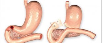
When early forms of cancer are detected, i.e. with cancer in situ, endoscopic treatment :
- mucosal resection (EMR) - removal of the tumor along with part of the gastric mucosa;
- submucosal dissection (ESD) - removal of the tumor along with part of the submucosal layer. It is carried out for adenocarcinoma up to 2 cm, allowing it to be removed in a single block, without incisions [8].
These operations are performed under general anesthesia. First, the boundaries of the tumor are determined and, using electrocoagulation, the resection edges are “marked” (cauterized), retreating 3 mm from the tumor border. An injection of saline and Voluven (Hydroxyethyl starch) is then injected into the submucosal layer to separate the tumor from the muscle layer. Then the adenocarcinoma is removed and its “bed” is examined: is there any bleeding or perforation, and was it possible to completely remove the tumor. After which the removed fragment is removed and sent for histological examination.
After surgery, patients are discharged 3–4 days later. The effectiveness of treatment, if all standards are met, is 98%. Recommended:
- a month after surgery, and then every 3 months, do FGDS;
- six months after surgery - CT scan;
- For a month, strictly follow a diet - eat liquid pureed food, exclude alcohol and hot food.
Surgical treatment in later stages
Surgery for gastric adenocarcinoma is carried out in several stages: first, the tumor itself is removed, then food passage is restored [9].
Basic radical operations for gastric adenocarcinoma:
- gastrectomy - complete removal of the stomach;
- subtotal proximal resection - removal of the upper part of the stomach along with the cardiac part;
- subtotal distal resection - removal of the lower 2/3 or 3/4 of the stomach.
All types of operations are performed both in an “open” manner (laparotomy) and through small incisions (laparoscopically), including using robotics. The choice of surgical approach and the extent of the operation depends on the extent of the process: the degree of damage to the walls of the stomach, involvement of the esophagus, duodenum and the presence of metastases.
After tumor removal, lymph node dissection . This is a standard operation to remove lymph nodes for stomach cancer. It is carried out to reduce the risk of relapse, since adenocarcinoma very often metastasizes through the lymphogenous route, i.e. with lymph flow.
For single metastases in other organs, simultaneous operations are performed, i.e., several interventions at once.
If the patient has many distant metastases, then surgical treatment is usually not performed . But when emergency life-threatening conditions develop, such as perforation of the stomach wall, bleeding or stenosis, the operation is performed to save the patient’s life. The volume of surgical intervention should be minimal.
Chemotherapy
The main treatment for tumors that cannot be removed is chemotherapy [12]. It is needed to kill tumor cells or significantly slow down their growth.
The doctor may prescribe monotherapy, i.e. treatment with one drug, or combination chemotherapy using several drugs. Combination chemotherapy can enhance the antitumor effect.
Combination treatments include:
- Perioperative chemotherapy is the preferred method, given before surgery to shrink the tumor and after surgery to fight remaining cancer cells [10][11];
- adjuvant chemotherapy - performed after surgery;
- adjuvant chemoradiotherapy - carried out after non-radical tumor removal, i.e. when cancer cells remain at the edges of the resection and continue to grow.
The choice of a specific combination depends on the patient’s condition, his age, as well as the severity and nature of concomitant diseases.
Indications for chemotherapy:
- spread of adenocarcinoma beyond the mucous layer;
- damage to the lymph nodes;
- presence of metastases.
The patient may refuse chemotherapy , but he must understand that without treatment the tumor can quickly reappear and metastasize.
Symptomatic treatment (palliative care)
Symptomatic therapy is carried out at stage IV of cancer, when special treatment methods are contraindicated. It helps relieve the symptoms of the disease through adequate pain relief.
It is recommended to gradually move from weak painkillers (for example, Ketoprofen) to stronger drugs, including narcotics (Tramadol or Morphine). In case of severe pain, they can be used on average every 4 hours.
Can the results be wrong?
There is a small chance that the results will be erroneous. Two factors lead to false-positive and false-negative results: errors by the medical worker performing the histological examination and malfunction of the equipment.
The main errors in the procedure leading to false results:
- incorrectly selected areas of the mucous membrane for sampling;
- insufficient biopsy volume;
- violation of the rules for storing biomaterial;
- non-compliance with the rules and stages of preparing biomaterial for research.
If the doctor has reason to suspect erroneous results, the procedure will be repeated.
What to do after a biopsy
Stomach cancer at certain stages is subject to surgical treatment. The size of the tumor and its characteristics determine whether the stomach will be removed completely (gastrectomy) or partially (resection). Why is a histological examination performed - incl. to select the optimal treatment tactics.
For stomach cancer, in some cases, chemotherapy and radiation therapy are indicated. They reduce the size of a large tumor to an operable size, and make it possible to remove the remains of malignant cells after a gastrectomy.
The prognosis for recovery depends on a number of factors: the patient’s age, the presence of concomitant diseases, the presence of metastases, and the degree of malignancy of the tumor. Late diagnosis of the disease does not mean that it is a hopeless case. Doctors may suggest immunotherapy, or palliative care.
has been organizing high-quality individual medical care for many years.
Over the years of work, we have accumulated statistics on leading foreign clinics and are ready to recommend to patients for pregnancy management after breast cancer only those medical centers where they will truly provide the most effective care.
Histology: what kind of analysis is this?
The collection of biological material and its careful histological analysis are very important in many branches of medicine. In order to identify pathology, for example, histology of a mole, cervix, digestive tract, endocrine system, and so on can be prescribed. Our questions about histology were answered by Sergei Viktorovich Ezhov, an oncologist-mammologist at the Expert Clinic Voronezh.
- Sergey Viktorovich, tell us what kind of analysis this is - histology? What is its essence?
— Histological analysis is a highly accurate research method that allows you to determine pathological deviations in the structure of tissue. This method is used in many areas of medicine, but its main essence is that using this method it is possible to diagnose the presence of malignant tumors, determine their structure, as well as the stage of the pathological process.
— What can be a biomaterial for histological examination?
— The biomaterial can be one or another suspicious tissue (skin, mucous membrane, muscle, bone) taken through a biopsy, or a preparation obtained as a result of surgery.
— When is histology prescribed?
— As I already said, histological examination can be used in many areas of medicine, but its special value lies in determining the nature and nature of changes in tissue at the slightest suspicion of an oncological process. This study is also carried out before prescribing antitumor therapy to develop a treatment plan and during treatment to monitor its effectiveness.
— What does histology show?
— Histological analysis makes it possible to detect inflammatory processes in tissue, establish the nature of a particular neoplasm (i.e., whether it is benign or malignant), determine the level of malignancy, and also identify the localization of the primary tumor focus. You must understand that only a specialist should decipher the results of histological examination. I would not recommend that patients do this on their own.
Biopsy
— How to get tested for histology? Is special training needed?
— Before conducting a histological examination, it is necessary to perform a biopsy and send the resulting material (biopsy) for analysis. As such, the biopsy does not require any preparation; there are only a number of restrictions and recommendations that the patient must adhere to the day before and during the collection of the biopsy. It is important to inform your doctor about all medications you are taking. On the eve of the study, you must stop taking medications that affect blood clotting (in consultation with the doctor who prescribed them), and also inform the doctor who will perform the biopsy about the presence of allergies. It is undesirable to carry out this manipulation in women during menstruation. Alcohol consumption is contraindicated.
— How is histology performed?
— The material obtained from the biopsy is placed in formalin and sent for histological examination to the pathomorphological center. There the material is filled with paraffin (most often). After cooling and hardening, thin sections are made, onto which various reagents are applied for a detailed study of the tissue under a microscope by a pathologist.
— How long does it take to perform a histological analysis?
— If the result is obvious and beyond doubt, the conclusion is issued on average in 7-10 days. If doubts or suspicions arise about the accuracy of the diagnosis, the drug may be studied longer.
— Are there errors in histology results?
— Unfortunately, especially in small settlements, problems may arise with the quality of histological analysis. This is due to various reasons (lack of doctors, equipment, consumables). But if doubts arise about the correctness of the histological conclusion, a review of the finished preparations is usually prescribed in another pathomorphological center.
Want to learn more about other types of tests? Read articles in our section You can make an appointment with an oncologist here ATTENTION: the service is not available in all cities
Interviewed by Marina Volovik
The editors recommend:
Precancer: to be afraid or not to pay attention? Fear has big eyes. Is fibroids really that dangerous? Breast cancer is not a death sentence!
For reference:
Ezhov Sergey Viktorovich
In 2007 he graduated from the medical faculty of Kursk State Medical University.
2007 - 2009 - clinical residency in the specialty “Oncology” at the Medical Radiological Research Center of the Russian Academy of Medical Sciences in Obninsk (today - MRRC named after A.F. Tsyb - a branch of the Federal State Budgetary Institution "National Medical Research Center of Radiology" of the Ministry of Health of Russia).
Currently, he is a doctor-oncologist-mammologist at the Expert Clinic Voronezh.
Reception is conducted at the address: st. Pushkinskaya, 11.