The article was prepared by a specialist for informational purposes only. We urge you not to self-medicate. When the first symptoms appear, consult a doctor.
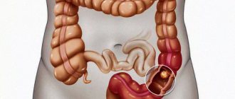
Teniarinhoz is a parasitic disease belonging to the group of biohelminthiases. The causative agent of teniahrynchiasis in humans is bovine tapeworm, which parasitizes the small intestine. Teniarinhoz most often has a chronic course and manifests itself in disorders of the digestive system, as well as in toxic-allergic reactions.
Bovine tapeworm is widespread throughout the world, but most people affected by this helminthic infestation live in Central and Southern Africa, Asia, South America, China, Mongolia and Australia. This is explained by the widespread development of cattle breeding. As for Russia, cases of teniarinhoz are registered in Dagestan, Tuva, the Republic of Sakha and Buryatia. There is evidence of human infection with bovine tapeworm in the Irkutsk, Novosibirsk, and Tyumen regions. Patients with a similar diagnosis are admitted to hospitals in the Perm, Altai and Krasnoyarsk regions.
The parasite spreads in patches, most often the disease is registered among rural residents. The population that suffers is mainly those who eat either raw or poorly cooked meat from cows. The danger is posed by salted and dried meat containing cysticerci of the worm.
The maximum number of cases of teniarhynchosis is recorded during periods of mass slaughter of livestock - winter and autumn. If we look at statistics by gender and age, adults get sick more often than children, and the overwhelming majority of patients are men. At the same time, workers from livestock farms and meat processing plants predominate.
What is a bull tapeworm?
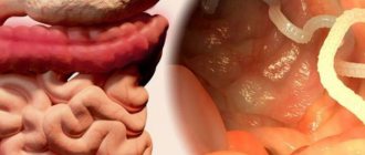
Bovine tapeworm is a helminth that parasitizes the small intestine of humans. It belongs to the class of tapeworms, a type of tapeworm. This worm is also called the unarmed tapeworm. Its body has a flat, ribbon-like shape and consists of a neck, scolex (head) and strobila. On the scolex there is a rudimentary proboscis and four suckers without hooks. The neck of the worm is short and extends into the strobila. The strobila itself may consist of 2000 (or less) proglottids (segments).
The bovine tapeworm is a very large worm that can reach more than 10 m in length. This helminth is a hermaphrodite, and its female and male reproductive systems are well developed. The genitals are located in the middle third of the strobila.
The uterus of the bovine tapeworm is closed, the eggs mature and accumulate inside it. As the number of eggs increases, the uterus begins to stretch, after which protrusions appear on its sides (from 18 to 32 pieces on each side). Other organs atrophy. The eggs do not mature evenly, so the terminal segments may contain from 50 to 150 thousand larvae ready for invasion. They do not need to ripen in the external environment.
The eggs of the bovine tapeworm are round in shape, covered on the outside with a thin transparent shell. Inside the mature egg there is an oncosphere - an embryo equipped with 3 hooks.
The segments located in the terminal part of the strobila begin to stretch and narrow, and then tear away from the strobila, moving forward. After separation from the strobila, the segments move through the human intestines and exit into the external environment through the anus along with feces. It is interesting that some segments passively move through the intestines along with digested food, and some can independently, and quite actively, make their way to the human anus. Every day the patient can secrete from 1 to 23 segments. At the same time, the worm itself does not become shorter, since new segments constantly grow from the neck. They gradually develop, mature and move towards the tail part of the worm, from which they are subsequently detached.
The definitive host of the bovine tapeworm is humans, and the intermediate host is cattle. Already 2-4 months after the invasion, the patient begins to release worm eggs into the environment. This can continue for 15 years or more.
Diagnostic methods
Diagnosis of taeniasis and cysticercosis is very difficult, since these diseases do not have specific manifestations. A detailed questioning of the patient is of no small importance. What matters is the person’s place of work, the nature of the food consumed, travel to the countryside in the next few months, and contact with sick people. Patients should also be asked about the appearance of helminth segments in the feces. To do this, doctors usually show on preparations what the worm segments look like.
If the survey results are positive, patients are asked to bring the discharged segments to the laboratory for a final diagnosis. It is worth keeping in mind that some people may hide the fact of segmental discharge. These include children and adolescents, elderly people and food industry workers.
Laboratory diagnosis of taeniasis includes macroovhelminthoscopy: mature proglottids of pork tapeworm are found in the stool of an infected person. Detection of only helminth eggs does not make it possible to establish a final diagnosis, because worm eggs are no different from the causative agent of another helminthiasis - bovine tapeworm.
Figure 7 - Comparative characteristics of bovine and pork tapeworms
A general blood test reveals eosinophilia and leukocytosis, however, such changes in blood parameters are usually characteristic of the initial stages of the disease. The diagnosis of cysticercosis is also based on interviewing the patient. Questioning the infected person, especially about the presence of taeniasis in the past, is important here.
Of the laboratory diagnostic methods, a general blood test is important when eosinophilia can be detected. Also, the diagnosis of cysticercosis is confirmed using serological research methods: the reaction of complement binding with an antigen from cysticerci; the indirect hemagglutination reaction and enzyme-linked immunosorbent assay are considered more specific.
For the diagnosis of cysticercosis of the central nervous system, CT and MRI diagnostics are of no small importance, with the help of which oval formations with a clearly defined membrane, sometimes calcified, are detected. The cerebrospinal fluid is also examined, in which lymphocytosis and eosinophilia are detected, sometimes increased protein, as well as single scolex.
Cysticercosis of the skin and skeletal muscles is diagnosed by biopsy of tumor-like formations, inside which a helminth larva is found. Sometimes radiography helps to identify calcified dead Finns.
Cysticercosis of the eye is determined using ophthalmoscopy. It is rare to find cysticercus in the fundus. As a manifestation of increased intracranial pressure in cysticercosis of the brain, congestive optic discs may appear in the fundus of the eye.
Cysticercosis of the lungs and heart can be diagnosed by chest x-ray. The radiograph reveals small rounded shadows with clear boundaries, often calcified. The size of the shadows varies from five to seven millimeters. As a rule, such shadows are scattered throughout all fields, their number varies from a few to dozens.
A positive result of the complement binding reaction of blood and/or cerebrospinal fluid with cysticercus antigen confirms the diagnosis of cysticercosis.
Symptoms of bovine tapeworm
Symptoms of bovine tapeworm may be absent at all, or they may appear brightly, causing severe problems with human health. Sometimes, for many years, the only symptom of infection is the presence of worm segments in the stool, or their independent crawling out of the anus. This process is always accompanied by unpleasant sensations and leads to the development of neurotic disorders in the patient. Patients compare what is happening to foreign bodies crawling in the anus, which cause severe itching.
The clinical picture of teniarhynchosis may look like this:
- Impaired motility and excretory function of the gastrointestinal tract as a whole.
- Catarrhal inflammation of the intestines, which occurs against the background of the traumatic effect of the parasite on the mucous membrane of the small intestine.
- Feeling of heaviness and pain in the epigastric region.
- The presence of heartburn and belching not associated with food intake.
- Increased salivation.
- Constant feeling of nausea, periodic urge to vomit.
- Abdominal pain that is not associated with any gastrointestinal diseases. The pain has no clear localization. When the worm moves through the valve connecting the small and large intestines, the pain becomes cramping in nature.
- Unstable stool, in which constipation is replaced by diarrhea, flatulence.
- Often, against the background of teniarhynchosis, the patient develops a duodenal ulcer or biliary colic.
- Intestinal obstruction occurs when the worm clumps into a ball and prevents the passage of feces.
- Insufficient absorption of minerals and vitamins due to the presence of a parasitic worm in the intestines leads to the fact that the patient’s nails and hair deteriorate, and the skin becomes dry and prone to inflammation. The body as a whole suffers.
- A person infected with bovine tapeworm experiences a constant feeling of hunger; he wants to eat all the time. However, despite the increased appetite, weight does not increase. Moreover, prolonged parasitism of the worm in the intestines leads to loss of body weight.
- During its life, the tapeworm releases toxic substances. They poison the human body, causing increased allergic reactions and eosinophilia.
- Patients often note worsening night rest, increased irritability and fatigue, periodic dizziness, headaches and weakness in the limbs. Naturally, people do not associate these symptoms with parasitic infestation. Meanwhile, such an asthenovegetative complex is the result of intoxication of the body with waste products of the bovine tapeworm. In rare cases, epileptiform seizures may develop.
- From the cardiovascular system, increased heart rate and decreased blood pressure are possible. Sometimes painful sensations occur in the heart area, tachycardia is noted, at such moments tinnitus appears, and spots may appear before the eyes. Nosebleeds develop less frequently.
- Cases have been described in which movable segments entered the respiratory tract and the middle ear through the Eustachian tube. They may be detected in vomit.
- A number of patients experience cracks in the tongue, its soreness, as well as an increase in the size of the tongue.
- In weakened patients, urticarial exanthema may occur, when a rash covered with crusts appears on the body.
Carrying a bovine tapeworm during pregnancy is dangerous, as it can lead to premature labor, miscarriage and severe toxicosis. In addition, people with bovine tapeworm in their bodies often suffer from anemia.
It should be noted that full symptoms rarely appear. They tend to increase, depending on how long the parasite lives in the human body.
Thus, in the chronic stage of the disease, 4 main symptom complexes of bovine tapeworm in humans are noted:
- Asthenovegetative (weakness, asthenia);
- Abdominal (abdominal pain);
- Dyspeptic (disorders of gastrointestinal tract functions);
- Food (increased appetite).
Ways of human infection with bovine tapeworm
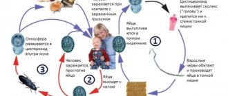
The mechanism of transmission of the parasite is fecal-oral, and the main route of infection is food. After a sick person begins to release helminth eggs into the environment, they end up in water, soil, grass, etc. After a certain time, the egg finds its intermediate host - cattle. The larva lives in his body for 4-5 months, during which time it becomes ready to invade the human body.
It should be noted that a human carrier of bovine tapeworm does not pose a threat to another person in terms of immediate infection. The worm larva, immediately after leaving the body of its main host, cannot infect another person. In order to prepare, it needs an intermediate host organism.
The main routes of human infection with bovine tapeworm:
- Eating undercooked or undercooked beef.
- Failure to comply with household and sanitary hygienic skills when working with raw meat and when performing economic activities.
- Taking a sample from raw minced meat. Housewives are often infected this way.
- It is also worth noting the culinary preferences of a particular nation. For example, people often become infected by eating shish kebab and stroganina. It has been established that when preparing shish kebab, the mass of pieces of which is 50 g or more, with the standard frying method, most of the cysticerci remain viable.
The natural susceptibility of people to infection with bovine tapeworm is high.
Epidemiology of teniarynchosis
Spread of teniarynchosis
Bull tapeworm is ubiquitous. Especially many cases of the disease are recorded in regions with developed livestock farming, the population of which traditionally eats semi-raw and raw meat. Important factors in the spread of helminthiasis are fecal contamination of soil and water bodies with helminth eggs, insufficient veterinary examination of meat, low sanitary level of settlements and livestock farms, as well as peculiarities of national food habits.
Rural residents get sick 3 times more often than urban residents. Of all the cases, up to 80% are adults.
Teniarinhoz is most common in the countries of South America, Africa and Australia, as well as in Mongolia, China and a number of countries in South and Southeast Asia. The incidence in Russia is registered in the Chechen Republic, Dagestan, the Altai Republic, Komi, the Yamalo-Nenets Autonomous Okrug, the Mari El Republic, the Novosibirsk and Orenburg regions.
One sick shepherd can infect an entire herd of cows.
At-risk groups
Bovine tapeworm is most often found among people working with cattle: livestock breeders, farm workers, meat processing plants and slaughterhouses, cooks, shepherds, milkmaids, calf workers, etc.
How animals become infected
Cattle, yaks, zebras, buffalos and possibly reindeer become infected with eggs or segments of the bovine tapeworm, which are excreted by a sick person in the feces into the external environment. They swallow them with hay, grass, water, soil, eggs and segments enter the mouth when licking urine located next to feces.
Bovine tapeworm eggs exhibit increased resistance in the external environment. At an ambient temperature of 10 - 300C, they remain viable for up to 150 days in grass, up to 70 days in liquid manure, and up to 33 days in water.
Rice. 2. Eggs and segments of a bovine tapeworm under a microscope.
How does a person become infected with bovine tapeworm?
- The source of infection for humans is cattle. Teniarinhoz develops when consuming meat products containing the parasite - insufficiently fried or cooked meat. This happens when the technology of culinary processing of meat products is violated, the habit of trying and eating raw minced meat, raw salted and dried meat, beef kebabs, rare steaks, etc.
- The things of a sick person, the towels he used and the food he prepared can serve as a source of helminthiasis.
- The danger comes from equipment, cutting boards, knives and other kitchen utensils used in the process of preparing contaminated meat.
- Danger to humans comes from raw water, unwashed fruits, vegetables and herbs, and dirty hands.
Rice. 3. Bovine tapeworm in meat. Finns in animal muscle tissue.
Why is bovine tapeworm dangerous?
In addition to the fact that bovine tapeworm is harmful to health in general, the following life-threatening complications may develop:
- Mechanical intestinal obstruction. This occurs when there are several worms in the intestines, or if a single individual becomes entangled and forms a ball.
- Cholecystitis and cholangitis. The worm can penetrate the bile ducts, blocking the natural outflow of bile, which leads to the development of these complications. In medicine, a case of blockage of the bile duct by a bovine tapeworm with subsequent development of fatty necrosis of the pancreas has been described.
- Pancreatitis. It is possible that the helminth may invade the pancreatic tissue.
- Appendicitis.
- Peritoneal abscess. A complication develops when the intestinal wall is perforated and the worm exits into the abdominal cavity.
Life cycle of bovine tapeworm development
The life cycle of the development of the bovine tapeworm is quite complex. It involves a change of two masters. The intermediate host is cattle, and the permanent host is humans. In addition to livestock, tapeworm larvae can choose wild yaks, buffalos, and deer as victims.
In humans, the worm can live and parasitize in the small intestine for 20 years. All this time, a person will be a carrier of tapeworm, as well as a source of contamination of the environment, releasing tapeworm eggs containing oncospheres into it along with feces. The terminal segments emerge from the human anus, subsequently entering the soil, water, pastures, and onto the grass during watering. In external conditions, helminth eggs can exist for a month.
Cattle consume contaminated water, grass, and hay and become infected with bovine tapeworm. After entering the gastrointestinal tract, the worm larvae are absorbed into the bloodstream and spread throughout the animal’s body. They settle in muscle tissue and can be found in connective tissue elements (in the heart, in the tongue), where they remain to mature. After 4-5 months, they turn into fins and cysticerci, which contain the protoscolex (the head of the larval form of taeniaids) of an adult bovine tapeworm. In the muscles of cattle, larvae can exist for 1-3 years.
When a person eats beef with infective larvae, they enter his stomach and then into his intestines. There, under the influence of gastric juice and bile, the protoscolex is freed from the fin, attaches itself to the intestinal wall with the help of suction cups and begins to grow.
An adult bovine tapeworm will form in the human body after 2.5-3 months. Most often, 1 worm is found in the patient’s intestines.
Helminthic nematodes are diseases caused by parasitic roundworms (helminths) and their larvae in the human body.
Hookworm infections include two types of helminthiases: hookworm disease, caused by Ancylostoma duodenale, and necatoriasis, caused by Necator americanus. Both helminths parasitize in the small intestine, mainly in the duodenum, sucking with hooks (hookworm) or cutting blades (necator). Infection occurs when larvae penetrate human skin or when they are ingested with contaminated vegetables, fruits, or water. Within 7-10 days, the larvae migrate through the lymphatic and blood vessels, then, after 1-1.5 months. After turning into sexually mature individuals in the small intestine, they begin to lay eggs. Their lifespan ranges from several months to 20 years.
During the period of larval migration, toxic-allergic reactions occur, small capillary ruptures and eosinophilic infiltrates appear in the lungs. Under the influence of hookworm secretions, hemolysis of red blood cells occurs, intestinal bleeding occurs, followed by the development of hypochromic anemia. When the larvae penetrate the skin, itching and burning of the skin (especially between the toes), swelling, and erythema are noted.
Ascariasis . The causative agent is the roundworm, which parasitizes the human small intestine. Infection occurs when invasive roundworm eggs are ingested from unwashed fruits, berries, vegetables, or water contaminated with sewage (Fig. 1.24).
Rice. 1.24. Ascariasis
A larva develops in the small intestine and migrates through the lymphatic and blood vessels within 7-10 days. Through the walls of blood vessels it penetrates into the lungs, then into the trachea, pharynx, is swallowed again and, when it re-enters the small intestine, develops into an adult helminth. The lifespan of roundworms is about a year.
In the migration stage, vascular damage occurs with the development of hemorrhages, eosinophilic infiltrates in various organs.
In the intestinal stage (two months from the onset of the disease), toxic-allergic and neuro-reflex manifestations of ascariasis are noted, the activity of the gastrointestinal tract is disrupted, nausea, unstable stools, pain in the epigastric region, dizziness, headaches, and increased nervous irritability occur.
Strongyloidiasis The causative agent is a nematode belonging to the genus Strongyloides stercoralis, a small filamentous parasite 2.2 mm long. It parasitizes mainly in the duodenum and upper parts of the small intestines, sometimes it can penetrate the ducts of the liver and pancreas. It enters the human body through the skin (when walking barefoot) and through the mouth (when consuming contaminated vegetables, fruits, water).
Strongyloides, unlike other helminths, can complete their life cycle in the human body without exiting into the external environment. Migrating with the bloodstream, the larvae damage the walls of blood vessels, leading to multiple hemorrhages in organs and ulcerations.
In addition to mechanical effects, they also cause toxic-allergic damage to organs and tissues.
Trichinosis . The causative agent of this type of helminthiasis is Trichinella. Human infection occurs by eating animal meat (domestic pigs, wild boars, badgers, etc.) infected with Trichinella. In the digestive tract, Trichinella are freed from capsules, quickly develop into adults, and after 3-4 days the females give birth to young Trichinella, which are carried throughout the body by the flow of blood and lymph, entering muscles rich in blood vessels (intercostal muscles, muscles of the diaphragm, tongue, eyes, pharynx ). Coiling into spirals, they are encapsulated and subsequently calcified, remaining viable for several decades. The intestinal stage lasts about 8 weeks.
Trichinella has a toxic-allergic effect on the body. By sensitizing the body, they lead to capillaropathy, edema, and muscle infiltrates. In addition, they cause trauma to the tissues in which they parasitize.
Trichostrongylosis . The causative agent is a small round helminth Trichostrongylidae. It parasitizes the small intestine of herbivores (cattle and small livestock). It is less common in humans. Infective larvae from animals enter the human intestine with contaminated water or food, where they develop into an adult parasite.
Trichuriasis is one of the most common helminthiases.
The causative agent of trichocephalosis is the whipworm (Trichocephalus trichiuris), which parasitizes the large intestine, mainly the cecum. Attached to the intestinal wall with a thin (hair-like) head end.
Human infection occurs when ripe whipworm eggs enter the mouth with unwashed vegetables, fruits and various food products, onto which they are carried by flies, dust or other means.
A larva develops in the intestines, turning into a mature helminth after a month.
The lifespan of a whipworm is about 5 years.
The whipworm, which fixes itself on the intestinal wall, injures the intestinal mucosa, causing inflammation, swelling of the mucous membrane, and reflex changes in other abdominal organs.
Being a hematophage, whipworm leads to the development of hypochromic anemia, sensitizes the body and has a toxic effect on vital organs and systems.
Enterobnoz . The disease is caused by pinworms, which parasitize the upper part of the large intestine. With intense infestation, pinworms can also be found in the lower part of the small intestine, in the appendix.
Eggs are laid not in the intestines, but in the perianal folds. They develop both in the external environment and on the human body, and therefore repeated self-infection is possible. From a pinworm egg that enters the human digestive tract, a larva is formed, which within 3 weeks turns into a mature pinworm.
Pinworms have a mechanical, toxic and allergenic effect on humans. Inflammatory processes occur in the intestinal tissues, since helminths promote the penetration of pathogenic microbes into the tissues. Inflammatory processes appear in the perianal area, urinary tract and genitals (especially in girls).
Opisthorchiasis . The causative agent is the cat fluke or Siberian fluke. It parasitizes the bile ducts of the liver, gall bladder and pancreatic ducts of humans, cats, dogs and some wild animals. It is characterized by focal distribution in the river basins of the Dnieper, Kama, Volga, Don, Donets, Northern Dvina, Pechora, Neman, Ob, Irtysh.
Infection occurs when eating raw thawed, frozen, lightly salted, insufficiently fried fish, mainly carp species (ide, chebak, dace, etc.). The cat fluke parasitizes the human body for a long time, often 20-40 years.
Opisthorchis suckers and spines covering their body injure the mucous membrane of the bile and pancreatic ducts, causing an inflammatory process. The accumulation of a large number of helminths impedes the outflow of bile and pancreatic juice.
Hyperplasia of the glandular epithelium in opisthorchiasis predisposes to the growth of tumors in the liver.
The toxic and neuro-reflex effects of the helminth can lead to dysfunction of a number of organs and systems (stomach, intestines, cardiovascular and nervous systems). In the initial period, severe allergization of the body is possible (eosinophilia in the blood).
Alveococcosis . The causative agent is the larval stage of alveococcus. The larvae, which are microscopic in size and resemble a bubble, most often parasitize the liver, sometimes in other organs in the form of large clusters. This type of helminthiasis, in addition to humans, affects rodents. The sexually mature stage of alveococcosis is observed in dogs, cats, foxes, arctic foxes, and wolves, from which humans become infected by drinking water from contaminated reservoirs or wild berries collected in the habitats of animals with alveococcosis.
The accumulation of alveococcal larvae in the liver, like tumor formations, infiltrates nearby organs and tissues, significantly impairs blood circulation, leading to degenerative changes and tissue atrophy. In addition, alveococcus larvae have a toxic and allergenic effect on the entire body.
Hymenolepidosis . The disease is caused by the dwarf tapeworm, which parasitizes the small intestine and passes through the human body as a larval and adult stage, i.e. man is its successively intermediate and final master. Human infection occurs by ingesting helminth eggs that come into contact with a patient or objects contaminated with his feces.
In the gastrointestinal tract, the helminth egg is freed from the shell, the oncosphere is attached to the intestinal villi with hooks and forms cysticercoids, which turn into sexually mature helminths. The development of the parasite ends within 3 weeks. In this case, inflammatory changes in the mucous membrane occur in the intestines, often with ulceration, and toxic-allergic reactions are also observed.
Rice. 1.25. Teniarinhoz. Pork tapeworm development cycle
Diphyllobothriasis . The causative agents of the disease are various types of tapeworms - wide, small, narrow, etc. The most common is the wide tapeworm, the length of which reaches 9 m, the life expectancy is tens of years. The definitive hosts are humans, dogs, cats, foxes, bears, the first intermediate hosts are Cyclops crustaceans, and the second hosts are freshwater fish (pike, burbot, ruffe, whitefish, ide). A person becomes infected by eating raw or half-raw fish or insufficiently processed caviar if they contain helminth larvae.
The tapeworm, attaching to the intestinal mucosa, injures it. Clusters of tapeworms can cause intestinal obstruction.
By absorbing vitamin B, the broad tapeworm leads to hypovitaminosis B12, a disruption of the biosynthesis of folic acid carried out by the intestinal microflora. This is the main reason for the development of anemia.
Teniarinhoz . The causative agent is bovine tapeworm. A person becomes infected by eating raw or insufficiently heat-treated cattle meat that contains bovine tapeworm larvae (Finns).
Infection is possible when tasting raw minced meat.
The development of taeniasis occurs with a change of hosts. The definitive host is humans, in whose small intestine the taeniids parasitize at the sexually mature stage; the intermediate host is cattle. In the small intestine of a person, a sexually mature helminth develops from swallowed finna within about 3 months, which has a toxic, mechanical and allergenic effect on the human body, injures the intestines, and inhibits the secretory function of the stomach.
Tennose . A disease caused by pork tapeworm (Fig. 1.25), which, unlike bovine tapeworm, can parasitize the human body in both the mature and larval stages. In the latter case, cysticercosis occurs. Infection with taeniasis occurs when eating raw or insufficiently heat-treated pork meat affected by Finns. An adult helminth develops in the small intestine from the finna.
Pork tapeworm has a mechanical, toxic-allergic and reflex effect on the human body. If oncospheres of a pork tapeworm enter the human stomach, which is possible during antiperistaltic movements of the intestine, if it contains sexually mature forms of pork tapeworm, as well as when consuming contaminated food products or from contaminated hands, cysticerci are carried by the bloodstream into various tissues and organs. Cysticerci can affect various parts of the brain, the organ of vision, the heart muscle, lungs, and diaphragm. The lifespan of cysticerci is calculated in years, sometimes decades.
Echinococcosis . The disease is caused by echinococcus in the larval stage. In the sexually mature stage, it parasitizes in the intestines of dogs, wolves, and jackals. The direct source of human infection with echinococcosis is dogs, which become ill by eating animal organs infected with echinococcosis. There may be helminth eggs on dogs' fur.
Echinococcus larvae have a mechanical, toxic-allergic effect on the human body, promote the development of immunological reactions, affect the liver, and less often the lungs, kidneys, and peritoneum. The bladder-shaped larva lives in the human body for many years. By squeezing tissue, blisters impair blood circulation and cause functional disorders. Suppuration and rupture of blisters with contamination of neighboring organs and anaphylactic shock are possible.
Diagnosis of bovine tapeworm
Diagnosis of bovine tapeworm presents certain difficulties. The fact is that the disease has few specific symptoms that would indicate the presence of a parasite in the body.
In this regard, a delicate questioning of the patient to establish the fact that cysticerci were creeping out of his anus is of particular importance. It is this symptom that is of utmost importance in terms of determining invasion. Very often, patients also notice segments in the feces after defecation.
If the segments cannot be detected, then provocation of their release is possible: the use of pumpkin seeds, garlic or a saline laxative.
If there is a suspicion of a parasitic disease, then a stool test is performed for eggs and fragments of strobila worms. This analysis is called “coproovoscopy”.
Additional examination methods are:
- Thick smear method (Kato method).
- Enrichment method (Fulleborn sedimentation method and Kalantaryan flotation method).
- Perianal-rectal scraping.
- Imprint on adhesive tape.
Since the above examination methods do not allow us to clarify which tapeworm is parasitic in the human body: pork or bovine, a thorough examination of mature segments is necessary. Thus, the lateral branches of the uterus of the bovine tapeworm number from 18 to 32 pieces. While the uterus of a pork tapeworm will have 8 to 12 lateral branches on one side.
Sometimes the worm can be detected during contrast radiography of the small intestine. It looks like light stripes.
As for the general blood test, it can detect an increase in the number of eosinophils, leukopenia and anemia. However, these indicators are transitory.
Signs of teniarinhoz
Heartburn is a symptom of a disease such as teniarhynchosis.
It is impossible to determine teniarhynchosis by external manifestations. Outwardly, a person simply looks tired, he may have a poor appetite, and he will lose weight. There are no other clinical manifestations.
To detect the presence of bovine tapeworm, a blood test must be performed. If eosinophilic anemia or leukopenia is found there, this will be a signal about the presence of a parasite. An x-ray can be taken. The photo will show the parasite and its size.
The size of the worm is important for adequate treatment. The doctor will prescribe special medications suitable for this case. The larger the worm, the higher the dose of medications. Only a doctor can correctly calculate the dosage.
A frivolous approach will lead either to an insufficient amount of medicine, which cannot kill the helminth, or to an excess of medicine, which can negatively affect health. If the worm has been living in the human body for more than one month, symptoms characteristic of this disease may appear. They are divided into 4 groups:
- dyspeptic, that is, from the gastrointestinal tract, such as nausea, diarrhea, heartburn, vomiting, and sometimes constipation;
- asthenovegetative – general weakness, irritability, sleep disturbances, high fatigue, headaches;
- abdominal – abdominal pain;
- taste.
But all the above symptoms also occur in many other diseases. Therefore, the success of diagnosis depends on the timeliness of contacting a specialist with complaints and on the doctor’s literacy.
But still, laboratory tests rarely detect tapeworm. Most often, people find moving proglottids in their stool. After this, they go to see a doctor, undergo an examination and receive appropriate treatment.
Treatment of bovine tapeworm
Treatment of bovine tapeworm comes down to taking antiparasitic drugs. Sometimes it is carried out in a hospital setting, although outpatient therapy for teniahrynchiosis is not excluded. Parasitological monitoring of the effectiveness of the therapeutic regimen is mandatory.
The patient is prescribed anthelmintic drugs, the main one is Fenasal, and the additional one is Biltricid. Fenasal is taken either in the evening after a light dinner, or in the morning on an empty stomach. The dosage is selected by the doctor, on average for an adult it is 2-3 g. Biltricide is also taken once.
After taking the drug, the parasite exits through the anus without any additional measures.
On the eve of treatment and during therapy, a gentle diet is indicated.
It is based on the following principles:
- Exclusion of fatty, fried, smoked, salty and sweet foods;
- The basis of the diet is low-fat soups, rice, buckwheat, fermented milk products, low-fat fish;
- Drinks – jelly, compotes, teas;
- An absolute ban is imposed on beets, cabbage, spinach, grapes, peaches, raspberries, gooseberries, chocolate, coffee, alcohol, apricots, and legumes.
You should eat in small portions, at least 5 times a day.
It is possible to supplement the main therapeutic regimen with herbal medicine. For this purpose, the doctor prescribes capsules with male fern extract and pumpkin seeds. During the treatment period, cleansing enemas and laxatives are indicated.
The criteria for cure are the absence of segments in the patient’s feces for 4 months after the therapeutic course. If segments are detected, then therapy is repeated with the same drugs.
The prognosis for recovery is most often favorable. Doctors try to rid patients of teniarynchosis as quickly as possible and observe them for another 3-4 months. In addition, people at risk, for example, workers on farms and livestock farms, are checked with particular care.