Hymenolepiasis is a type of helminthic infestation that is caused by the parasitism of the dwarf tapeworm (Hymenolepis nana, less commonly Hymenolepis Dimunita) in the human body.
The frequency of occurrence of this helminthiasis is estimated at 1-20% among the population of the Russian Federation, 0.1-58% among residents of the whole world. The main patients are children under 10 years of age.
The geographic distribution of hymenolepiasis includes almost all countries, including the entire territory of Russia (data from 2012 indicate that the disease was detected in 34 constituent entities of the Russian Federation). But most often helminthiasis is recorded in areas with a warm, dry climate:
- 1southern regions of Russia (Caucasus, Krasnodar region) and Siberia;
- 2 southern CIS countries;
- 3Sicily and other Mediterranean countries;
- 4Argentina, Brazil, Ecuador and other countries of South America;
- 5countries of Southeast and Central Asia;
- 6North Africa.
The percentage of children infected in these regions can reach 25%.
General information
Hymenolepiasis is a helminthiasis and belongs to the group of cestodiases , since its causative agent is tape flatworms ( cestodes ), or more precisely, the dwarf tapeworm .
The disease was assigned an ICD-10 code: B71.0. Hymenolepidosis is most often found in urban areas of southern climatic regions with a dry and hot climate, including in the Russian Federation, post-Soviet countries, especially in the southern regions, as well as in Latin America, North Africa, Italy, Pakistan, Iran, etc.
Prevention of infection in children and adults
Individual prevention for hymenolepiasis includes a set of measures:
- Regular, thorough hand washing, introducing children to the rules of personal hygiene.
- Thorough washing and processing of fruits, vegetables, herbs, if possible, it is advisable to remove the peel and peel.
- Annual medical examination with copro-ovoscopy (preferably 3 times).
- If you have allergic diseases or gastrointestinal dysfunction, perform a targeted search for helminthiases.
Public prevention consists of identifying and deworming infected people, examining all contact persons, sanitary education work in endemic areas, among tourists, and in children's clinics.
If hymenolepidosis is detected in persons working in the food industry, the person is suspended from work until the end of treatment and dispensary observation. If a disease is detected in one of the family members, everyone, including parents, is subject to examination, since helminthiasis is a contact disease.
Pathogenesis
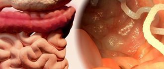
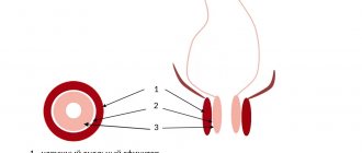
The parasitizing stage of the life cycle occurs in dwarf tapeworms in the human small intestine, where the worm egg is released from the shell and penetrates the mucous membrane of the wall (tissue phase, which can also take place in lymphoid follicles, liver tissue, mesenteric lymph nodes). The cysticercoid falls into the lumen and turns into an adult in approximately 2 weeks during the process of strobilation. It can reach sizes of 1.5-3 cm in length. Its movable head (scolex) is equipped with four suckers, a retractable proboscis and a corolla of chitinous hooks. The body of the parasite is divided into 200-300 tender segments - starting from the head from the immature, hermaphroditic ones and the last one containing the uterus with eggs. After destruction or separation of the segments, the eggs are released. They contain mature embryos, they are released during the act of defecation - the eggs are released along with the feces.
Model of dwarf tapeworm
Parasites feed on the entire body surface of neodermis thanks to pinocytosis . Usually, a person is both an intermediate and final host of the parasite, since both the tissue and intestinal phases of development take place in the body. One generation of parasitism takes approximately 2 months, then repeated oral invasion or intraintestinal autosuperinvasion is possible. In some cases, with age, there were cases of spontaneous self-liberation from invasion.
Helminthiasis
Parasitic worms found in the body of humans, animals and plants cause diseases called helminthiasis. Human parasitic worms disrupt the functioning of internal organs and the entire body as a whole, causing poor absorption of nutrients, weakness and diseases of the host body. There are three groups of worms that are classified as helminths:
- Nematodes or roundworms (pinworms, roundworms, Trichinella, Filaria, Intestinal eel);
- Trematodes or flatworms (Opisthorchis, Chinese fluke, Paragonimus, Schistosomes);
- Cestodes or tapeworms (Bovine and pork tapeworms, broad tapeworms, echinococcus, alveococcus).
Helminths have a similar morphology and are multicellular organisms that are visible to the naked eye. In their life cycle, helminths necessarily go through a number of stages: egg, larva and adult. During their life cycle, some worms change hosts. Such helminths are called biohelminths. Geohelminths are parasitic worms that do not require a change of hosts, and their life cycle occurs in the body of a single host. NEMATODOSIS Ascariasis Etiology, pathogenesis .
The causative agent is roundworm, which parasitizes in the adult stage in the small intestine. The lifespan of roundworms is about a year. In the migratory stage (the first 6–8 weeks after infection), roundworm larvae have a mechanical and sensitizing effect, causing hemorrhages and eosinophilic infiltrates in the tissues of various organs. In the intestinal phase (8 weeks after infection), adult roundworms cause general toxicoallergic and neuroreflex reactions of the body and a variety of local mechanical effects.
Symptoms, course
. The migration phase often occurs under the guise of acute respiratory infections and bronchitis; volatile eosinophilic infiltrates in the lungs are possible. In the intestinal phase, a gastrointestinal form is distinguished (salivation, nausea, loss of appetite, cramping pain around the navel, sometimes disorders of stool and gastric secretion); hypotonic (decreased blood pressure, weakness) and neurological (dizziness, headache, fatigue, sleep disturbance, vegetative-vascular disorders) forms.
Complications
. Ascariasis intestinal obstruction, a characteristic feature of which is the release of roundworms with vomit and palpation of a moving “tumor” - a ball of helminths; ascariasis appendicitis; perforated peritonitis; ascariasis of the liver with the development of jaundice, purulent angiocholitis, liver abscess, subphrenic abscess; ascariasis of the pancreas with symptoms of acute pancreatitis; crawling of roundworms into the respiratory tract with the development of asphyxia.
Diagnostics.
The diagnosis of the early phase of ascariasis is based on the detection of nematode larvae in the sputum and antibodies in the blood, and the late intestinal phase - ascaris eggs in the feces.
Prevention
.
Mass examination of the population and treatment of all those infected with ascariasis. Protection of the soil of vegetable gardens, orchards, and berry fields from contamination with feces. Thorough washing and scalding of vegetables and fruits with boiling water. Personal hygiene measures .
Hookworm disease (hookworm and necatoriasis)
Etiology, pathogenesis.
The causative agents are hookworm and necator, parasitic in the small intestines of humans, most often in the duodenum. Infection occurs when the larvae actively penetrate the skin or when they are ingested with contaminated vegetables, fruits, or water. The larvae migrate through the systemic and pulmonary circulation, lasting 7–10 days. In the small intestine, the larvae turn into sexually mature individuals and after 4–6 weeks begin to lay eggs. The lifespan of hookworms ranges from several months to 20 years. During the migration period, the larvae cause toxic-allergic phenomena. Adult helminths are hematophagous. When fixed to the intestinal mucosa, they injure tissue, lead to the formation of hemorrhage, erosion, cause bleeding, anemia, support the state of allergies, gastrointestinal dyskinesia and dyspepsia.
Symptoms, course
. Soon after infection, skin itching and burning, urticarial rashes, asthmatic phenomena, fever, and eosinophilia occur. In the late stage, nausea, drooling, vomiting, abdominal pain, intestinal dysfunction (constipation or diarrhea), and bloating appear. Sometimes pseudoulcer syndrome occurs (hunger pain in the epigastric region, hidden bleeding). The development of hypochromic iron deficiency anemia is characteristic; its severity depends on the intensity of invasion, nutritional deficiency, and individual characteristics of the organism. With low intensity, the invasion can occur subclinically, manifesting itself as eoeinophilia.
Diagnostics
. The diagnosis is confirmed by the detection of eggs in the stool and occasionally in the duodenal contents.
Prevention
. In areas of hookworm infection, you should not walk barefoot or lie on the ground without bedding. It is necessary to thoroughly wash and scald fruits, vegetables, and berries with boiling water; before eating them, you should not drink unboiled water.
Strongyloidiasis
Etiology, pathogenesis.
The causative agent is the intestinal eel, parasitizing in the intestinal wall (mainly the duodenum), sometimes in the ducts of the liver and pancreas, and during the migration period - in the bronchi and lung tissue. The penetration of Strongyloides larvae into the human body can occur actively through the skin (when walking barefoot, etc.) and through the mouth (when eating contaminated fruits, vegetables, and also when drinking water). Adult parasites, localized in the intestinal wall, injure the intestinal crypts (Lieberkühn's glands), solitary follicles and contribute to ulceration of the mucous membrane, and the larvae, migrating, damage the tissue of the liver, lungs and other organs. Of great importance is the toxic-allergic effect of adult parasites and their larvae on the human body, as well as secondary infection.
Symptoms, course
. Signs of allergy often occur: skin itching, burning, urticaria, eosinophilia, sometimes “volatile infiltrates” in the lungs, etc. In the late stage, the symptoms of gastroduodenitis, enterocolitis, and sometimes cholecystitis predominate. The typical triad is chronic recurrent urticaria, persistent enterocolitis and long-term high eosinophilia. Dizziness, headache, insomnia, and increased excitability are common. Sometimes the disease manifests itself only as eosinophilia and skin itching. The course is long, many years (until specific treatment), since with this helminthiasis (especially with constipation), autoreinvasion is possible. There are indications of a connection between hyperinvasion and the use of corticosteroids and immunosuppressive drugs. In severe forms of the disease, ulcerative enterocolitis develops with dehydration, cachexia, anemia and death.
Diagnostics
. The diagnosis is based on clinical data and the detection of acne larvae in feces (when examined using the Berman method) and in duodenal contents, less often in sputum and urine.
Trichocephalosis
Etiology, pathogenesis.
The causative agent is the whipworm, which parasitizes the human colon. The lifespan of the parasite is about 5 years. Whipworms injure the intestinal mucosa, are hematophagous and promote inoculation of microflora, causing reflex reactions in other organs of the abdominal cavity. Their metabolic products sensitize the body.
Symptoms, course
. Worries include drooling, decreased (less often increased) appetite, pain in the right half of the abdomen and epigastrium, nausea, constipation or diarrhea. There may be inflammatory phenomena in the intestines. Headache, dizziness, restless sleep, irritability are often observed; moderate hypochromic anemia and slight leukocytosis are possible. With intense invasion in children, cases of rectal prolapse (due to persistent diarrhea), epileptiform seizures, and Meniere's syndrome have been described. At low intensity, whipworm infestation is asymptomatic.
Diagnostics.
The diagnosis is made when whipworm eggs are found in the stool.
Prevention
. Mass examination of the population and treatment of all infested forms. Protection of the soil of vegetable gardens, orchards, and berry fields from contamination with feces. Thorough washing and scalding of vegetables and fruits with boiling water. Personal hygiene measures.
Trichostrongylidosis
The causative agents are small helminths from the family Trichostrongylidae. The main source of invasion is sheep, goats, and cattle. A person becomes infected through contaminated food or water.
Symptoms, course
. Characterized by nausea, abdominal pain, unstable stools, dizziness, and headache. Sometimes hypochromic or normochromic anemia and loss of strength develop. The diagnosis is based on the detection of trichostrongylid eggs in the stool.
Prevention.
Compliance with personal hygiene measures.
Trichinosis (trichinosis)
Etiology, pathogenesis
. The causative agent is Trichinella. In the larval stage, it enters the human body with the meat of pigs or wild animals (boar, bear, badger, etc.). After digestion of meat, muscle Trichinella are released from capsules, penetrate into the villi tissue, quickly grow, develop, and already 80–90 hours after infection, females lay numerous larvae, which are spread throughout the body. The place of further development of young Trichinella are the striated muscles, especially the intercostal, masticatory, muscles of the diaphragm, tongue, pharynx, and eyes. Here, after 2–3 weeks, trichinella coil up, become encapsulated, and some of them become calcified. The duration of the intestinal stage of Trichinella is about 2 months. Muscular trichinella remain viable for up to 20 years or more. The breakdown and metabolic products of Trichinella sensitize the body, causing eosinophilia, capillaropathy, muscle infiltrates, edema and other painful phenomena. The mechanical effects of adult Trichinella in the intestines and their larvae in muscles and other tissues, as well as secondary infection, are important.
Symptoms, course
. The incubation period is from 3 to 45 days. Early symptoms are characteristic: facial puffiness, accompanied by conjunctivitis, fever, muscle pain, eosinophilia. Various skin rashes are common, as well as gastrointestinal disorders, hypotension, muffled heart sounds, palpitations, headache, and insomnia. In mild forms, only eyelid swelling and eosinophilia are noted. The duration of the disease is from 2–3 days to 8 weeks or more. Sometimes a recurrent course of trichinosis is observed. Complications: myocarditis, meningoencephalitis, thrombosis of arteries and veins, pneumonia, nephritis, etc.
Diagnostics.
The diagnosis is based on epidemiological (consumption of undercooked pork and other meat, group diseases), characteristic clinical data (eosinophilia, swelling of the eyelids, fever, muscle pain). Serological reactions with trichinosis antigen are used. Confirmation of the diagnosis can be the detection of Trichinella and characteristic infiltrates in biopsied pieces of muscle (trapezius, deltoid, gastrocnemius) or in the remains of meat that caused the disease. A biopsy is performed on the 9th–10th day of illness.
Prevention.
You should not eat raw or undercooked pork, as well as the meat of wild boar, bear, badger, etc.
TREMATODOSIS Clonorchiasis
Clonorchiasis is a helminthiasis of the liver and pancreas caused by the fluke fluke. It is found among the population living in the Amur basin. Pathogenesis, clinical picture, diagnosis, treatment and prevention are the same as for opisthorchiasis.
Metagonimiasis
Metagonimiasis is a helminthiasis caused by a small trematode. The pathogen parasitizes in the small intestine of humans, dogs, cats, and pigs. Human infection occurs through consumption of raw fish. In the intestine, larvae hatch from the metacercariae of Metagonimus, which reach sexual maturity in the thickness of the mucous membrane after 2 weeks and emerge into the intestinal lumen. As a result of mechanical and toxicoallergic effects, enteritis develops.
Symptoms, course
. Abdominal pain, drooling, persistent diarrhea. Sometimes it occurs subclinically.
Diagnostics
. The diagnosis is based on the detection of Metagonimus eggs in the stool.
For prevention, see Diphyllobothriasis.
Nanophyetosis
Nanophyetosis is an intestinal helminthiasis that occurs among the population of the Far East. The causative agent is nanophyetus, which parasitizes the intestines of humans, dogs, cats, minks, and badgers. Infection occurs by eating raw fish. Helminths have a mechanical and toxic-allergic effect on intestinal tissue.
Symptoms, course.
Pain and rumbling in the abdomen, diarrhea, constipation, drooling and nausea at night.
Diagnostics
. Diagnosis is made based on the detection of nanophyetus eggs (resembling diphyllobothrid eggs) in the stool.
Treatment
. Male fern extract in a dose of 3–3.5 g.
Prevention
. You should not eat raw, uncooked or insufficiently salted and dried fish, as well as “live” pike caviar.
Opisthorchiasis
Etiology, pathogenesis
. The causative agent is the cat fluke, or Siberian fluke, which parasitizes the bile ducts of the liver, gallbladder and pancreatic ducts of humans, cats, dogs, etc. The parasite lives in the human body for 20–40 years. Infection occurs when eating raw (thawed, frozen), lightly salted and insufficiently fried carp fish (ide, chebak, dace, etc.). Opisthorchises injure the mucous membranes of the pancreatic and bile ducts, create obstacles to the outflow of bile, and contribute to the development of cystic enlargements and liver tumors. They have toxic and neuro-reflex effects. In the early stage, pronounced allergization of the body is observed (eosinophilic-leukemoid blood reactions).
Symptoms, course
. The incubation period is about 2 weeks. In the early period there may be fever, pain in muscles and joints, vomiting, diarrhea, pain and enlargement of the liver, less often the spleen, leukocytosis and high eosinophilia, allergic skin rashes. In the chronic stage, the most common complaints are pain in the epigastric region, right hypochondrium, radiating to the back and left hypochondrium. Sometimes there are attacks of pain such as gall bladder colic. Frequent dizziness and various dyspeptic symptoms. Muscle resistance in the right hypochondrium, liver enlargement, occasionally icteric sclera, enlarged gallbladder, and symptoms of pancreatitis are detected. Most often, with opisthorchiasis, the phenomena of angiocholitis, cholecystitis, biliary dyskinesia, chronic hepatitis and pancreatitis develop, and less often - symptoms of gastroduodenitis and enterocolitis. In some cases, a picture of cholangitic cirrhosis of the liver occurs. Opisthorchiasis can be asymptomatic.
Diagnostics
. The diagnosis is based on the detection of helminth eggs in the stool and duodenal contents.
Prevention
. Explaining to the population the dangers of eating raw, thawed and frozen (stroganina), lightly salted and insufficiently fried fish.
Fascioliasis
Etiology, pathogenesis.
Pathogens: liver fluke and giant fluke. The main source of human invasion is various farm animals. Human infection usually occurs in the warm season when fasciola larvae are ingested in water, sorrel, lettuce and other greens. The lifespan of helminths in the body is about 10 years. Trauma and toxic-allergic damage to the hepatobiliary system are important. Fasciolae can be carried into other tissues and organs.
Symptoms, course
. The disease is characterized by eosinophilia, allergic phenomena, disorders of the liver and gall bladder, reminiscent of the symptoms of opisthorchiasis (jaundice and attacks of gall bladder colic are more common).
Diagnostics
. Diagnosis of the early stage of fascioliasis is difficult, since helminth eggs are released only 3–4 months after infection. Immunological methods are used. In the late stage, the diagnosis is based on the detection of fasciola eggs in the duodenal contents and feces.
Prevention
.
Prohibition of drinking water from stagnant bodies of water; Thoroughly washing and scalding the greens with boiling water. CESTODOSES Alveococcosis Etiology, pathogenesis.
The causative agent is the larval stage of alveococcus. Infection occurs after oncospheres enter the mouth after contact with contaminated skins of foxes, arctic foxes, dogs, with the water of stagnant reservoirs and by eating wild berries collected in endemic areas. Clusters of larvae (usually in the liver) infiltrate and grow into tissues, disrupt the blood supply to organs, cause tissue degeneration and atrophy, and have a toxic-allergenic effect.
Symptoms, course
. The disease develops slowly and remains asymptomatic for a long time. There is a progressive enlargement of the liver, heaviness and pressure appear in the right hypochondrium, and a dull aching pain. After a few years, the liver becomes lumpy and very dense. Jaundice may develop. The spleen often becomes enlarged. Possible ascites. When the nodes disintegrate, body temperature rises, sweating, leukocytosis, eosinophilia, and increased ESR are observed. Hyperproteinemia and hypergammaglobulinemia are characteristic. Necrotization and invasion into the inferior vena cava can lead to profuse bleeding. With metastases to the lungs, symptoms of pneumonia, bronchitis, and hemoptysis may occur. Brain metastases mimic the clinical picture of a brain tumor.
Diagnostics.
Diagnosis is based on clinical data. Serological tests with alveococcal antigen are performed. To clarify the localization, X-ray and ultrasound examination, liver scanning, peritoneoscopy, and computed tomography are used. Test puncture is strictly prohibited due to the risk of contamination of other organs. Differentiate from tumors, echinococcosis and cirrhosis of the liver.
Hymenolepiasis
Etiology, pathogenesis
. The causative agent is the dwarf tapeworm. Infection occurs through ingestion of parasite eggs that come into contact with the patient and with contaminated feces, household items (chamber pots, toilet seats, etc.) and restroom walls. The local effect of larvae and adult forms on the intestinal mucosa is expressed in the destruction of villi, proliferative inflammation (sometimes with ulceration) of the mucous membrane with copious secretion of mucus. Toxic-allergic effects are also observed.
Symptoms, course
. Decreased appetite, nausea, abdominal pain, diarrhea or constipation, dizziness, headache, irritability, increased fatigue, restless sleep. Sometimes weight loss, moderate anemia, and eosinophilia are observed. Hymenolepidosis can be asymptomatic.
Diagnostics _
. The diagnosis is made based on the detection of dwarf tapeworm eggs in the feces.
Prevention
. Maintaining hygiene of the body, clothing, home, and public hygiene. Examination of all members of the patient’s family for helminths and simultaneous treatment of all infested ones.
Diphyllobothriasis
Etiology, pathogenesis
. The causative agent is the broad tapeworm. Its lifespan is tens of years. Human infection occurs when eating fresh, insufficiently salted caviar and raw fish (pike, perch, omul, grayling, etc.). The tapeworm, attaching to the intestinal mucosa with its bothria, injures it. Large accumulations of the parasite can clog the intestinal lumen. Helminth metabolic products sensitize the body. Absorption of vitamin B12 by the broad tapeworm directly from the digestive tract leads to hypovitaminosis B12 and the development of anemia.
Symptoms, course
. Characterized by nausea, unstable stools, and the release of fragments of strobili during defecation. Patients sometimes complain of weakness, dizziness, and abdominal pain. Occasionally, pernicious anemia develops, close to Addison-Birmer anemia.
Diagnostics
. The diagnosis is made by detecting tapeworm eggs and strobila fragments in the feces.
Prevention
. You should not eat raw, uncooked or insufficiently salted and dried fish, as well as “live” pike caviar.
Taeniasis
Etiology. The causative agent is the pork tapeworm, which can parasitize humans not only in the sexually mature stage, but also in the larval stage, causing the disease cysticercosis. The adult helminth parasitizes the small intestine for many years. Humans become infected with taeniasis by eating raw or half-raw meat containing taeniasis.
Symptoms, course.
The same as with teniarhynchosis, however, the segments of the parasite do not actively come out of the anus (occasionally, tender segments come out with feces, which the patient usually does not notice).
Diagnostics
. The diagnosis is made on the basis of repeated examination of stool for the presence of helminth segments and mucus from the perianal folds (by scraping) for the presence of tapeworm eggs.
Prevention.
You should not eat undercooked or undercooked pork.
Causes
Infestation of the tapeworm Hymenolepis nana occurs through contact (through dirty hands, food, water and household items, such as toys, pots, door handles) and children are 4-5 times more susceptible than adults. This is due to age characteristics and lack of necessary hygiene skills. Infection occurs fecal-orally - from sick people (in more rare cases, from rats and mice, infection occurs with helminths Hymenolepis diminuta, while the intermediate hosts are insect larvae). Most often, infection occurs when hygiene rules are violated, for example, as a result of untidiness.
Ways of infection with hymenolepiasis
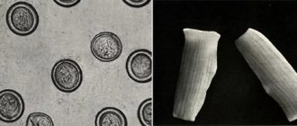
The eggs are spherical or ellipsoid (40 × 50 µm), with a shell, transparent, double-circuited, they contain a larva in the form of an oncosphere with 6 hooks. They can remain viable for up to 1 day, or more so for up to a month in water or a humid environment. Not resistant to drying, exposure to light rays, temperature 3°C, boiling for 15 minutes at 60°C.
Types of disease
If you suspect that this disease occurs, you should consult a doctor.
Features of the development of helminths and the possibility of autoinvasion require deworming, symptomatic therapy and active preventive measures. For specific treatment of hymenolepiasis, anthelmintic drugs praziquantel (one dose) or phenasal (in the form of 3 seven-day, 4 five-day or 6-7 two-day cycles or according to another scheme), and male fern extract are used. In between deworming cycles, restorative therapy (multivitamins, calcium supplements) is carried out. Control stool tests are performed 15 days later and then monthly for six months after the end of the main course of treatment for hymenolepiasis. If helminth eggs are detected, a second course of therapy is carried out.
During treatment of hymenolepiasis, it is important to adhere to a nutritious diet, observe a sanitary and hygienic regime (disinfect care items, promptly change underwear and bed linen, regularly perform personal hygiene). Patients with negative results of control stool tests within 6 months after the end of the course of therapy for hymenolepiasis are considered cured.
Peptic ulcer of the stomach or duodenum can be a consequence of hymenolepiasis, in other cases against its background it takes a more severe course. Hymenolepidosis can be complicated by mesenteric lymphadenitis.
Symptoms of dwarf tapeworm
Hymenolepidosis causes both dyspeptic disorders (development of ulcers, enteritis ) and negative reactions from the central nervous system. Patients develop the following symptoms:
- general malaise or weakness;
- increased fatigue and memory impairment;
- changes in appetite and weight loss;
- attacks of heartburn and frequent belching;
- nausea with vomiting;
- severe abdominal pain, even acute abdomen;
- the presence of blood and mucus in the stool;
- stool instability;
- moderate bloating and pain on palpation;
- hepatomegaly;
- deterioration of mood and development of tearfulness, irritability;
- headache;
- dizziness;
- sleep disturbances and even episodes of insomnia ;
- epileptimorphic seizures .
Infested individuals experience allergotoxic and immunosuppressive effects of waste products of the larval stage and sexually mature parasites, as well as products formed during the breakdown of host tissues. allergosis that develops causes eosinophilia , skin itching and rashes, and an asthmatic condition.
Epidemiology
The main source of G. is a sick person. Humans can become infected from mice and rats. The main mechanism of transmission is fecal-oral. The most important transmission factors are dirty hands, as well as toys, food, and household items contaminated with feces. In untidy subjects, helminth eggs may linger on the perianal folds, which may cause epidemiol. significance, especially in cases of G.’s combination with enterobiasis (see). One of the transmission factors may be flies. The role of insects as intermediate hosts of helminths in the epidemiology of helminths on the territory of the USSR is negligible. Ascariasis has an indirect effect on the spread of G. In areas where the latter is widespread, G. is almost never found or not found at all.
G. affects mainly children of preschool and primary school age. The maximum incidence occurs between the ages of 4 and 9 years. Large outbreaks can occur in children's institutions with unsatisfactory hygiene. condition. Diseases of adults are also common.
Pathogenesis. A person's susceptibility to primary and repeated infections of G. depends on nutritional conditions, immunol, condition, concomitant diseases, climate, etc. The leading pathogenetic factors in G. are allergies and the mechanical effect of cysticercoids and mature tapeworms on intestinal tissue, as well as the effect on the host body products of their metabolism and decay. Allergies and constant intestinal microblood loss underlie the pathogenesis of anemia in G. Intestinal dysbiosis and allergic reactions are associated with disturbances in fermentation and absorption processes in the intestine. These same factors underlie hypovitaminosis B2, C and PP, observed in G. The long course of G. is caused by superinvasions, since the lifespan of one generation of helminth, as a rule, does not exceed 2 months. Superinvasion can be exogenous and intraintestinal autosuperinvasion.
Tests and diagnostics
To confirm the diagnosis of hymenolepiasis, in addition to studying the clinical picture, it is necessary to conduct a study to identify eggs in the masses of feces. For this purpose, the scatological methods of Kalantaryan, Fulleborn and Kato are used.
Serological tests in patients can reveal mild or more severe anemia , eosinophilia, leukopenia, lymphocytosis, monocytopenia .
How to get tested for hymenolepiasis?
Since the release of dwarf tapeworm eggs occurs cyclically and depends on the strength of the invasion, in vitro tests should be taken three times, every 5 days.
In order to most correctly pass the test for hymenolepidosis for the pool on the eve of the tests, prescribe Fenasal (0.5 -1) and then take Purgen , which increases detection by about 50%. Since the dwarf tapeworm is not resistant to environmental influences, stool examinations should be carried out no later than 3 hours after defecation.
What you need to know about the analysis
In order for a specialist to correctly establish a diagnosis, he first relies on the patient’s medical history (patient complaints) and the results of the tests obtained. Microscopic examination of stool three times with an interval of 2 weeks.
The biomaterial is collected in the morning using a native smear. This method will be effective if there are a large number of parasite eggs. The enrichment method involves the study of stool using Fenasal at a low dosage before delivery the next morning.
The method has a number of contraindications, so it is necessary to consult a doctor before using it. The examination itself is divided into specific (examination of stool for the presence of helminth eggs) and nonspecific (blood test, examination by a specialist).
Diet for hymenolepiasis
Diet for worms
- Efficacy: healing effect
- Terms: 21-30 days
- Cost of products: 1300-1400 rubles per week
The diet during the treatment of this helminthiasis should, first of all, be fortified and complete - capable of fully providing the body with proteins, fats and carbohydrates.
It is best to give preference to dairy and plant foods, and also limit the consumption of animal fats and roughage. It is important to adhere to the rules of a healthy diet:
- cook steamed dishes, eat them fresh, stewed or boiled;
- give up fatty, smoked, too hot, spicy and salty foods;
- It is best to switch to proper bread, rye bread and give up refined white flour and confectionery products;
- use healthy vegetable oils (olive, pumpkin, sesame), herbs, seeds and nuts;
- replace sugar with natural sweets and fruits.
Pathological anatomy
The villi of the small intestine affected by cysticercoids are thickened, the stroma is shifted towards the epithelium.
As the larvae mature, the phenomena of necrobiosis and necrosis intensify, the stroma is saturated with exudate and infiltrated with segmented leukocytes, and the epithelial cells are partially desquamated. In places where the head of the tapeworm is fixed, the villi are destroyed, and the resulting ulcers reach the submucosal layer. Characterized by plethora of all layers of the small intestine, hyperemia of lymphoid follicles, degeneration of nerve cells of the intestinal plexus. G. is accompanied by damage to other organs. Characterized by focal dystrophy of the myocardium and liver; proliferation of connective tissue of the liver, lungs, pancreas, myocardium; lymphoid and lymphoidoeosinophilic infiltrates in the lungs, liver, pancreas, and in severe cases - in the pia mater.
Immunity
Apparently, most people have a short-term course of G., since sufficiently strong immunity develops that prevents the occurrence of superinvasions. Under unfavorable conditions, relative immunity occurs, which is insufficient to prevent re-infections. In these cases, the disease takes a long course. At the age of 10-14 years, most children usually get rid of the invasion without treatment, which is explained by both the development of age-related immunity and the acquisition of gigabytes. skills.
It is possible to defeat parasites!
Antiparasitic Complex® - Reliable and safe removal of parasites in 21 days!
- The composition includes only natural ingredients;
- Does not cause side effects;
- Absolutely safe;
- Protects the liver, heart, lungs, stomach, skin from parasites;
- Removes waste products of parasites from the body.
- Effectively destroys most types of helminths in 21 days.
There is now a preferential program for free packaging. Read expert opinion.
Read further:
Dwarf tapeworm (hymenolepis nana): development cycle, symptoms and treatment, routes of infection
Why take an enterobiasis test for a swimming pool certificate: rules, deadlines and instructions
Causative agents of anthrax: who is the causative agent, characteristics, carriers
Bovine tapeworm - treatment and control of bovine tapeworm, what methods and medications to use
Staphylococcus aureus in the stool, nose, throat and intestines: symptoms, degrees of infection and treatment
How the roundworm uses oxygen in the process of breathing, roundworm breathing