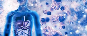
Every year, more than thirty thousand residents of Russia are diagnosed with gastric carcinoma, which means the appearance of a malignant tumor in the tissues of the organ. As a rule, it develops from glandular cells designed to secrete gastric juice. The disease is in second place in the frequency of detection among oncological pathologies, and men are affected more often than women. In recent years, the total number of patients has been decreasing, but the proportion of women among them is increasing: today, for every four male patients, there are three women with stomach cancer.
Kinds
The disease is classified according to several main characteristics. Based on localization, neoplasms of the anterior or posterior wall, the fundus of the stomach, the pyloric part, and the cardiac region (the junction of the stomach and the esophagus) are distinguished. According to the method of development they distinguish:
- mushroom-shaped or polyp-shaped, characterized by slow growth and extremely weak metastasis;
- ulcerated - saucer-shaped, with a clear demarcation from healthy tissue and late metastasis;
- ulcerative-infiltrative – with germination into the tissue and the absence of a clear pathological boundary;
- diffuse-infiltrative - in the form of numerous ulcers, with a tendency to grow into the tissue.
According to the histological form, i.e. type of malignant cells, distinguished:
- signet ring cell carcinoma of the stomach - one of the most aggressive types of squamous cell tumor, the development of which is associated with hormonal imbalances, affecting people in the age ranges of 40-50 and 60-70 years;
- adenocarcinoma - with long-term development (over 15-20 years), developing from glandular cells of the mucosa and accounting for 96% of all clinical cases;
- undifferentiated gastric carcinoma, characterized by rapid growth, developing in epithelial tissues and prone to active metastasis;
- glandular-squamous form, which is characterized by a fragmented structure of fragments of adenocarcinoma and squamous cell tumor;
- an unclassifiable form formed from multilayered epithelial tissue.
Based on biological activity, a distinction is made between intestinal-type neoplasms, consisting of highly differentiated, clearly structured cells, and diffuse or discogesive gastric carcinomas with a loose structure, which are characterized by a high degree of aggressiveness, rapid growth and active metastasis in the early stages.
Stomach cancer
One of the important aspects of the work of any doctor is the manifestation of oncological vigilance, since there are a number of tumors that in the early stages of development do not give a pronounced clinical picture, but remain a fairly common phenomenon in the population. One of these neoplasms is stomach cancer, which ranks second in the structure of mortality from malignant neoplasms in men and third in women.
Fungal carcinoma of the stomach with ulceration, located in the antrum.
Gastric cancer is a malignant tumor arising from the gastric mucosa. The mucous membrane is represented by a single-layer columnar epithelium, lamina propria and muscle, forming folds, gastric fields and gastric pits into which the excretory ducts of the gastric glands exit. Gastric cancer in 90% of cases is adenocarcinoma.
Papillary adenocarcinoma of the gastroesophageal junction with tumor infiltration into the submucosa.
Etiology
At the moment, there is no clear answer to the question of the etiology of the disease. Chronic infection with Helicobacter pylori is of primary importance. Other causes include infection with the Epstein-Barr virus, genetic mutations, obesity and environmental factors. Among environmental factors, smoking, nitrate-containing foods, and pickled foods have the greatest influence.
Therefore, much attention in the doctor’s practice is paid to the treatment of diseases underlying the development of cancer, such as chronic atrophic hyperplastic gastritis associated with Helicobacter pylori, adenomatous polyps, pernicious anemia, conditions after gastrectomy, Menetrier’s disease (hypertrophic gastropathy, hyperplastic giant-fold gastritis), as well as treatment of precancerous changes in the mucous membrane: intestinal metaplasia and dysplasia.
Tubular adenocarcinoma
Pathogenesis
Among the theories of gastric cancer carcinogenesis, the theory of the P. Correa cascade, proposed by the author back in 1988, dominates. At that time, this model did not include H. pylori; it was included only two years later. There is no information reliably confirming the synthesis or secretion of mutagenic or carcinogenic substances by the bacterium. Therefore, according to the theory, chronic persistence of H. pylori causes only a violation of the cellular renewal of the gastric mucosa, which consists in the accelerated movement of cells from the generative zone without full differentiation into the zones where mature specialized epithelial cells are located. This occurs due to the production of cytokines and oxygen metabolites by the cells of the inflammatory infiltrate. The inability of the gastric glands to fully function develops and, as a consequence, atrophy, metaplasia, dysplasia and carcinoma.
Among the causes of diffuse cancer are genetic mutations in the CDH1 and CTNNA1 . A mutation in the TP53 causes Li-Fraumene syndrome, and in the APC gene - familial adenomatous polyposis.
Diffuse infiltrative gastric cancer (linitis plastica)
Clinical picture and symptoms
The tumor in the early stages does not have pathognomonic symptoms. As the disease progresses, the following symptoms appear:
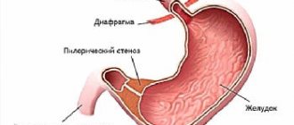
- Local may be represented by pain in the epigastric region . As the disease progresses, the character changes to shingles when it grows into the pancreas, and angina when it grows into the diaphragm. When located in the cardia region, dysphagia occurs, and over time, a feeling of heaviness in the chest, nausea, and vomiting develops. With the development of a tumor in the pyloric region, there is a violation of the evacuation of food and, as a result, heaviness after eating in the epigastrium, vomiting of food eaten the day before, belching of rotten fluid, splashing of liquid, cramping pain due to increased peristalsis of the stomach. When a tumor grows into the transverse colon, bloating, rumbling, and stool retention occur.
- Common symptoms include: loss of appetite, aversion to food or lack of satiety, weight loss, increasing weakness, increased fatigue. This symptom complex A.I. Savitsky combined it into the “small sign syndrome”.
- A tumor complicated by bleeding is manifested by weakness, dizziness, palpitations, fatigue, fainting, vomiting of unchanged blood, or “coffee grounds”, melena (tarry feces). When perforation occurs, “dagger” pain occurs, intensifying with movement, severe weakness, cold sweat, and sometimes loss of consciousness. Fever with intoxication syndrome occurs when the tumor becomes infected.
- When metastases appear in the liver, compression of the extrahepatic bile ducts occurs, jaundice occurs, and skin itching occurs. Peritoneal carcinomatosis or compression of the portal vein is manifested by ascites and obstruction. With a significant increase in Schnitzler's metastasis in the perirectal tissue, defecation disturbance occurs. Dysfunction of the ovaries occurs with bilateral damage to the ovaries (Krukenberg cancer). By palpation, Irish metastases are determined in the axillary lymph nodes, Virchow metastases in the left supraclavicular node and Sister Mary Joseph metastases in the navel along the round ligament of the liver.
An infiltrative-ulcerative type of stomach cancer, located in the body of the stomach along the lesser curvature.
Treatment
It is performed using surgical technologies (including endoscopic), combined chemotherapy, and radiation methods.
Endoscopically operated:
- early cancer in patients at high risk for surgery;
- carcinoma located within the mucous membrane;
- tumor size not exceeding 2 cm and without ulceration;
- neoplasm with no metastatic lesions of regional and distant lymph nodes;
- cancer with no lymphovascular invasion.
There are two options for endoscopic operations: resection of the mucosa and resection of the mucosa with dissection of the submucosal layer. If, after a routine histological examination of the removed part, invasion of tumor cells into the submucosal layer, a poorly differentiated form of cancer or lymphovenous invasion is revealed, the patient is subject to surgical treatment.
If the patient has submucosal invasion, large extent, poorly differentiated forms, ulcerated tumors, then subtotal gastrectomy or gastrectomy with lymphadenectomy is performed.
Early stomach cancer. A: type I (elevated). B: type IIa (elevated). C: IIc (in-depth). D: IIb+IIc (flat, recessed)
Planning and implementation of surgical treatment includes several stages: choice of surgical approach, choice of the scope of surgery on the organ, lymph node dissection, choice of reconstruction method:
The choice of surgical approach depends on the level of tumor spread. If there is no involvement of the cardioesophageal junction and the esophagus, then the access is midline laparotomy. With limited damage to the distal esophagus and cardia, a thoracolaparotomy access on the left or a wide diaphragmotomy from a laparotomy access. In case of total damage to the stomach with transition to the esophagus or body cancer with proximal spread to the esophagus - abdominomediastinal (in some cases, with a high intersection of the esophagus, in order to safely form an esophageal-intestinal anastomosis, it is possible to use a left thoracotomy along the VI intercostal space).
The choice of the extent of the operation includes options such as gastrectomy, subtotal distal and subtotal proximal gastrectomy:
Radical surgery includes a one-time removal of the affected area of the stomach with two omentums, tissue, and regional lymph nodes; the organ is intersected at a distance of at least 5 cm from the visible edge of the tumor with a limited type of growth and at least 6–7 cm for cancer of ulcerative-infiltrative and diffuse types. Confirmation of complete excision of the tumor occurs by conducting an urgent histological examination along the line of intersection.
- Distal subtotal gastrectomy is performed for cancer of the antrum of the stomach without foci of severe dysplasia and cancer in situ in the proximal part of the stomach or in patients with low functional reserves when an exophytic or mixed tumor has spread to the lower third of the body of the stomach. The left paracardial lymph nodes and nodes in the hilum of the spleen are not removed, as they are rarely affected in this type of cancer.
- Proximal subtotal gastrectomy is indicated for cancer of the cardioesophageal junction, for small tumors of the upper third of the stomach of exophytic or mixed growth.
- Gastrectomy with removal of all regional lymph nodes is performed in all other cases. For resectable gastric cancer of the linitis plastica type (diffuse cancer), undifferentiated forms of cancer, diffuse type hereditary gastric cancer syndrome, only gastrectomy is performed.
- Lymph dissection. Lymphogenous metastasis in gastric cancer occurs in 47.7% of cases and depends on the depth of tumor invasion. The standard procedure is to remove 1st and 2nd order lymph nodes. In the presence of distant metastases, palliative interventions are performed.
Combined adjuvant chemotherapy is carried out according to XELOX (CAPOX) regimens 4–6 weeks after surgery in the absence of severe complications and after normalization of clinical and laboratory parameters. The regimens include the drugs capecitabine and oxaliplatin.
Neoadjuvant chemotherapy is carried out with 3 courses of regimens CF (Cisplatin, 5-FU), ECF (Epirubicin, Doxorubicin, Cisplatin, 5-FU), ECX (Epirubicin, Doxorubicin, Cisplatin, Capecitabine) or EOX (Epirubicin, Doxorubicin Oxaliplatin), then if the tumor resectable, then an operation is performed, after which an additional 3 cycles of similar chemotherapy are prescribed. Perioperative chemotherapy is an alternative to postoperative chemoradiotherapy for resectable gastric cancer with lymph node dissection, according to the results of studies, as it helps to increase five-year survival.
For locally advanced unresectable and disseminated cancer of the stomach and esophagogastric junction, systemic chemotherapy is prescribed. The choice of a specific combination depends on the patient’s condition, the nature and severity of concomitant diseases. In particular, the CF, CX, XELOX, IF (1a), FOLFOX regimens demonstrated a statistically significant increase in patient survival, in contrast to monocomponent treatment.
Symptomatic therapy is aimed at relieving complications. In case of bleeding, endoscopic bleeding control is performed.
Tumor stenosis is eliminated by bougienage or recanalization, balloon dilatation, and installation of a self-expanding stent in the area of stenosis. If these procedures are not possible, gastrojejunostomy, palliative gastrectomy, percutaneous endoscopic or interventional gastrostomy in patients with dysphagia, endoscopic or surgical jejunostomy in patients with stenoses at the level of the middle and lower third of the stomach are performed. The pain is relieved by external beam radiation or drug therapy. It is also possible to use regional anesthesia. Ascites is relieved with diuretics, laparocentesis, and intra-abdominal administration of cisplatin.
The highest five-year survival rate is observed after radical operations for non-invasive tumors of high differentiation and those that do not involve metastases in regional lymph nodes.
Thus, the absence of pathognomonic symptoms for early forms and the wide prevalence of H. pylori in the population allow gastric cancer to remain in the leading positions in morbidity and mortality in Russia, despite the emergence of more modern treatment methods.
Polypoid gastric cancer with hemorrhage in the center of the tumor. The cancer is located in the antrum along the lesser curvature of the stomach.
Sources
- Chissov V.I. et al. Malignant neoplasms in Russia in 2010 (morbidity and mortality) // M.: FSBI “MNIOI im. PA Herzen" Ministry of Health and Social Development of Russia. – 2012. – P. 12.
- Maev I.V., Zairatyants O.V., Kucheryavyi Yu.A. Intestinal metaplasia of the gastric mucosa in the practice of a gastroenterologist: a modern view of the problem //Ros. magazine gastroenterol., hepatol., coloproctol. – 2006. – T. 16. – No. 4. – pp. 38-47.
- Kharnas S. S., Levkin V. V., Musaev G. Kh. Stomach cancer (clinic, diagnosis, treatment): Textbook. village for students //M.: Publishing house. house “Russian doctor. – 2006.
- https://vk-cc.com/FPDGQC
- https://vk-cc.com/pDw45jl
- https://vk-cc.com/Pnyovkr
- https://vk-cc.com/skU7LdXw
- https://vk-cc.com/Pfu36JGn
Symptoms
At the early stage of gastric carcinoma, symptoms are either completely absent or do not differ from the signs of a normal inflammatory process. Subsequently, the patient develops:
- decreased appetite and weight loss;
- anemia, pale skin;
- constant heartburn and bloating;
- nausea, occasional vomiting after eating;
- stomach ache;
- swelling of the supraclavicular lymph nodes;
- with a tumor in the cardia area - difficulty swallowing;
- stomach bleeding;
- at the last stage – cancer intoxication.
Causes of stomach tumors
Unfortunately, it is impossible to say that research has identified all possible causes of stomach cancer. However, some factors predisposing to the appearance of a tumor are known.
- Frequent consumption of foods containing large amounts of animal fats.
- Consumption of vegetables grown with nitrates and nitrites. You should know where exactly in which types of vegetables harmful substances accumulate, and try not to eat these parts.
- Alcohol consumption - modern research shows that with frequent alcohol consumption, the likelihood of malignant stomach tumors increases.
- Smoking.
- Cosmetics, like smoking, are also a source of nitrates and nitrites in the body, so excessive use of them also increases the risk of stomach cancer.
- Abuse of smoked and dried food, since this type of food also contains nitrates and nitrites.
- “Risk products” also include hard cheeses and beer, mostly pasteurized.
The presence of so-called “precancerous diseases” also significantly increases the risk of stomach tumors. These include:
- chronic stomach ulcer;
- gastric polyps;
- chronic gastritis with low acidity;
- pernicious anemia (with B12 deficiency).
Also at risk for stomach cancer are people who have had part of their stomach removed due to some other disease.
The risk of cancer depends on the severity of concomitant atrophic gastritis, since it reduces the acidity of gastric juice, which entails an increase in the number of microbes, and, as a consequence, an increase in the formation of nitrogenous compounds.
Regarding gastric ulcers, until recently it was believed that in every eighth case it turns into cancer, but modern research has shown that in most cases the disease was not an ulcer, but was gastric cancer at an early stage with ulceration. However, in some cases, malignancy of the stomach ulcer still occurs.
Forms of stomach cancer
A malignant tumor of the stomach, like tumors of any other hollow organs, has three types of growth:
- Exophytic type. The tumor grows into the lumen of the organ. This type of tumor growth includes the following varieties: plaque-like
- saucer-shaped - is the most common type of growth of stomach cancer (accounts for about 35% of diseases); This is an ulcerated cancer; its edges are raised and clearly defined; It is quite difficult to visually distinguish it from a stomach ulcer; has a relatively benign course
- polypoid - is the rarest type of growth of stomach cancer (only about 5% of cases); the tumor does not have ulceration and is well limited from healthy tissue; of all varieties has the most favorable prognosis.
- infiltrative-ulcerative
Histological types of stomach cancer tumors
Today there are 6 main histological types of stomach tumors:
- adenocarcinoma (glandular cancer);
- mucous cancer (also called colloid);
- solid cancer;
- fibrous cancer (scirrh);
- small cell carcinoma;
- squamous cell carcinoma.
Adenocarcinoma is the most common form of stomach cancer, while squamous cell carcinoma is the least common.
Adenocarcinomas come in several varieties:
- Papillary adenocarcinoma. This tumor is characterized by the formation of finger-like (narrow or wide) epithelial outgrowths with a fibrous base. Tumor cells are most often polarly oriented (this orientation has a pronounced character).
- Tubular adenocarcinoma. This tumor consists of tubular structures that are located in the fibrous stroma. The lumens of the glands become cystically dilated due to the mucus content.
- Mucinous adenocarcinoma (mucinous adenocarcinoma). Such a tumor is characterized by the presence of extracellular mucin in large quantities (more than half of the tumor). Tumor cells are arranged either in chains or randomly, and are surrounded by “lakes” of mucus.
- Signet ring cell adenocarcinoma. In such a tumor, more than half of it is represented by cells that contain mucin in the cytoplasm. Intracytoplasmic mucin compresses the nuclei of tumor cells and displaces them to the periphery; therefore the cells have a signet ring shape. Such cells tend to diffusely infiltrate. They can be combined with severe fibrosis (this tumor is called scirrhous carcinoma). They do not form tubules, but functionally they are glandular, and an admixture of the glandular component in such tumors is very often found - this is what allows them to be classified as adenocarcinomas.
Based on the degree of differentiation of tumor cells, adenocarcinomas are divided into 3 types:
- well-differentiated - such tumors consist of regular glandular structures reminiscent of non-tumor glands of the stomach, especially those lined with metaplastic intestinal epithelium;
- moderately differentiated adenocarcinomas have a structure “intermediate” between the structure of highly differentiated and poorly differentiated tumors;
- poorly differentiated adenocarcinomas consist of individual cells and clusters; The glandular structures of such tumors are difficult to determine.
Colloid (mucosal) gastric cancer is characterized by the spread of the tumor mainly in the submucosal layer or between the layers of the muscular layer; the tumor forms in the form of layers of mucous masses from cells containing mucus. This type of cancer is characterized by a noticeable enlargement of the stomach. The stomach wall, saturated with such mucus, can be greatly thickened. When it is cut, mucus flows out of it.
Fibrous cancer (scirrhus) of the stomach is characterized by a significant degree of development of connective tissue, among which small cancer cells with a cubic shape form small cells and cords. Sometimes the number of cancer cells is so small that the nature of the tumor can only be determined by examining regional lymph nodes. The ulcerative disintegration of such a tumor can cause gastric bleeding, sometimes quite profusely.
Solid cancer (or brain cancer) is a type in which the tumor tissue is highly anaplastic; it is rich in small polygonal cancer cells; On the contrary, there is very little connective tissue stroma in such tumors.
Small cell gastric cancer is a fairly rare form (approximately 0.6% of gastric cancer cases). The tumor consists of small cells similar to lymphocytes. These cells form solid structures or sheets. Many cells contain neuroendocrine granules containing serotonin, gastrin or other peptides. According to clinical and morphological characteristics, small cell tumor of the stomach is very similar to small cell tumor of the lung.
Squamous cell carcinoma of the stomach is extremely rare. It originates from the metaplastic glandular epithelium of the stomach.
Mixed forms of stomach cancer are quite common. In addition, some tumors can change from one state to another as they grow, for example, adenocarcinoma can turn into solid or colloid cancer, and solid into fibrous.
Causes and risk factors
Oncologists and gastroenterologists identify a number of factors contributing to the development of a malignant tumor:
- presence of Helicobacter pylori bacteria in the stomach;
- inflammation of the mucous membrane;
- the presence of a similar disease in close relatives;
- exposure to the gastric mucosa of toxins or chemical irritants;
- long-term smoking and excessive drinking;
- the presence of neoplasms in other organs of the digestive system;
- mature and old age;
- bad eating habits - addiction to fried, spicy foods, smoked foods, fatty foods.
Causes of neoplasms
The exact reasons for the development of the infiltrative form of oncopathology have not been reliably elucidated. It is known that the prerequisites for the appearance of adenocarcinoma are an unhealed ulcer and progressive atrophic gastritis. Tissue malignancy can occur at any stage of the disease.
Factors that provoke cell malignancy include:
- the patient lives in a region with poor ecology;
- poor nutrition with insufficient consumption of fiber, vitamins, minerals and an excess of foods in the diet that destroy the mucous membrane. Also, serious harm to the digestive tract is caused by too hot or cold food, regular overeating, and strict mono-diets;
- long-term work in hazardous work;
- hormonal disorders;
- weakened immune system;
- constant stress;
- smoking, alcohol abuse.
There is a genetic predisposition to the infiltrative form of oncopathology. If among the patient’s closest relatives there were people with malignant neoplasms of the cavity of the digestive organ, the disease develops before the age of 30 years.
Who is at risk
People who have been diagnosed with:
- atrophic gastritis;
- polyps with a wide base;
- alcoholism, drug addiction;
- chronic ulcer (large ulcerations are especially dangerous);
- congenital or acquired immunodeficiency conditions.
People with poor heredity, as well as those who work in the production of nickel, asbestos, chromium, rubber and other “harmful” enterprises, need to undergo regular medical examinations.
According to statistics, cancer pathology is diagnosed 15% more often in men. Patients of any age have a risk of developing malignant neoplasms, but the disease is more often diagnosed in people over 45 years of age.
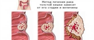
Stages
- The neoplasm does not extend beyond the mucous membrane, there are no metastases. The patient develops anemia, weakness, and fatigue.
- Malignant tissue grows into the muscle tissue of the gastric wall, and 1-2 metastases appear in the lymph nodes. Minor abdominal pain appears and the temperature rises slightly.
- The tumor penetrates the stomach wall into the abdominal cavity and metastasizes to 7 or more lymph nodes. The patient suddenly loses weight, experiences frequent vomiting with blood in the vomit, and the digestive process is disrupted.
- Cancerous tissue grows and gives abundant metastases to other organs of the abdominal cavity, lungs, bone tissue, and brain. Abdominal pain intensifies, the volume of the abdomen increases due to the accumulation of fluid in the peritoneum, the skin and eye sclera turn yellow. The condition is deteriorating sharply.
Stages of development
Like most malignant neoplasms, infiltrative cancer in the gastric cavity goes through four stages of development. Each of them has features:
- I - is primary in nature and responds well to therapy. Pathogenic cells are not aggressive and spread only to the mucous and submucosal layers of the organ cavity. There are no metastases or lesions of regional nodes;
- II - adenocarcinoma begins to grow, malignant cells penetrate into the lymph nodes closest to the main focus. The neoplasm is located within the gastric cavity, grows into the serous tissue, and initial metastases are observed;
- III - the muscles of the organ are affected, malignant cells are found in ten or more lymph nodes. Ulceration extends beyond the boundaries of the affected cavity, metastases form in nearby organs (spleen, liver, diaphragm and others);
- IV - characterized by the detection of metastases in distant organs and lymph nodes.
If at stages I and II the development of pathological changes can be stopped, then stages III and IV have an extremely unfavorable prognosis and are characterized by high mortality. The sooner you see a doctor, the greater the chance of cure.
Diagnostics
To identify gastric carcinoma, a gastroenterologist or oncologist prescribes a series of diagnostic studies and tests;
- EGDS - using a probe with a miniature video camera, the diagnostician examines the inner surface of the stomach to detect a tumor or ulcer and take a tissue sample for examination;
- endoscopic ultrasonography - ultrasound examination from the inside of the stomach using a sensor built into the endoscope to determine the depth of tumor growth into the wall;
- CT – layer-by-layer examination of stomach tissue to obtain a three-dimensional image of its structure;
- diagnostic laparoscopy – allows you to examine the tumor in detail and determine the possibility of surgery;
- blood test for tumor markers - to identify specific antibodies to cancer cells;
- general blood test - to assess hemoglobin levels and other characteristics;
- biochemical analysis - to determine important indicators of blood composition.
Second histological classification of stomach tumors
The histological classification according to Lauren has also become widespread, distinguishing two types of gastric carcinoma:
- Intestinal type of stomach cancer. Another name for it, intestinal cancer, means that the tumor is microscopic in size and consists of intestinal-type cells. This is usually a well-differentiated adenocarcinoma. The form is most often polypoid or mushroom-shaped. Most often, such cancer develops against the background of chronic gastritis with intestinal epithelial metaplasia. Diagnosed in patients over 50 years of age (in men - twice as often as in women).
- Diffuse type of stomach cancer. Most often it is signet ring cell undifferentiated cancer, sometimes it is poorly differentiated adenocarcinoma. Intestinal metaplasia outside the tumor is almost always absent, and epithelial-like cells are also absent in the tumor itself. This cancer is diagnosed in young patients, in men and women - with the same frequency.
Localization of stomach tumors
Most often, a stomach tumor is localized in its antrum and pylorus (about 70% of all cases). In second place is the lesser curvature of the body of the stomach (up to 15% of cases). In third place is cardia - up to 10%. Most rarely, tumors are localized on the anterior and posterior walls of the stomach - up to 5% of cases.
Treatment
The success of oncologists’ efforts is determined by a number of factors, and primarily by the type of tumor.
Thus, for signet ring cell carcinoma of the stomach, treatment with chemotherapy and irradiation of the tumor gives rather weak results. Surgery is the most effective, but in the later stages, unfortunately, it does not guarantee the patient’s recovery.
For all types of cancer, the treatment regimen depends on the stage of the disease.
- If a tumor is detected early, a small operation using an endoscope is sufficient, after which the patient returns to normal life without additional procedures.
- For patients with stage two, surgical removal of the affected tissue is indicated.
- At the second or third stage of the disease with deep growth in the tissue, there is often a need for complete removal of the stomach, after which the patient is prescribed a course of chemotherapy to destroy metastases and residual particles of malignant tissue. The risk of relapse is quite high, so the patient is under observation for a long time.
- Patients with stage 4 are classified as inoperable. They are prescribed courses of chemotherapy to slow tumor growth and prolong life. Surgeon intervention is possible in critical conditions - complete blockage of the digestive tract by a tumor, severe bleeding, etc.
Forecasts
For gastric carcinoma, the survival prognosis depends on the stage of tumor development. Patients whose treatment is started at the first stage recover in 95% of cases, at the second stage - in 56% of cases. Patients with stage 3 have a 5-year survival rate of 38%, but only 26% recover completely. With an advanced tumor, the prognosis is unfavorable: only 5% achieve five-year survival. However, the chances of recovery remain in any case: in clinics well equipped with modern equipment, the recovery statistics are significantly higher than average.
Rehabilitation
Recovery after gastrectomy requires enormous effort from the patient and medical staff. The patient is given special catheters to drain the postoperative wound, introduce nutrients into the intestines and remove waste products. In the first stages, anesthesia is used.
As the patient recovers, he begins to eat normally. First, he is given clean water, then liquid food is introduced into the stomach through a catheter or he is transferred to independent feeding. A strict diet must be followed for a long time, often until the end of life.