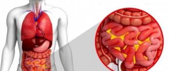
The peritoneum is a shell in the form of two sheets. The spaces they form contain serous fluid. The main function of the peritoneum is to create partitions between the internal organs and muscles, as well as to fix the organs inside the abdomen in a suspended state with the help of mesenteries and ligaments. The peritoneum protects the internal organs in another way. When it encounters microbes, substances are produced that lead to the death of harmful microorganisms. Peritonitis is an inflammation of the peritoneum, leading to disruption of the functioning of all systems and organs located in this area. What are the symptoms of the pathology?
Reasons for the development of peritonitis
Peritonitis - inflammation of the peritoneum
Peritonitis begins to develop when the peritoneum cannot cope with the huge number of invading and multiplying microbes. In this situation, the peritoneum becomes a source of infection. This disease is life-threatening and can end sadly if adequate measures are not taken to localize the inflammation and normalize the condition.
The introduction and spread of infection in the abdominal cavity most often develops as a result of trauma to the organs of this area, a violation of their integrity. The cause may be a disease of the internal organs. Rarely, peritonitis can develop when microorganisms are carried into the area by blood or lymph.
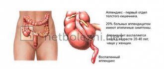
In most cases, peritonitis does not occur as an independent disease, but as a complication of diseases of the abdominal organs. For example, appendicitis, intestinal obstruction, perforated ulcer of the stomach, as well as the duodenum, if measures are not taken in a timely manner, end in peritonitis. Inflammation of the peritoneum is caused by the destruction of the organ after the disintegration of the tumor. Necrosis of a fragment of the intestine due to a hernia, trauma to the abdominal cavity, accompanied by injury, rupture of an organ, partial destruction of the wall of the stomach or intestines by a foreign body can also result in peritonitis.
Sometimes, with heart disease, fluid accumulates in the abdominal cavity, which, in the event of unfavorable developments, suppurates. This becomes another cause of peritonitis.
Not all types of peritonitis are caused by pathogenic microorganisms. For example, blood entering the abdominal cavity due to a violation of the integrity of the vessel also leads to peritonitis. In this case, the type of disease is called aseptic or germ-free. The disease lasts at this level for no more than 6 hours. After this period, microbes from the intestinal area penetrate into the hematoma. After this, peritonitis becomes common.
results
When assessing the level of leukocytes in patients upon admission to the hospital, its increase was revealed in all comparison groups ( p
≤0.5).
A study of the dynamics of leukocyte levels showed a statistically significant decrease in this indicator on the 3rd day in the main group of patients (10.3·109±1.2) compared to the control group (16.4·109±1.5) with p
=0.006 ( Fig. 1) and on the 5th day in the main group (before normal: 6.3·109±0.6) compared with the control (13.6·109±1.1) at
p
=0.001.
Rice.
1. Dynamics of leukocytosis in children with widespread purulent peritonitis. Results of the analysis of the dynamics of the LII indicator according to Ya.Ya. Kalf-Kalifa in children with widespread purulent peritonitis showed a significant increase in both the main (up to 3.8±0.4) and control (3.7±0.3) groups, p
≤0.5.
After 1 and 3 days, a more significant significant decrease in LII was found in patients of the main group (up to 2.1 ± 0.2; p
= 0.01 and 1.52 ± 0.3, respectively) compared to children in the control group (up to 2. 2±0.3 and 3.23±0.2, respectively).
After 5 days, the results of comparison of LII in patients of the main group reliably confirmed a decrease in this indicator almost to normal (1.13±0.2), in contrast to LII (1.83±0.2) in children of the control group ( p
≤0.5 ) (Fig. 2).
Rice.
2. Dynamics of LII with widespread purulent peritonitis. When studying changes in ESR after 1 and 3 days, a reliable significant decrease in this indicator was found in children of the main group (up to 15.3 ± 1.2 and 12.7 ± 0.9 mm/h, respectively) compared with children in the control group (up to 21 .7±0.9 and 16.4±1.1 mm/h, respectively), p
≤0.5.
After 5 days, the results of the analysis of ESR revealed a significant decrease almost to normal in children in the main group (5.4±0.7 mm/h), in the control group up to (13.4±0.9 mm/h), p
=0.01 (Fig. 3).
Rice.
3. Dynamics of ESR in children with widespread peritonitis. Based on the dynamics of the temperature reaction, significant normalization was determined in the main group compared to the control group with widespread peritonitis after 3 days ( p
≤0.5).
A statistical study of the dynamics of regression of SCI (intestinal paresis) revealed a significantly significant relief of it in patients of the main group on the 2nd day, compared with the control group (on the 3rd-5th day), on average by 1.5 days ( p
≤0 ,5) (Fig. 4).
Rice.
4. Dynamics of restoration of intestinal motility in children with peritonitis. Analyzing the dynamics of the TCA indicator, we determined a statistically significant ( p
≤0.5) restoration of this indicator in children of the 2nd subgroup (Reamberin + Remaxol) of the main group after 1 and 3 days (up to 38.4±2.3 and 41.3±1.5 g/l, respectively) and on 5th day (up to 44.3±1.9 g/l) compared to the control group (Fig. 5).
Rice.
5. Dynamics of OKA in children with widespread peritonitis. Data from a study of the dynamics of transaminases (aspartate and alanine aminotransferase - AST and ALT) showed statistically significant ( p
≤0.5) decrease in the level of AST and ALT after 3 days in patients of the 2nd subgroup of the main group (Reamberin + Remaxol therapy) compared with children in the control group.
Signs of peritonitis
The symptoms of peritonitis are very characteristic, so every surgeon can diagnose it!
Manifestations of peritonitis are due to the reasons that provoked its development. But the main signs of one stage or another are the same in any case.
Reactive stage
This is the first stage, it occupies the first day of the disease. The lesions are local in nature. Patients first feel sharp pains that appear unexpectedly. In this case, you can accurately determine the place where the pain comes from. Some compare the pain at this stage to the blow of a dagger.
The localization of pain is associated with the organ that became the source of the disease. For example, with appendicitis, pain will be felt in the inferolateral zone on the right. If this is a perforation of a stomach ulcer, then pain will appear in the left hypochondrium or in the epigastric region. The pain is felt strongly, and it gradually spreads.
How does a woman feel with pelvioperitonitis?
The disease develops quickly. In the first hours, the condition may be relatively normal, so patients often attribute mild manifestations of peritonitis to poisoning or other causes. Then the high temperature rises (up to 39–40 degrees), the woman shudders and has a headache. Sharp cramping or twisting pain in the abdomen is often observed.
The condition is getting worse every hour. Due to poisoning of the body, nausea and vomiting begin. There is a white coating on the tongue. The heartbeat quickens (tachycardia), the pulse reaches 100 beats/min or more. Possible loss of consciousness.
It is impossible to diagnose pelvioperitonitis on your own, since similar symptoms can also occur in other serious conditions: ectopic pregnancy, acute endometritis, tubo-ovarian abscess, appendicitis, intestinal obstruction, ruptured ovarian cyst, renal and hepatic colic. All these diseases are also very dangerous, so if you have symptoms even remotely similar to those mentioned above, you should urgently seek medical help!
Treatment of peritonitis
Surgery. Peritonitis
If the diagnosis is established, emergency surgery is performed. During the operation they act according to the circumstances. After examining all the tissues, the affected areas of the organs are sutured, tumors are removed, and bleeding is stopped. Existing purulent foci are brought back to normal, they are washed with antiseptic solutions. It is advisable to use Ringer's solution.
If the inflammation has affected large areas, then washing is carried out over several days. After surgery, antibiotics are administered in large quantities. They also direct actions to eliminate dehydration.
The famous surgeon S.I. Spasokukotsky noted back in 1926 that if the operation was performed in the first hours after the development of inflammation, then 90% of patients recover. The operation results in recovery in 50% of cases within the first 24 hours. And only 10% have a chance of survival if the operation is performed after the third day.
In our time, approximately the same trend continues. Surgical intervention ends with recovery on the first day. At the second stage, the success of treatment is already doubtful. Recovery occurs if the organs and systems are not severely affected. At the third stage, it is not possible to correct the condition, because irreparable changes occur to the internal organs.
Material and methods
The material for the study was our experience of treatment from 2001 to 2014 in the pediatric surgical departments of the State Clinical Hospital No. 1 named after. N.I. Pirogov and SOKB named after. V.D. Serdavin, 232 children aged 1 to 15 years with widespread purulent peritonitis, who, in addition to standard surgical tactics and modern antibacterial therapy, received optimized pathogenetic therapy. In the main group (148 patients), in which optimized pathogenetic therapy was used, 2 subgroups were identified: in the first (64 patients), they received the antihypoxant Reamberin at a dosage of 10 ml per 1 kg of body weight per day (intravenously, drip with a daily dose distributed over 2 administration), in the second (84 children) - Reamberin and the hepatoprotector Remaxol in daily dosages of 5 ml per 1 kg of body weight (Reamberin was used in one administration, Remaxol was used in the second, 12 hours later). The drugs were prescribed in preoperative preparation and postoperative therapy for 4-5 days (after stabilization of the child’s condition). The use of the hepatoprotector Remaxol in children has received permission from the bioethical committee (decision of the National Educational Institution of Higher Professional Education "Reaviz Medical Institute" dated 04/15/11).
All children of the main group received nutritional support (trophic nutrition) with specialized drugs (Peptomene, Alfare, Izosur, etc.). Surgical treatment involved laparoscopic sanitation of the abdominal cavity. A comprehensive examination included studying the dynamics of leukocyte levels, leukocyte intoxication index (LII) according to Kalf-Kalif, erythrocyte sedimentation rate (ESR), temperature, total albumin concentration (TAC), and transaminase levels.
To treat widespread purulent peritonitis, 95 children underwent midline laparotomy, sanitation and drainage of the abdominal cavity. In 137 cases, laparoscopic sanitation of the abdominal cavity was performed. After introducing trocars at typical points, the abdominal cavity was inspected. If appendicitis was detected, appendectomy was performed using a ligature method.
The cause of widespread peritonitis in 8 patients was destructive changes in Meckel's diverticulum (video-assisted wedge resection of the small intestine was performed), in 3 patients it was perforation of the small intestine by foreign bodies (magnets), the foreign bodies were removed and the perforation was sutured. After eliminating the cause of widespread purulent peritonitis, sanitation of the abdominal cavity was performed, followed by drainage through a suprapubic trocar. In 47 cases, the right or left lateral canals were additionally drained through a counter-aperture.
In the comparison (control) group, 84 children with widespread purulent peritonitis received standard pathogenetic therapy - the use of solutions of crystalloids and colloids in infusion treatment: glucose (5 and 10%), sodium chloride (0.9%), albumin (10%), voluven (6%).