The liver is the largest human gland; its functions are diverse and necessary. The two most important ones include detoxification (the liver cleanses the blood of toxins and breakdown products) and digestion (the liver produces bile enzymes and fatty acids).
In addition, the liver participates in the metabolism of proteins and fats, maintains blood glucose levels, synthesizes a number of vitamins and biologically active substances, regulates water-salt metabolism, and fights antigens that penetrate the bloodstream due to active phagocytosis by astrocytes of the liver capillaries. It is not surprising that any disruption to the functioning of such an important organ leads to a deterioration in a person’s well-being, and often to various diseases.
Ultrasound examination provides information about the liver in both children and adults. The child’s liver has sonographic features, which will be discussed below.
Brief anatomy and diagnostic methods
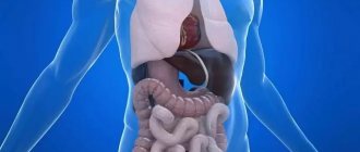
The liver is a vital organ that is located under the diaphragm, in the right hypochondrium. The liver has a visceral (lower) and diaphragmatic (upper) surface. This organ has a bilobed structure: the left and right lobes are distinguished. The left lobe, in turn, includes the caudate and quadrate lobes). The structure of the liver is granular.
The study of liver pathologies is carried out by many methods:
- clinical and anamnestic (by questioning the patient),
- biochemical,
- ultrasonic,
- immunological,
- radiological,
- by puncture biopsy.
It is necessary to understand what the advantages and disadvantages of ultrasound examination are.
Advantages and disadvantages
The advantages of the ultrasound method for diagnosing the liver are:
- non-invasiveness,
- safety,
- multidimensionality of research
- the ability to assess vascular blood flow in Doppler mode,
- relative speed and low cost of the procedure.
Disadvantages include deterioration in image quality in people with developed subcutaneous fat and patients with severe intestinal bloating, and lower spatial resolution compared to radiological methods (CT, MRI).
Ultrasound of the liver: preparation for the procedure
During an ultrasound of the liver, as well as the gallbladder, there is a possibility of some difficulties arising. There are several reasons for this. Firstly, the patient’s excess weight may become an obstacle to the study, since the fat layer impedes the passage of ultrasound waves. Secondly, the reason for the difficulty or even impossibility of conducting an examination is often excessive gas formation in the intestines (flatulence).
Therefore, it is very important to properly prepare for the procedure, strictly following all the doctor’s recommendations. This is especially true for obese people and people suffering from flatulence or constipation.
Indications
Why is it necessary to do such a study? It is usually necessary in the following cases:
- the presence of subjective complaints indicating a possible disease of the liver and biliary tract: pain in the abdomen, right hypochondrium, yellowness of the skin, the appearance of an enlarged venous network in the umbilical region, dyspeptic disorders - nausea, vomiting, frequent belching;
- availability of laboratory test data (blood, bile, etc.) indicating liver damage;
- ascites, hepatomegaly, splenomegaly established during an objective examination;
- suspicion of one or more formations in the liver;
- the need for surgical intervention for the purpose of diagnosis or treatment;
- Ultrasound for abdominal injuries;
- monitoring dynamic changes in the liver.
Ultrasound of the liver: preparation for the study
You should definitely tell your doctor about taking medications, as some of them can affect the size of the liver, and the data obtained will be interpreted incorrectly. The doctor should keep this in mind when making a diagnosis.
At least three days before the examination, the doctor prescribes a special diet. Drinking alcohol and smoking are also prohibited. If necessary, special medications may be prescribed to help prepare for the examination.
What medications help prepare for a liver ultrasound?
For some digestive problems, the patient is prescribed medications so that nothing interferes with studying the structure of the liver during diagnosis.
These drugs include the following:
- Carminatives (Espumizan) and enterosorbents (activated carbon, Polysorb and others). Prescribed for flatulence and bloating in order to eliminate increased gas formation. They begin to take them 2-3 days before the ultrasound with meals.
- Enzyme preparations (Mezim, Festal, Creon and others), which help ease digestion and cope with constipation. You can also use mild laxatives or a cleansing enema the night before the test.
Diet and eating rules
The doctor prescribes a diet that excludes foods that contribute to the formation of gas and constipation. These products include: legumes, raw vegetables and fruits, sweets, yeast-based flour products, whole milk, carbonated drinks, sweet juices, alcohol. Preference should be given to dietary foods: soups, lean meat, cereals. Depending on the condition of the patient’s gastrointestinal tract, the diet can last from three to seven days. Since the examination is carried out strictly on an empty stomach, you should refuse food 6-8 hours before the ultrasound procedure. Patients with diabetes are allowed to eat a cracker or a piece of bread. Dinner on the eve of ultrasound diagnostics should be as light as possible.
Norms and anomalies
The diagnostician evaluates the size, shape, echogenicity and echostructure of the liver. Additionally, the relative position of the liver with other organs and structures is assessed.
To assess the echogenicity of the liver parenchyma, the doctor compares it with the echogenicity of the kidney and spleen: normally, the liver parenchyma is somewhat more echogenic than the renal cortex, as well as the parenchyma of the spleen and pancreas.
On an ultrasound machine, the liver is normally fine-grained, which is due to point and linear formations distributed throughout the organ.
The norm for the right hepatic lobe along the midclavicular line is about 130 mm, and in asthenics this parameter can reach up to 140 mm. In cross-section, the thickness of the right lobe reaches 110–125 mm. The size of the liver from the edge of the right lobe to the most distant point of the diaphragmatic dome is up to 149 mm.
The norm of the left lobe of the liver varies within the following limits: vertical size - up to 60 mm, thickness - no more than 100 millimeters. The angle of the lower edge of the left lobe is less than 30°.
The gallbladder is a pyriform organ with anechoic contents. The wall of the gallbladder does not exceed 4 mm in thickness. Normally, the contents of the gallbladder are homogeneous, anechoic, the internal contour is clear and even, the presence of a physiological inflection is allowed in tall patients.
Ultrasound function during puncture
Using the device, the location of the needle insertion is determined. After the needle penetrates the node, the doctor watches for changes in the image. The disappearance of the formations suggests that these were hemangiomas and not malignant tumors.
The cost of the study and where it is carried out According to the medical direction, echography of the liver is carried out in state medical institutions. The price of the service is minimal or provided free of charge. In general centers and private clinics, the price of the study will range from 700-1200 rubles.
Liver enlargement in children and adults
Ultrasound signs of hepatomegaly (liver enlargement)
- the sum of the craniocaudal size (height) and thickness of the right lobe exceeds 260 mm,
- the sum of the craniocaudal size (height) and thickness of the left lobe exceeds 160 mm,
- the angle of the lower edge of the right lobe becomes rounded, more than 75°.
Enlarged liver (hepatomegaly) in adults usually indicates different stages of liver fibrosis (up to cirrhosis), benign and malignant neoplasms, hepatosis, etc.
In a child, the situation with liver enlargement is somewhat different: for children, the size of the liver is determined according to special age tables. Moderate enlargement of the liver in a child in some cases is an individual feature. In other cases, such a situation in the child’s body may reflect the presence of a nonspecific reaction of the hepatobiliary system to various pathological processes.
A significant increase in liver size in a child may be a sign of the following:
- liver tumors,
- fatty hepatosis,
- nodular hyperplasia,
- in a child - fetal hepatitis.
Thus, the study of the liver in children is somewhat different from the study of this organ in adults.
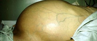
This patient's liver is enlarged and hyperechoic.
Liver granularity on ultrasound
The structure of the liver is essentially granular. In this case, there are fine-grained, medium-grained and high-grained.
It is necessary to understand that the structure of a healthy liver is fine-grained. However, if the liver structure becomes medium-grained, this may indicate liver pathology (for example, chronic viral hepatitis or fatty infiltration). In addition, it must be borne in mind that a medium-grained liver often occurs with a simultaneous increase in the density (or echogenicity) of the liver. If the structure is highly granular, then we can talk about dystrophic pathologies or inflammation.
Lump on ultrasound, “light” or “bright” liver
Typically, pathological changes represent changes in the condition of the liver parenchyma. Increased liver density (increased echogenicity) is usually a sign of diffuse liver disease. On a sonographer's screen, this increase in density may appear as a “white” (or bright) liver, which may also indicate fatty liver disease or hemochromatosis.
A dense liver may also indicate:
- acute hepatitis,
- chronic hepatitis,
- metabolic diseases,
- various infectious diseases,
- congestive liver,
- hematological diseases,
- cirrhosis of the liver,
- liver granuloma,
- diffuse liver metastases.
This image shows a liver with increased echogenicity, which in this 64-year-old patient is caused by steatosis
Outbreaks
Foci in the liver can be formations of different echostructure: dense or mixed, hyperechoic or hypoechoic. Hyperechoic areas are the same as areas of increased echogenicity; they are displayed on the device screen as light areas. Hypoechoic – respectively, areas of reduced echogenicity, are displayed as dark areas.
Most often, focal formations on an ultrasound machine are represented by:
- Cysts,
- Liver abscess (formation of infectious-inflammatory origin),
- Cellular adenoma,
- Hemangiomas,
- Cellular adenoma (benign formation, most often found in women of reproductive age),
- Malignant neoplasms in the liver and metastases.
It should also be taken into account that the echogenicity of foci sometimes does not differ at all from the echogenicity of the liver parenchyma.
The patient, a woman, was admitted to the doctor with complaints of pain in the right hypochondrium. The examination revealed a hyperechoic inclusion in the liver - an adenoma.
Metastases
Unfortunately, the first place in occurrence among focal liver lesions is occupied by metastases. They are distinguished by a significant variety of echographic signs, given their origin from carcinomas of various structures (most often it is cancer of the stomach, colon, and ovaries).
Hyperechoic metastases are fairly dense three-dimensional objects with clearly visible boundaries, an almost homogeneous or heterogeneous structure, the vascular pattern around the formation is disrupted due to compression of the growing tissue of the vessels.
Isoechogenic formations are very similar in their indicators to parenchymal tissue in terms of echogenicity. However, they can be indicated by an abnormal vascular pattern and (or) bulging of the capsule in the case of a subcapsular location; their detection requires high quality equipment and the professionalism of the researcher.
Hypoechoic metastases are homogeneous space-occupying formations with a clear, simple contour, usually small to medium in size. It is not often possible to find anechoic metastases, which resemble the structure of cysts in their shape and echogenicity, but they do not have the effect of distal enhancement, the contour is usually uneven, and the contents are heterogeneous.
Metastases should be distinguished from some similar anomalies, such as:
- hepatocellular cancer,
- cholangiocellular carcinoma,
- liver hematomas,
- foci of fatty infiltration,
- hemangiomas (moles on the liver).
“Red moles” are often visible on ultrasound. These can be hemangiomas, which are benign formations of epithelial cells and vascular smooth muscles, usually no more than 3 centimeters in size (capillary) or more (cavernous, which can reach impressive sizes), hyperechoic.
The structure of hemangiomas is finely cellular with clearly defined contours that can be easily distinguished from the surrounding tissue. If the diagnosis of hemangioma is confirmed, the patient requires regular monitoring (every 3-6 months).
Metastatic inclusion in the liver. The red arrow is the aperture. Yellow – metastatic node. Blue – mirror image. Diagnosis: clear cell carcinoma.
Cysts and hematomas
Echinococcal cysts associated with parasitism of granulosa echinococcus appear as simple anechoic cystic formations. Cysts may also appear as multicystic lesions with thick, layered septa.
Traumatic cysts (hematomas) arise as a result of aseptic development of the hemorrhage site.
Traumatic cysts are visualized as a round or oval cavity with anechoic contents, as well as hyperechoic linear inclusions of blood clotting products. Subsequently, the hematoma transforms into a hyperechoic formation, which can most often be found in the VI and VII segments of the right hepatic lobe.
Diffuse liver changes
Diffuse changes in the liver may indicate the following pathological processes:
- about the inflammatory process, hepatitis: there is a medium-grained structure of the parenchyma, hyperechogenicity of the organ (increased echogenicity), an abnormal vascular pattern;
- diffuse fatty hepatosis (at the same time also medium-grained organ and its increased echogenicity), cirrhosis, in which the echostructure becomes heterogeneous due to areas of fibrosis, edema and regeneration of hepatocytes, the contour of the liver is tuberous, the size is increased in the early stages, reduced in the later stages. There are also signs of increased pressure in the portal vein system (portal hypertension) - dilation of the main veins, ascites, splenomegaly (enlarged spleen).
Each ultrasound “finding” should be assessed dynamically and taking into account the conclusion of the attending physician and test results, it is important not to immediately panic in the face of a disappointing conclusion, but to remember that an ultrasound specialist can accurately describe the size, shape, localization and echographic features of the pathological focus, but not can always establish its morphological identity.
Hyperechogenicity of the liver, a typical picture of steatosis. A 75-year-old female patient complains of pain in the right hypochondrium.
Liver spots
These types of areas on the liver look different from other areas on ultrasound. Spots on the liver may indicate the following pathologies:
- infections
- hemangiomas
- adenoma
- granuloma
- inflammatory processes
- various types of tumors of benign and malignant origin.
To diagnose this type of object, it is necessary to undergo additional procedures and tests.
Thus, an ultrasound examination of the liver allows doctors to obtain a sufficient amount of information for diagnosis about both the liver of a child and an adult. At the same time, the array of data that can be obtained during this study is huge: it allows you to diagnose major liver pathologies, be it hepatitis, cirrhosis and fibrosis, hemangiomas, hematomas and much more. The analysis is based mainly on the size of the organ and indicators of the liver parenchyma (echogenicity, granular structure, etc.), as well as the clarity of the contours of the organ structures.
How is the procedure done?
During an ultrasound scan of the liver, the patient is only required to lie quietly on the couch in the position in which the doctor says (usually lying on his back or on his side). It takes no more than 15 minutes. A gel is applied to the area of study, which helps the ultrasonic waves pass better, and the diagnostician moves a special sensor over it, receiving the necessary data on the monitor. At the end of the procedure, the patient can print out pictures of the liver from the most important angles.
Ultrasound of the liver with contrast
Ultrasound diagnostics of the liver using a contrast agent is a relatively recent, but very informative research method. The patient is injected intravenously with a special harmless suspension with microbubbles, the size of which is similar to the size of red blood cells, which allows them to penetrate even the smallest vessels. Thanks to the echo contrast agent, benign and malignant formations can be accurately diagnosed. The drug is eliminated within 15 minutes after administration.