Indications for ultrasound diagnostics
Ultrasound diagnostics may be prescribed if unpleasant symptoms occur. It is imperative to undergo ultrasound diagnostics if:
- discomfort or severe sensations in the liver area;
- observing regular pain or swelling in the right hypochondrium;
- yellowing of the skin and mucous membranes, the white membranes of the eyes;
- symptoms of nausea;
- a feeling of bitterness in the mouth;
- stone disease;
- abdominal injury;
- poisoning;
- bad tests;
- overweight;
- poor nutrition.
In addition, the procedure is mandatory if an operation has been performed or the patient’s health is being monitored.
For children, the reasons for ultrasound diagnostics may be:
- vomit;
- weight loss;
- frequent diarrhea;
- vague pain in the abdomen;
- poor appetite.
How to prepare for an ultrasound of the gallbladder
There is nothing difficult in preparing for an ultrasound of the gallbladder. You just need to implement the requirements:
- take the last meal eight hours before the start of the study;
- clear the intestines of gases.
This will give the organ enough time to fill with bile.
To achieve maximum diagnostic accuracy, you need to follow a certain diet for several days before the procedure. You need to know what to eat before an ultrasound of the gallbladder.
Here is a list of what you should not eat before an ultrasound of the gallbladder: drink alcohol and eat food that accumulates gases. This should include: sparkling water, fruits and vegetables (fresh), legumes and dairy foods, bread, yeast-based treats. Patients diagnosed with diabetes are allowed a small snack with crackers and tea.
What can you eat before an ultrasound of the gallbladder? In addition to flour and sweet products, products that contain fiber, are made from milk and cause bloating, you can eat any food.
Prohibited foods and drinks
Preparation for an ultrasound diagnostic procedure does not cause any particular difficulties for people. As a rule, it begins 3-4 days before visiting the ultrasound room. Of course, no one forces a person to completely give up food and water. It is enough to follow a few general recommendations:
- minimize gas-forming foods - for example, legume dishes, raw vegetables, black cereal bread;
- give up carbonated drinks, coffee, alcohol;
- do not consume bakery and confectionery products - they often provoke putrefactive, stagnant processes in the intestines.
The diet recommends the presence of light, low-calorie dishes that do not require increased work from the digestive organs. It is imperative to monitor the timeliness of bowel movements. If difficulties arise with defecation, it is better to perform a cleansing enema.
If a person is prone to flatulence, the doctor will prescribe a short course of carminative medications. Motilium or Espumisan have proven themselves to be excellent.
What actions are carried out on the eve of diagnosis?
How to properly prepare for an ultrasound of the gallbladder immediately before the study:
- have a light, nutritious dinner before 7 p.m.;
- remove everything from the intestines in a natural way or take auxiliary laxatives (without hay);
- do not smoke or chew anything (this will prevent excess bile from appearing).
Ultrasound of the gallbladder no matter how much you eat: adults may not eat for about eight hours before diagnosis; infants can finish breastfeeding within a few hours.
Ultrasound of the pelvic organs of women
Usually the study is carried out on days 5-7 of the menstrual cycle. In some cases, the day of the test is determined by your attending physician.
Transvaginally (through the vagina): you must empty your bladder before the test.
Transabdominal (through the abdominal wall): onto a full bladder. An hour before the test, do not urinate, drink up to 1 liter of any liquid without gas (water, tea, fruit juice) and walk around. There are no restrictions on food or drink on the day of the test. The study is not carried out on an empty stomach.
Types of ultrasound and the specifics of their implementation
There are two types of ultrasound diagnostics: simple and functional. They differ in technique, duration and results.
How an ultrasound scan of the gallbladder is performed depends on the type of examination.
- Simple. Use an external sensor. The patient needs to lie down with his stomach up. The part being examined is lubricated with a gel, which improves contact between the body and the sensor, and ultrasonic waves pass into the tissue. Sometimes the specialist asks the patient to hold his breath, stand in a knee-elbow position, or turn to the left side to straighten intestinal loops that interfere with the examination. In order to identify sand or stones in the organ, the patient will have to stand up and bend forward.
- The functional type examination includes routine fasting diagnostics. Then the patient has breakfast and repeats the examination in three stages, after several time intervals. This serves as an assessment of the functionality of the bile duct and canals.
Dynamic echo-choledochography
A similar procedure with a choleretic breakfast to assess the functional activity of the common bile duct - common bile duct. After waiting a half hour after breakfast, the examination is carried out again to determine the difference in tone that has arisen.
At the same time, all incoming patient complaints regarding:
- intensity of pain attacks;
- features of the disease state;
- increasing pain shock;
- duration of the attack.
Ultrasound with gallbladder function, what is it and how to prepare for it
This procedure helps to accurately determine the characteristics of the motor function of the organ and its pathways. In this way, the doctor will be able to determine the content of the gallbladder, its size, structural parameters and ability to contract the walls. This type of examination is carried out using choleretic food on an empty stomach. That is, in real time it becomes clear how the motor and contractile functions of the organ work. Bile volumes are measured on an empty stomach and for an hour after eating breakfast.
The principle of preparation for ultrasound of the gallbladder with determination of function:
- undergoing a routine ultrasound scan on an empty stomach;
- eating a choleretic breakfast in the form of two boiled eggs, a tablespoon of sour cream and full-fat yogurt.
Indications for liver examination
Ultrasound is prescribed for the following pathologies:
- painful sensations in the hypochondrium on the right side;
- abnormalities in blood test results that indicate the presence of liver disease;
- diseases of the gallbladder or pancreas at any stage;
- yellowing of the skin and whites of the eyes;
- drinking alcohol or taking medications for a long time;
- various injuries to organs located in the peritoneal cavity;
For chronic liver diseases, ultrasound should be performed systematically.
A list of pathologies and their symptoms that will help you understand what an ultrasound of the liver and gallbladder shows:
- Acute or chronic hepatitis.
With this disease, one or two lobes of the organ in question enlarge, the edges become rounded, the liver darkens, and the veins expand.
- Tumors.
They appear as an unusually shaped area with unclear boundaries. At the same time, the lymph nodes become enlarged and the gallbladder is displaced.
- Cirrhosis.
It is characterized by an increase in the size of the entire organ or its left lobe, while the tissue density increases, the structure becomes heterogeneous, and tuberosity of the sidewalls is formed. At a late stage of development of the pathology, the liver decreases in size.
- Cysts.
They appear in the form of rounded convex formations with a thin wall and smooth edges. They can be single or multiple. At the same time, individual areas of the liver increase in size.
- Giardiasis.
Manifested by the formation of darkened or light spots on the liver.
- Fatty degeneration.
Characterized by enlargement of the entire organ and lower lobe. In this case, the outline of the liver becomes unclear, the edges become rounded.
Ultrasound of a removed bladder
This procedure is called dynamic echocholedography. For this, a routine ultrasound is performed. Next, the patient needs to take food in the form of a sorbitol solution. After an hour and a half, the procedure is repeated.
This technique is used for patients who have undergone surgery on an organ, but complain of the following symptoms:
- pain persisted or appeared after removal;
- there is a bitter taste in the mouth;
- there is nausea and vomiting;
- the stool is broken.
Why are drinks prohibited before an ultrasound?
In the rules for preparing for a diagnostic examination of the liver, water is indicated as a factor that affects the reliability of the information. Doctors explain that when drinking even pure drinking liquid, the intestines mistake it for food and begin to actively work. Seething appears and gases are formed. The image on the ultrasound machine screen will be unclear.
You will have to demonstrate willpower for no more than 1–1.5 hours. It is for this period that the intestines should remain calm. If thirst still torments a person, you should blot your lips with a damp cloth.
The optimal time for studying internal organs using ultrasound methods is in the morning, when the body has already rested and the digestive system has had time to absorb the food consumed the day before. In cases where the study of the condition and functional activity of the liver is scheduled for the afternoon, a light breakfast and weak drinks, for example, green tea, pure water without gas, are allowed.
High-quality preparation is the key to the success of ultrasound examination of the liver. You can drink water, but follow the rules.
Principles for interpreting research results
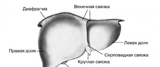
This organ is located on the right side of the liver - in its lower zone. The gallbladder consists of three sections:
- bottom;
- immediate body;
- neck.
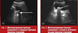
Many patients wonder what the gallbladder looks like on ultrasound? In the results of ultrasound diagnostics, the organ has the shape of an ellipse, similar to a pear.
The standard volume of the gall bladder in an ordinary person is less than 50 milliliters. If the organ is deformed, folds may form in the mucosa that bend inward. To know how to decipher an ultrasound of the gallbladder, you need to understand what indicators are assessed during diagnosis, as well as their norm.
The study evaluates the following parameters:
- size and shape;
- Wall thickness;
- amount of bile;
- the presence or absence of pathological abnormalities;
- contour structure;
- changes in bile duct parameters;
- wall structure.
The results are interpreted immediately after the procedure. Deviation from normal indicators allows the specialist to identify a particular disease in the patient.
How often should an ultrasound of the gallbladder be performed for patients with diagnosed disease? Repeated procedures must be completed as many times as necessary to clarify the data and monitor the condition of the organ.
Normative characteristics of the liver and gall bladder in an adult
| Liver | width | 23-27 cm |
| length | 14-20 cm | |
| diameter | 20-22.5 cm | |
| shares | left – 6-8 cm; right – 12.5 cm | |
| hepatic duct | D = 5 mm | |
| vein | D = 15 mm | |
| Gall | width | 5 cm |
| length | 10 cm | |
| diameter | 3.5 cm | |
| wall thickness | 4 mm | |
| duct | D = 6.8 mm | |
| lobar ducts | D = 3 mm |
How can you check the gallbladder, other than ultrasound?
In addition to ultrasound diagnostics, several other methods for studying this organ are used:
- X-ray;
- CT;
- research using radionuclides;
- endoscopic and laparoscopic examinations;
- probing with biopsy.
These methods have some contraindications and place different stress on the body. Therefore, it is recommended to resort to their use in extreme cases. Some patients are afraid that ultrasound diagnostics are also harmful to the body and ask how often can an ultrasound of the gallbladder be done? But it is worth noting that the ultrasound diagnostic procedure is absolutely harmless to the human body, so it can be performed an unlimited number of times.
Where to do an ultrasound of the liver?
To get an accurate liver ultrasound result, it is recommended to choose a clinic responsibly. MedCenterService patients receive a number of benefits:
- Informative and accurate research, thanks to the use of the latest generation medical technology.
- Individual approach and competent transcription. The examination is carried out by highly qualified doctors.
- Comfortable conditions and no queues. Patients are accepted by appointment. In addition, the clinics are equipped to meet the needs of patients.