Pain syndrome
The appearance of pain is typical for large cysts, as they compress the surrounding tissues, including the nerve plexuses. Small cysts do not exert such pressure, therefore, as a rule, there are no complaints of pain. This symptom is especially characteristic for the period of formation of false cysts during acute and exacerbation of chronic pancreatitis, and it is caused to a greater extent by destructive processes. Over time, the intensity of the pain decreases, it is characterized as “dull” or rather “discomfort”. A characteristic symptom is the “lucid interval” (temporary improvement and absence of pain after acute pancreatitis or injury). The most severe pain is caused by cysts located on the posterior surface of the gland and compressing the solar plexus area. Impact on this nerve plexus creates in patients a very intense, long-lasting burning pain that radiates to the back. Movement, compression by clothing, a belt, or a belt increase its intensity. The condition is somewhat alleviated when taking the knee-elbow position (“on all fours”). A pronounced increase in pain - “dagger” pain can indicate the occurrence of complications (for example, rupture of a cyst), gradual progression of pain along with an increase in body temperature and the appearance of intoxication - about its suppuration.
Dyspepsia
Other manifestations of pancreatic cysts may include dyspeptic disorders: nausea, vomiting (which may result in an attack of pain), and stool instability. As a result of the decrease in the amount of pancreatic juice entering the intestines, the digestion of food and the absorption of nutrients are disrupted. As a result, the patient loses weight, loses weight, and becomes weak.
Intestinal obstruction
Sometimes large pancreatic cysts cause compression of neighboring organs, impairing their patency. When a cyst is located in the head of the gland, obstructive jaundice may occur (yellowishness of the skin, sclera, and itchy skin appears); if the portal vein is compressed, swelling in the legs or ascites develops. Very rarely, large pancreatic cysts compress the intestinal lumen (duodenum), which interferes with the passage of food. In these cases, incomplete high intestinal obstruction may form.
Diagnosis of pancreatic cyst
When examining the abdomen, its asymmetry is possible: the appearance of a bulge or protrusion in the area where the formation is located.
Laboratory tests show a slight increase in leukocytes in the blood, an increase in ESR, and sometimes the level of enzymes in the blood increases: amylase, phosphatase and bilirubin, which is associated with exacerbation of pancreatitis and damage to working pancreatic tissue.
More specific and meaningful information can be obtained by using instrumental diagnostic methods.
- Ultrasound examination
allows not only to detect a formation, see its location, measure its size, evaluate its contents, but also to identify indirect signs of complications: for example, in the case of suppuration of a cyst, ultrasound can see the unevenness of the echo signal against the background of the cavity; in case of malignancy (malignancy), heterogeneity of the contours . - Magnetic resonance imaging and computed tomography
provide more detailed information about the size, localization of the cyst, and its connection with the bed of the ducts. Contrast tomography helps to recognize cystic tumors. - Endoscopic retrograde cholangiopancreatography
(ERCP) has a unique place in the diagnosis of pancreatic cysts. This endoscopic examination provides detailed information about the connection of the cyst with the ducts of the gland, which determines treatment tactics. However, this examination requires the injection of a contrast agent directly into the pancreatic duct, which causes ductal hypertension and is accompanied by a high risk of infection. Therefore, at present, ERCP is performed according to strict indications: for example, if an obstructive nature of the cyst is suspected, in order to remove the stone from the pancreatic duct or install a stent in the obstruction area. - Endosonography
(a modern diagnostic method that combines the advantages of endoscopy and ultrasound) allows not only to clearly visualize the location, size of the cyst and the nature of its contents, but also to take a targeted biopsy if the presence of a cystic tumor is suspected.
Publications in the media
Incidence: 9.1 per 100,000 population in 2001. Pancreatic cancer • Epidemiology . Pancreatic cancer is the third most common among gastrointestinal tumors and the fifth cause of death from malignant neoplasms. The incidence of adenocarcinoma is constantly increasing, especially among men aged 50–60 years. Adenocarcinomas account for more than 90%. • Etiology. Pancreatic cancer may be associated with tobacco smoking, diabetes, and exposure to asbestos. The risk increases significantly in patients with hereditary forms of pancreatitis. Mutations have been described that increase the incidence of malignant neoplasms, incl. pancreas (for example 600185, 13q12.3, BRCA2 gene, ; *601916, 3p21.1, ARP gene, ). • Pathological anatomy . In most cases, these are adenocarcinomas of varying degrees of maturity, giving early and extensive metastases to regional lymph nodes (parapancreatic, mesenteric, etc.).
• Clinical manifestations •• Early symptoms are nonspecific - pain in the epigastric region, weight loss, aching back pain •• Symptoms depend on the location of the tumor in the pancreas ••• Head of the gland. Most often, cancer develops in the head of the pancreas (50–80%). With this localization, in 75% of patients, the main symptoms (weight loss and obstructive jaundice) appear without a painful attack. Approximately 25% of patients with a tumor localized in the head of the pancreas experience girdle pain and vague discomfort in the epigastrium. Since the pancreas is located retroperitoneally, detection of its tumors in the early stages during physical examination is difficult and becomes possible with significant tumor sizes (a tumor-like formation is palpated in the epigastric region) or with metastasis. If there is a palpable tumor of the head of the pancreas in approximately 20% of cases, we can already talk about its inoperability. An enlarged, painless gallbladder (Courvoisier's sign) indicates tumor obstruction of the pancreatic and/or bile ducts ••• Cancer of the body or tail of the pancreas is detected less frequently, and it manifests itself in later stages, since tumors of this location cause obstructive jaundice only in 10% of cases.
• Diagnosis. It is rarely possible to detect pancreatic cancer in the early stages •• Non-invasive diagnostic techniques ••• CT and ultrasound can detect tumors measuring 2-3 cm ••• If pancreatic tumors are large enough and displace the duodenum (late stages), they can be detected with radiography of the upper gastrointestinal tract •• Invasive diagnostic techniques ••• Percutaneous aspiration biopsy of a tumor with a thin needle under ultrasound or CT control followed by cytological examination of the punctate with high accuracy and practically without complications allows you to diagnose a malignant neoplasm of the pancreas ••• Endoscopic retrograde cholangiopancreatography with using a fiber duodenoscope to cannulate the pancreatic duct. After introducing a radiopaque contrast agent into the duct, a series of photographs are taken. Using this technique, small pancreatic tumors can be diagnosed. In addition, cytological examination of the epithelium and contents of the pancreatic duct is possible •• Laboratory studies. In 80% of patients, serum alkaline phosphatase activity is increased, which is due to compression of the pancreatic part of the common bile duct. Elevated levels of carcinoembryonic Ag (CEAg), LDH, and serum glutamate oxaloacetate transaminase are often noted. Jaundice is found in 65% of patients, and 25% have high serum amylase levels. A relationship has been identified between the tumor marker CA19–9 and pancreatic carcinoma (the sensitivity of this test for pancreatic cancer is 80%, and the specificity is 90%) •• Angiography can detect displacement or compression of the pancreatic or duodenal artery. The venous phase can be especially informative in case of blockage of the superior mesenteric or splenic veins •• A secretin stimulation test reveals a decrease in the volume of pancreatic secretion with normal levels of enzymes and bicarbonate.
• TNM classification (see also Tumor, stages) •• pTis - cancer in situ •• T1 - tumor up to 2 cm in greatest dimension, limited to the pancreas •• T2 - tumor more than 2 cm in greatest dimension, limited to the pancreas • • T3 - tumor grows into any of the following structures: duodenum, bile ducts or peripancreatic tissues •• T4 - tumor grows into any of the following structures: stomach, spleen, colon, adjacent large vessels •• N0 - no metastases in regional lymphatics nodes •• N1 - there are metastases in regional lymph nodes. • Grouping by stage • Stage 0: TisN0M0 • Stage I: T1–2N0M0 • Stage II: T3N0M0 • Stage III: T1–3N1M0 • Stage IV •• T4N0–1M0 •• T1–3N0–1M1.
• Treatment •• Pancreaticoduodenectomy (Whipple's operation) for resectable tumors is the standard method of surgical treatment for cancer of the head of the pancreas ••• Resectability of the tumor is established on the operating table according to several criteria: absence of metastases to the liver; the tumor does not invade the hilum of the liver, the portal vein behind the pancreas, the area of the superior mesenteric artery and other abdominal organs ••• Histological confirmation of tumor malignancy can be obtained using a needle aspiration biopsy performed before or during surgery ••• The Whipple procedure involves removal of the head pancreas, duodenum, distal part of the common bile duct, gallbladder and distal stomach. Restoration of gastrointestinal tract patency is carried out by the formation of gastrojejunostomy, choledochojejunostomy and pancreaticojejunostomy. The mortality rate is 15%. The most common complications are bleeding, abscess formation and failure of the pancreaticojejunostomy •• Left hemipancreatectomy with splenectomy and lymphadenectomy is performed when the tumor is localized in the midbody and in the tail of the pancreas •• Pancreatectomy is not widespread. Potential benefits of surgery: Multifocal tumor removal is possible (40% of pancreatic cancers). The survival rate after this operation is not much higher than after pancreaticoduodenectomy. In addition, after pancreatectomy, a particularly severe form of diabetes occurs, which worsens the patient’s quality of life after surgery •• Palliative operations for pancreatic cancer are performed more often than radical ones due to late diagnosis ••• Palliative operations are aimed at eliminating the obstruction to the outflow of bile, for which a decompressive anastomosis is applied between the gastrointestinal tract and the gallbladder, or the common bile duct ••• Almost 20% of patients require a repeat operation aimed at restoring patency between the stomach and duodenum, if such a shunt operation is not performed earlier. Therefore, in many clinics, choledochojejunostomy is supplemented with gastrojejunostomy. ••• Sometimes, to resolve obstructive jaundice and ensure internal drainage of bile, percutaneous transhepatic biliary drainage is used, which avoids traumatic surgery. •• Chemotherapy is used quite widely in the treatment of pancreatic cancer. Complex regimens for the use of drugs, including fluorouracil, cause a temporary reduction in tumor size, but do not increase life expectancy. • Combined treatment (intraoperative radiation therapy and implantation of radioactive sources into the intestine) is used to suppress the growth of the primary lesion and prevent the development of metastases. Preliminary results are quite encouraging (in inoperable cases, the average life expectancy is 13 months) •• Radiation therapy reduces the size of tumor formation in 60–70% of patients; it can be used as a palliative method.
• The prognosis for pancreatic cancer is extremely unfavorable •• 5-year survival rate does not exceed 5%. Most patients die within 1 year after surgery •• The average life expectancy of patients with unresectable cancer is 6 months •• Even in patients with operable pancreatic tumors, the effectiveness of surgical treatment is low. Only 10% of patients after pancreatic resection live more than 5 years.
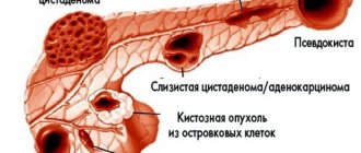
Pancreatic cystadenocarcinoma. Serous and mucous cystadenomas are characterized by multiple small (serous) or large (mucous) cavitary formations. The cysts do not communicate with the pancreatic ducts, do not contain amylase, the content of serum amylase is usually normal (increased in 60–75% of cases of pseudocysts). Angiography reveals hypervascularization. Lesions are more often observed in women; in typical cases, weight loss is observed, and there is no history of pancreatitis. Treatment is surgical - excision, often with complete cure.
ICD-10 • C25 Malignant neoplasm of the pancreas •• D01.7 Cancer in situ of other specified organs of the digestive tract.
Treatment of pancreatic cyst - surgery
The clear answer is surgery only! Modern technologies make it possible in many cases to avoid major surgery and limit ourselves to minimally invasive endoscopic or endovideosurgical intervention.
The following methods of treating cysts are possible:
- Removal of the cyst itself or a cystic part of the pancreas.
- Internal drainage of the cyst.
- External drainage of the cyst.
With the first method, as a rule, the cyst is removed along with a section of the pancreas. The volume of intervention depends on the size of the formation, on the location where the cyst is located, on the condition of the tissues adjacent to it. Distal resection, distal or pancreatoduodenal resection, are quite complex and require appropriate technical and medical support. Currently, these operations are included in the list of types of high-tech medical care performed under quotas from the Ministry of Health of the Russian Federation or under compulsory medical insurance policies. These operations can be performed in our Clinic both traditionally and laparoscopically.
Drainage operations are considered the most physiological and less traumatic, which are aimed at creating an outflow from the cyst into the stomach, duodenum or small intestine using internal drainage. An anastomosis is created, which ensures the delivery of gland juice to food, which relieves the pain reaction and rarely leads to relapses. Currently, it is also possible to perform these operations using endoscopic techniques, which are performed endovideosurgically or endoscopically with ultrasound guidance.
External drainage of cysts in planned surgery has recently been rarely used. The choice of this type of intervention is most often forced in emergency situations. Indications for this method:
- if the process of cyst formation has not completed;
- serious condition of the patient;
- suppuration of the cyst.
Such interventions are called palliative, they do not solve the problem, but can lead to relapse, to fistulas. They are used as one of the stages of patient treatment. Drainage operations can be carried out only after confirmation of a non-tumor cause of the formation.
Conservative treatment of the underlying disease is mandatory. For pancreatitis, it is imperative to follow a diet, the purpose of which is to reduce the secretion of pancreatic juice as much as possible.
Enzyme replacement drugs and analgesics are used; secretion suppressants. It is imperative to monitor the level of glycemia and, if necessary, correct it.
Benign pancreatic tumors
Symptoms
The pancreas is a mixed secretion gland.
Exocrine tissues are located in the area of the larger head adjacent to the duodenum, where pancreatic digestive enzymes are excreted through special ducts. The endocrine islets of Langerhans are concentrated mainly in the caudal part of the gland; The pancreas secretes insulin, glucagon, ghrelin, pancreatic peptide, somatostatin, gastrin and, possibly, some other bioactive regulatory substances into the systemic circulation. In general, this organ plays a key role in the life of the body and performs many functions, any of which can be affected depending on the location and nature of the neoplasia. Tumors in the exocrine regions, supporting tissues, or stroma (the scaffolding tissue for functional cells) usually develop asymptomatically, at least until they reach a sufficiently large size and begin to exert noticeable and sometimes very painful mechanical pressure on the tumor. adjacent structures and organs. This type of neoplasia includes hemangiomas, lipomas, neuromas, leiomyomas, fibromas, growing, respectively, from vascular walls, adipose tissue, neuronal membranes, muscle fibers, and connective tissue.
A large (over five centimeters) benign tumor of the pancreas can cause nonspecific pain of a girdle or radiating nature; in rare cases, mechanical compression of the bile ducts or other lumens of the gastrointestinal tract can cause jaundice, dyspepsia, or even intestinal obstruction.
A different picture is observed in cases where a benign tumor develops in the endocrine structures of the gland and produces hormones. This hormone-producing neuroendocrine neoplasia is named depending on which cells form its basis and what hormone they secrete. In particular, there are insulinomas, glucagonomas, gastrinomas, etc. About three quarters of all such cases are insulinomas and are accompanied by increased secretion of insulin, which causes a cascading hormonal imbalance with the predominant symptoms of hypoglycemia: hyperhidrosis, total weakness, tachycardia, emotional instability, irritability, feelings of hunger, anxiety, fear. Patients with such tumors are characterized by a rapid “unreasonable” increase in body weight. In the most severe cases, hypoglycemia can result in a coma.
Accordingly, with glucagonoma, the opposite, hyperglycemic symptoms are observed: patients lose weight, an erythematous rash appears on the flaky skin, the mucous membranes become inflamed - in general, the clinical picture of diabetes mellitus develops.
Gastrinomas accelerate the secretory activity of the stomach and are characterized by therapeutically resistant ulcerations of the small intestine, pain, dyspepsia, disorders of intestinal motility and absorption function.
Forecast
The prognosis for pancreatic cysts is quite favorable. It depends both on the cause of the disease and on the timeliness of diagnosis and surgical treatment.
In our clinic you can undergo a full examination. The clinic’s specialists have all the necessary equipment for diagnosing and treating cystic formations of the pancreas. The full range of necessary surgical (both traditional and minimally invasive) interventions is performed. Most of them are included in the list of types of high-tech medical care (HTMC), performed under quotas of the Ministry of Health of the Russian Federation or compulsory medical insurance policies.