One of the leading places in the number of diseases occurring in people is occupied by diseases of the gastrointestinal tract. And if most of them arise not through human fault, but due to the penetration of bacteria, viruses or metabolic disorders into the body, then steatosis is precisely that disease, the fault for the appearance of which lies entirely with the person. What is pancreatic steatosis and how to treat it?
Pancreatic steatosis - what is it?
Steatosis is a metabolic disorder that causes fat to accumulate in organs. When talking about a disease, experts most often mean the pancreas and liver. They are the ones who suffer the most from the pathological process.
It is important to understand here that fat does not just cover the organ, but begins to replace its cells. For the time being, a person may not even know about his illness, because the functionality of the organ is not impaired. However, as soon as a certain point is passed, the body will give an SOS signal.
Other names for the disease: fatty infiltration of the pancreas, fatty degeneration of the pancreas, fatty replacement, “fatty” pancreas, lipomatous pseudohypertrophy, non-alcoholic fatty disease, non-alcoholic fatty steatopancreatitis.
Do liver steatosis need to be treated?
Very often, routine ultrasound diagnostics of the liver reveal diffuse changes in the liver such as steatosis (fatty hepatosis, fatty infiltration of the liver).
Liver steatosis is a common phenomenon for older people, most often accompanying the aging process, however, recently liver steatosis is also characteristic of young people, and even perhaps without accompanying excess weight? Of course, steatosis is a disease that is expressed in the fact that adipose tissue is formed inside the liver instead of liver tissue , which does not perform the function of the liver and differs from liver tissue in its structure by its high density. With steatosis, the amount of healthy liver gradually decreases and over time, the remaining healthy part will not be able to ensure the vital functions of the body. This stage of the disease is called cirrhosis of the liver and is difficult to treat.
The problem is complicated by the fact that the liver does not hurt, and even if it is seriously damaged, the patient does not have any complaints, with the exception of a possible feeling of heaviness in the right hypochondrium . Most often, heaviness and discomfort in the right hypochondrium with steatosis, as with other liver pathologies, are caused by an increase in its size.
Causes of steatosis
The main reason for the onset of steatosis is an unhealthy lifestyle. This includes:
- excessive consumption of fatty foods and alcohol;
- lack of treatment for diseases of the gastrointestinal tract, hepatobiliary and endocrine systems;
- incorrect dose of antibiotics or their non-systemic use;
- non-medical diets or deliberate changes in diet not agreed with the doctor;
- hereditary predisposition;
- lack of physical activity or insufficient amount of it.
Under the influence of these factors, organ cells begin to die. The body, trying to correct the situation and prevent the organ from dying, restores its functionality at the expense of fat. That is why in the initial stages the disease is asymptomatic. Gradually, adipose tissue, which cannot perform the functions assigned to the organ, prevails and inflammation begins, subsequently leading to necrosis.
Note!
The risk group includes people over 45 years of age who are obese or suffer from various addictions. These people may develop not only steatosis, but also hepatitis.
Steatosis - what kind of disease is it?
Under the medical term “steatosis” lies a certain process of transformation of pancreatic cells, from specific cells to fat cells.
Like all other diseases, this pathology does not awaken on its own. The desired disease is provoked by leading an incorrect lifestyle, for example:
- smoking;
- unhealthy diet;
- constant consumption of strong alcohol.
The importance of an organ such as the pancreas should not be underestimated. Citizens ignorant of medicine may think that it has only an indirect effect on the processes occurring in the body, however, this idea is completely wrong.
Despite the fact that food is not digested inside the pancreas, without it this process is virtually impossible, since it is it that secretes a set of enzymes - digestive secretions necessary for the decomposition of dietary fiber into microelements and their absorption.
Unfortunately, in conditions of impaired functioning of the desired organ, complications manifest themselves in the work of not only the gastrointestinal tract, but also other body systems.
Cell replacement process
Let's look at what stages the process of transformation of organ tissue goes through, during which the pancreas loses some of the cells that secrete secretions.
- During the first stage, various harmful factors influence the organ. The required stage may last for months, or even years. The lower the body’s resistance, the sooner potential consequences will manifest themselves.
- When the body's resistance decreases, the pancreatic cells responsible for the production of digestive juices die.
- As the organ tissue dies, each cell affected by necrotic processes is eventually replaced by fat. Just as connective tissue heals damage in the intestines, for example, or heals wounds on the surface of our epithelium, adipose tissue replaces the special structures of the gland.
It would seem, what is the danger if the organ ultimately retains its integrity due to the neglected natural healing process? Here's the thing. The fat cells that replaced the previously working areas of the organ thus healed the wounds and healed the gland as a whole, however, they are not able to take over and perform the functions of their predecessors. In this case, the following happens:
- the work of pancreatic tissues that produce secretions is carried out in extreme mode;
- the body tries to make up for the lack of working tissues of the organ, however, it oversaturates it with fat, as a result of which even more of the gland loses its ability to function.
Due to the ongoing processes described in the list above, the following consequences may occur:
- the pancreas will gradually die completely;
- instead of it, another organ is formed, which will consist of 100% fat
As a result, the renewed “fatty” gland will not be able to perform the functions of the pancreas, which means that the food entering the gastrointestinal tract will no longer be able to be processed. Nutrients will not enter the blood, organs, etc. As a result, the entire human body will gradually begin to die.
In the process of proliferation of fat cells, not only the pancreas can be destroyed, but other organs can also be damaged and affected by the process. That is why it is very important to understand what kind of disease has affected your body and prevent its further development.
Symptoms of the disease
Steatosis develops over a long period of time, months or even years. It all depends on a person’s lifestyle. At first there are no signs of the disease, but gradually the problem grows and the following arise:
- nausea,
- weight loss,
- heartburn,
- vomit,
- intolerance to fatty foods, alcohol and nicotine,
- pressure under the ribs
- diarrhea,
- light-colored stool, which may contain particles of undigested food,
- flatulence,
- sudden allergies to certain foods,
- skin itching,
- loss of appetite,
- weakness.
Cherepenko Lyudmila Vikentievna
doctor - therapist • doctor - cardiologist
When symptoms begin to appear, the disease is already in an advanced stage and requires prompt treatment. You can get an initial remote consultation from our specialists. They will collect complaints, draw up a clinical picture and tell you which treatment is right for you.
Online consultation
Diagnosis of pancreatic steatosis
To diagnose steatosis, the doctor needs a complete picture of the pathology. To do this, he asks the patient to go through:
- general clinical and biochemical tests,
- ultrasound examination of the diseased organ, as well as the liver and spleen,
- computed tomogram,
- magnetic resonance imaging.
In rare cases, a tissue biopsy is required for testing. All this is also done in order not to confuse steatosis with similar diseases.
State of the pancreas in metabolic syndrome in children
Metabolic syndrome is a syndrome complex that includes arterial hypertension, abdominal obesity, atherogenic dyslipoproteinemia, in particular hypertriglyceridemia and hypoalphacholesterolemia, type 2 diabetes mellitus (DM), impaired glucose tolerance and impaired fasting glycemia, gout and hyperuricemia with a single pathogenetic mechanism - tissue insulin resistance and hyperinsulinemia [3, 11].
MS has become a typical feature of civilized society, and its prevalence varies with increasing wealth and decreased physical activity in both adults and children [1, 4, 6, 8, 9]. In 2007, the International Diabetes Federation (IDF) proposed common criteria for unifying the diagnosis of metabolic syndrome in children in different countries.
It is generally accepted that in children aged between six and ten years, with an abdominal circumference equal to or greater than the 90th centile for sex and age, MS can be assumed if there is a family history of MS, arterial hypertension, coronary artery disease or obesity. For the age group of ten to fifteen years, the following diagnostic criteria are accepted: association of abdominal circumference ≥ 90th centile for sex and age with one or two of the following criteria: increased triglyceridemia (> 150 mg/dL), decreased high-density lipoprotein levels (≤ 40 mg/dL) dl), systolic blood pressure ≥ 130 mmHg or diastolic ≥ 85 mmHg, fasting blood glucose ≥ 100 (mg/dl).
For adolescents aged sixteen years and older, it is recommended to use the same criteria as for adults [14]. Some authors also suggested including non-alcoholic steatohepatosis as indicators of insulin resistance in both adults and children [5, 17].
According to various authors, the prevalence of MS among adolescents ranges from 4% to 10%, and among obese adolescents it is about 30% [5]. The most significant predisposing factors for MS in childhood include obesity, fasting hyperglycemia, arterial hypertension, and a third of obese children have insulin resistance [15, 16]. In studies by Cruz ML, Weigensberg MJ, Huang TT et al. (2004) demonstrated that the degree of insulin resistance in children aged 8–13 years correlates with low levels of high-density lipoproteins, triglyceridemia, and 40% of children with high systolic and diastolic blood pressure. At the same time, a number of studies have noted that 2/3 of children with primary arterial hypertension have insulin resistance (Ferrznnini E., Buzzigoli G., Bonadonna R. et al., 1987; Pollare T., Lithell H., Berne C. , 1990).
The gastrointestinal tract plays one of the most important roles in the formation of MS [2, 11]. The digestive tract is a kind of battle arena, where the main pathogenetic mechanisms of the formation of MS components are deployed in the early stages. In support of this, a connection between lipid and carbohydrate metabolism disorders caused by insulin resistance has recently been established with peptic ulcer disease, chronic gastroduodenitis, pancreatitis, fatty hepatosis, gastroesophageal reflux disease, and irritable bowel syndrome [11].
According to the results of studies in adult patients, obesity is a predisposing factor in the pathogenesis of acute or chronic pancreatitis [19]. Tsai CJ (1998) refutes the opinion of Sibert JR (1978) and believes that obesity in children is not a reliable factor in the development of acute pancreatitis, but it does affect the occurrence of complications [18]. In obesity, dysfunction of the beta-cell apparatus of the pancreas is often detected and, as a consequence, the formation of non-insulin-dependent diabetes mellitus [7, 10, 12, 13].
In the pediatric literature, information about the state of the pancreas in MS in children is rare.
In this regard, the purpose of our study was to study the condition of the pancreas in children and adolescents with MS.
Materials and methods. To achieve this goal, we conducted a case-control study, which included 70 children and adolescents aged 8 to 18 years who underwent a routine examination at LOGUZ Children's Clinical Hospital for chronic gastroduodenitis in remission. The average age of the children was 13.76 ± 1.4 years, the ratio of girls to boys was 1.1:1. Exclusion criteria from the study were the presence of hereditary diseases, severe organic diseases of various organs and systems or their complications, systemic connective tissue diseases, diseases requiring treatment with steroid hormones, type 1 diabetes, and secondary obesity.
Based on clinical, anthropometric and biochemical examinations, two groups of children were formed: Group 1 - 30 children with MS, verified according to IDF criteria, and Group 2 - 40 children with normal anthropometric indicators without signs of MS. The differences in body mass index (BMI) between the two groups were statistically significant (p < 0.01). Both groups were comparable by gender and age.
Examination methods included a clinical examination by a pediatrician, an endocrinologist, a study of ALT, AST, total protein, amylase, lipid profile, glucose levels, insulin in the blood serum, determination of the HOMA index, fibrogastroduodenoscopy, study of coprogram and elastase-1 in feces. Ultrasound of the abdominal organs and retroperitoneal space and echocholecystography were performed in all children using the Sonolina SL-1 apparatus according to generally accepted methods.
Mathematical and statistical data processing was carried out using the program Stat Soft Statistica 6.0 and Microsoft Exel 7.0 for Windows XP.
Research results
The studies showed that pain in the left hypochondrium bothered the examined patients rarely and with the same frequency in both groups (10% and 7.5%, p > 0.05). At the same time, complaints of nausea (10% and 5%, p < 0.05) and unstable stool (30% and 10%, p < 0.01) were significantly more common in the group of children with MS. They also more often complained of fatty stools (10% and 2.5%, p < 0.01).
During an objective examination, children with MS were significantly more likely to have pink striae (80% and 0%, p < 0.001), acanthosis nigricans (40% and 0%, p < 0.01), coated tongue (90% and 40%, p <0.001), hepatomegaly (20% and 0%, p <0.001). Palpation of the abdomen in the projection of the pancreas was painful in 10–12.5% of the examined children, without a significant difference between the groups.
Functional and structural changes in the pancreas in children with MS and chronic gastroduodenitis according to ultrasound data are presented in Table. 1.
As follows from table. 1, in children with chronic gastroduodenitis and MS, changes in the echostructure of the pancreas and an increase in its size were detected significantly more often than in children with chronic gastroduodenitis without metabolic disorders; they also had significantly larger sizes of the tail of the pancreas. These features, together with ultrasound data from the examination of the biliary tract, made it possible to diagnose dysfunction of the sphincter of Oddi of the pancreatic type in 23.3% of children with concomitant MS, which is significantly more often than in 10% of children with only chronic gastroduodenitis (p < 0.05). The results obtained may also indicate pancreatic steatosis.
When assessing laboratory parameters characterizing the functioning of the pancreas, the following data were obtained (Table 2). Coprologically, pancreatic syndrome was detected in 26.6% of children with MS and in 7.5% of children only with chronic gastroduodenitis, p < 0.05. Positive correlations were revealed between an increase in the size and change in the structure of the pancreas and pancreatic syndrome (r = 0.44, r = 0.68, p < 0.05). When assessing the activity of fecal elastase, it was revealed that the indicators in children with MS are significantly lower than in children only with chronic gastroduodenitis (302.7 ± 16.2 μg/g and 348.2 ± 44.22 μg/g, p < 0. 05).
Clinical, ultrasound, and scatological data in the group of children with MS in combination with chronic gastroduodenitis clearly indicate signs of exocrine pancreatic insufficiency, which requires optimization of therapy in children of this group. For this purpose, modern pancreatic enzymes in the form of minimicrospheres with an enteric coating (Creon) were included in complex therapy, which led to a significant reduction in children’s complaints of nausea, unstable and fatty stools.
Creon contains the following enzymes in physiological proportions: lipase, amylase, phospholipase, trypsin, chymotrypsin, elastase, carboxypeptidase A, carboxypeptidase B and collagenase.
Creon is characterized by a rapid and uniform distribution of the active substance in the stomach with its complete protection from inactivation by gastric acid. This is achieved by filling a gelatin capsule with a known amount of microspheres with a pancreatin preparation (diameter of microspheres from 0.8 to 1.25 mm), coated with an enteric coating. Dissolving in the stomach in a few minutes, the capsule releases microspheres that remain resistant to the action of highly acidic gastric juice (pH 1) for 2 hours. Microspheres are evenly mixed with gastric chyme and evacuated into the small intestine, where they quickly dissolve in an alkaline environment, releasing enzymes. This ensures rapid onset of the drug's action in the small intestine.
The pancreatic enzymes included in the drug Creon facilitate the digestion of proteins, fats, carbohydrates, which promotes their more complete absorption in the small intestine, stimulates the release of the pancreas, stomach and intestines’ own enzymes. The use of the drug leads to an improvement in the functional state of the gastrointestinal tract and normalization of the digestion process. Numerous publications convincingly indicate that treatment with Creon is very effective and should become standard for insufficiency of exocrine pancreatic function.
Conclusion
- In children with chronic gastroduodenitis in combination with MS, signs of both a violation of the exocrine function of the pancreas (complaints of nausea, unstable and fatty stools, coprological pancreatic syndrome, decreased fecal elastase activity and amylase levels in the blood) and its intrasecretory function are significantly more often observed. functions (acanthosis nigricans, as a sign of insulin resistance, changes in sugar levels, insulin and HOMA index) against the background of an increase in size and changes in the echostructure of the pancreas.
- Ultrasound signs of changes in the pancreas and biliary tract in children with concomitant MS significantly more often reveal dysfunction of the sphincter of Oddi of the pancreatic type, but may also indicate pancreatic steatosis.
- The identified changes require the use of pancreatic enzymes in the treatment of MS in children. The optimal form of delivery may be an enteric drug in Creon minimicrospheres.
Literature
- Bolotova N.V., Lazebnikova S.V., Averyanov A.P. Features of the formation of metabolic syndrome in children and adolescents // Pediatrics. 2007. No. 3. P. 35–39.
- Lazebnik L. B., Zvenigorodskaya L. A. Metabolic syndrome and digestive organs. M.: Anaharsis, 2009. 184 p.
- Metabolic syndrome. Ed. Corresponding member RAMS G. E. Roitberg. M.: MED press-inform, 2007. 224 p.
- Metabolic syndrome in children and adolescents. Ed. L. V. Kozlova. M.: GEOTAR-Media, 2008. 96 p.
- Novikova V.P. Fatty hepatosis in the structure of metabolic syndrome in children // Preventive and clinical medicine. St. Petersburg, 2010. No. 3–4. pp. 33–41.
- Odud E. A., Borodina O. V., Timofeev A. V. Metabolic syndrome in children and adolescents with obesity: possibilities for diagnosis, prevention and treatment // Farmateka. 2003. No. 8. P. 18–21.
- Peterkova V. A., Remizov O. V. Obesity in childhood // Obesity and metabolism. 2004 No. 1.
- Rovda T. S. Arterial hypertension in adolescents with different physical constitutions. Metabolic syndrome: abstract. dis. ...cand. honey. Sci. Kemerovo, 2000. 23 p.
- Sinitsyn P. A., Shcherbakova M. Yu., Larionova V. I., Petryaykina E. E. Metabolic syndrome in children // Pediatrics. 2008. No. 5. P. 124–127.
- Stroev Yu. I., Churilov L. P., Chernova L. A., Belgov A. Yu. Obesity in adolescents. Ed. 2nd. St. Petersburg: ELBI-SPb, 2006. 216 p.
- Uspensky Yu. P. Metabolic syndrome in patients with diseases of the digestive system. The place of therapeutic nutrition in the complex therapy of digestive diseases associated with metabolic syndrome // Clinical Dietetics. St. Petersburg, 2004. T. 1, No. 1.
- Ehtisham S., Barrett T.G. The emergence of type 2 diabetes in childhood // Ann Clin Biochem. 2004. Vol. 41 (Pt 1). R. 10–16.
- Entrada E. // Childhood obesity. Series editors Feld LG, Hyams JS R. 13–24.
- IDF (International Diabetes Federation). The IDF consensus definition of the metabolic syndrome in children and adolescents. Brussels: IDF, 2007. 24 p.
- Rosenberg B., Moran A., Sinaiko AR Insulin resistance (metabolic) syndrome in children // Panminerva Med. 2005. Vol. 47, No. 4. R. 229–44.
- Shoelson SE, Herrero L., Naaz A. Obesity, inflammation, and insulin resistance // Gatroenterol. 2007. Vol. 132. P. 2169–2180.
- Schwimmer JB, Deutsch R., Rauch JB, Behling C., Newbury R., Lavine JE Obesity, insulin resistance, and other clinico-pathological correlates of pediatric nonalcoholic fatty liver disease // J. Pediatr. 2003. Vol. 143. P. 500–505.
- Tsai CJ Is obesity a significant prognostic factor in acute pancreatitis? // Dig Dis Sci. 1998. Vol. 43. No. 10. R. 2251–2254.
- Sibert JR Pancreatitis in childhood // Postgrad Med J. 1979. Vol. 55. R. 171–175.
E. I. Aleshina*, Candidate of Medical Sciences, Associate Professor M. M. Gurova**, Candidate of Medical Sciences, Associate Professor V. P. Novikova*, Doctor of Medical Sciences, Professor M. Yu. Komissarova*, Candidate of Medical Sciences E. A Usychenko***, Candidate of Medical Sciences
*St. Petersburg State Pediatric Medical Academy, ***Leningrad Regional Children's Clinical Hospital, St. Petersburg **SMU, Kursk
Contact information for authors for correspondence
Treatment of pancreatic steatosis
Pathology can be treated in two ways: conservative therapy and surgery.
Conservative treatment
Therapy is possible when the patient’s condition is relatively normal and the organ is still performing its functions. Treatment includes:
- getting rid of excess weight,
- ban on alcohol and smoking,
- adherence to a medical diet - it is prescribed differently in each case,
- daily physical activity,
- taking medications.
Important!
Losing weight by even 5-7% of total body weight can improve the patient’s condition!
Surgical intervention
The operation is performed strictly according to indications, and such indications appear in extreme cases, when lymph nodes have formed in the pancreas and there is a threat of developing acute pancreatitis or necrosis.
Cherepenko Lyudmila Vikentievna
doctor - therapist • doctor - cardiologist
Do not neglect the doctor’s recommendation to have surgery. There are situations when pills will no longer help. If you doubt the doctor’s words, get an independent expert opinion from our specialists by phone or video call.
Online consultation
Treatment of NAFLD
Medications can help with liver disease, but it is also important to follow your doctor's recommendations for lifestyle changes.
- Play sports. It sounds scary, but you can start by walking briskly for 30 to 40 minutes every day. Then gradually add aerobic (dancing, running), and later strength exercises to strengthen the muscles. Reducing weight by at least three to five percent can already reduce the severity of steatosis3,5. Just make sure that your body weight does not decrease too rapidly; It is enough to lose weight by 0.5–1 kilogram per week3.
- Adjust your diet. Avoid sweet, fatty, fried foods and baked goods. It is best to adhere to the principles of the Mediterranean diet. Add to your diet foods with a high content of plant fiber, fresh vegetables and fruits (taking into account their calorie content), focus on vegetable fats and proteins (nuts, legumes, grains), fish and seafood 3.5.
- Stop smoking, alcohol, and sweet carbonated drinks3.5. Drug therapy includes taking hepatoprotectors based on essential phospholipids (EPL). They are embedded in the membranes of liver cells, restore them and protect them from damaging influences8. In addition, EPLs are involved in the processes of normalizing metabolism and help slow down the progression of the disease8.
Weight loss, proper nutrition, and medications can all help with steatosis. But remember that the most important thing when dealing with a disease is its timely detection. Pay close attention to your health and do not forget to get examined regularly.
Prevention from disease
The most effective prevention against fatty infiltration is to normalize your lifestyle. This means that you need to adjust your diet, get rid of bad habits and addictions, and also devote time to physical activity every day.
Steatosis is an insidious disease, but it can still be cured.
Cherepenko Lyudmila Vikentievna
doctor - therapist • doctor - cardiologist
Do not ignore the symptoms that appear and be sure to go to the doctor for consultation and diagnosis. If you are unable to make an appointment with a doctor, get a remote consultation from our specialists. After you pass the tests, doctors will develop a treatment plan or adjust it.
Online consultation
The main factors provoking the development of steatosis
Steatosis provokes diffuse changes in the pancreatic parenchyma, most often it is acquired. Moreover, in most cases, the culprit for the development of this disease is the person himself, who has tobacco or alcohol addictions, and also abuses high-calorie and fatty foods.
After all, it is these factors that are irritants for the pancreas and provoke the development of pathological processes in its tissues, which leads to the death of cells and the activation of natural protective reactions in the body.
But it should be noted that other diseases, including congenital and hereditary ones, can provoke diffuse steatosis. For example, cholelithiasis, cholecystitis, pancreatitis, autoimmune diseases, etc.
Only in isolated cases is congenital steatosis diagnosed. As scientists suggest, in this situation its development is provoked by various disorders at the gene level, which, unfortunately, cannot be eradicated.
Most often, this pathology is detected in people of mature age, and it is diagnosed equally in both men and women over 60 years of age, since it is at this age that the majority already have a whole “bouquet” of diseases, the development of which is associated with age-related changes in the body.
The main reason for the development of steatosis is bad eating habits.
However, as statistics show, recently cases of detection of steatosis in people aged 20-30 years have become more frequent. And the reason for this is leading an incorrect lifestyle and neglecting medical help in the development of other diseases.
Based on this, we can conclude that steatosis can develop in a person at any age. And in most cases, he himself becomes the cause of this due to his poor diet and negligence towards his health.
FAQ
What causes steatosis?
+
Steatosis is a disease that appears due to poor nutrition, various addictions and lack of proper physical activity on the body. If you lose excess weight even by a few percent of your total body weight, the patient’s condition will noticeably improve.
Why do we need the pancreas?
+
The pancreas is an organ of the digestive system. It produces special enzymes that are involved in the digestion of proteins, fats and carbohydrates. In addition, the pancreas regulates blood glucose levels using the hormone insulin, which it produces itself.
Can pancreatic steatosis be cured?
+
Yes, today the disease is successfully treated both with conservative methods and with surgery, which is required only in the most extreme cases. In the first case, it is important to adjust your diet, change your lifestyle to a more active one, get rid of bad habits, follow the prescribed diet and take medications in a timely manner.
How to treat the disease?
The pancreas is one of the most important glands in the body, which is entrusted with a number of key functions; accordingly, any deviation in its functioning can trigger the appearance of many serious diseases. Faced with pancreatic steatosis, many are interested in the question of whether it is possible to get rid of the pathology. Fatty infiltration cannot be eliminated; it is only possible with timely treatment to stop its development and maintain the functioning of the gland.
You should not self-medicate, as it can often lead to irreparable consequences. You can only find out how to treat a disease accompanied by debilitating symptoms from a specialist. When visiting a doctor, the patient undergoes a full examination, including anamnesis, tests and instrumental diagnostics of internal organs (ultrasound, MRI, etc.). Only after receiving the results, the patient is given a final diagnosis and appropriate therapy is prescribed.
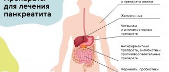
Drug treatment consists of prescribing drugs such as:
- enzymes (support the functioning of the pancreas and help normalize the digestion process);
- antisecretory drugs or proton pump inhibitors (block the formation of hydrochloric acid in the stomach);
- fat blockers (prevent the absorption and absorption of fats in the gastrointestinal tract);
- painkillers;
- antibiotics;
- antiemetics and antidiarrheals;
- vitamin complexes.
In case of extensive damage to the gland by fatty lesions that interfere with its normal functioning, surgical treatment is performed. Surgical intervention can be performed in 2 ways. Their fundamental difference is the type of operational access. In accordance with this, laparotomy and laparoscopic surgery are distinguished. Compared to classic abdominal surgeries, endoscopic manipulations are much easier and the recovery period after them is quick and painless.