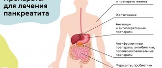
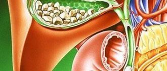
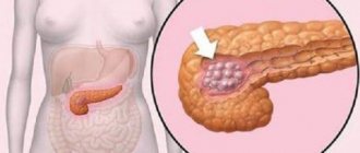
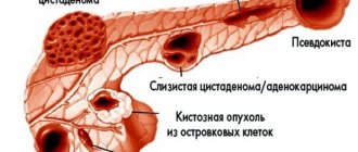
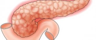
Inflammation of the pancreas is accompanied by constant or periodic pain of various types. This is the main symptom of pancreatitis, which can develop in acute and chronic forms. At the first manifestation of girdle pain around the abdomen, you should consult a doctor for examination. In most cases, treatment with medications and long-term diet is indicated.
Major diseases of the pancreas
Most pathologies of the pancreas begin to develop with pancreatitis. It can have 2 forms - acute and chronic. In turn, acute pancreatitis is of 3 types:
- Edema – accompanied by severe swelling of the gland, which disappears within 5-10 days.
- Sterile pancreatic necrosis - some of the gland cells die, which is not associated with infection.
- Infected pancreatic necrosis - in this case, cells die due to infection. This is the most dangerous condition, leading to death in 80% of cases.
Chronic pancreatitis develops over a long period of time and is not accompanied by acute pain. This disease most often occurs in the age group of 35-50 years. The pain can be either constant or paroxysmal (around the abdomen). This symptom is the most characteristic.
Clinical case of diagnosing pancreatic cancer
For citation. Vinnik Yu.S., Serova E.V., Bobkova A.V. and others. Clinical case of diagnosis of pancreatic cancer // Breast Cancer. 2016. No. 8. pp. 525–527
Pancreatic cancer (PCa) is a malignant tumor that develops from the ductal epithelium - ductal (81% of cases) or from parenchymal cells of the pancreas - acinar (14% of cases).
There is also unclassified prostate cancer. PCa is one of the most common malignant diseases. This type of disease in developed countries is in 5th place among the causes of death in the overall structure of cancer. PCa accounts for about 10% of all gastrointestinal tumors [1, 2]. The incidence in the USA is 11 cases per 100 thousand population annually, in Japan and England - 16, in Italy and Sweden - 18. In Russia, the incidence of prostate cancer is 8.6 people, and in Moscow - 11.4 per 100 thousand inhabitants. Over the past 50 years, the incidence has increased 4 times. Men get sick 1.5 times more often than women; the highest incidence occurs at the age of 60–70 years. At the same time, chronic pancreatitis of any etiology increases the risk of developing prostate cancer [2]. As with tumors of other locations, early diagnosis is very important for prostate cancer, which allows choosing the necessary tactics for patient management and increasing his life expectancy. Since this disease remains asymptomatic for a long time, up to 80% of patients die within a year after diagnosis. The leading role in the diagnosis of prostate cancer belongs to ultrasound, CT and MRI [1]. Malignant tumors can form in different parts of the pancreas: in 56-74% of cases - in the head, in 10-18% - in the body, in 6-8% - in the tail, in 6-28% of patients there is total damage to the pancreas. The localization of small cancer in the tail region is more favorable, which is associated with the possibility of performing distal resection of the pancreas with a more optimistic outcome [3]. Clinical case
Under our observation was patient R., 73 years old, who was sent to the gastroenterology department of the Krasnoyarsk Interdistrict Clinical Hospital No. 7 with a diagnosis of exacerbation of chronic recurrent pancreatitis of moderate severity.
From the anamnesis: in September 2013, pain began to bother me in the epigastric region and left hypochondrium. The patient sought medical help at the clinic at his place of residence. He was examined on an outpatient basis: fibroesophagogastroduodenoscopy showed cardial insufficiency, diffuse atrophic gastritis, and gastric xanthomas. Ultrasonography of the abdominal cavity revealed diffuse changes in the liver, pancreas, ductal changes in the liver, and an echo suspension in the gallbladder. At the outpatient stage, I received hyoscine butyl bromide, omeprazole, algeldrate + magnesium hydroxide, pancreatin - without a positive effect. Upon admission, the patient complained of aching pain in the epigastric region and left hypochondrium, worsening at night, and periodic bloating. An ultrasound examination (SonoScapeSSI-8000 device) of the abdominal cavity and kidneys revealed: liver dimensions 147×67×91 mm, smooth, clear contour, homogeneous structure, normoechoic, bile ducts are not dilated. Common bile duct – 4 mm. The diameter of the portal vein is 11 mm. The diameter of the aorta is 22 mm, with many calcified atherosclerotic plaques. The gallbladder measures 61×18 mm, pear-shaped, the wall is not thickened, there are no additional formations in the lumen, echo suspension is not visualized. Pancreas: head – 22 mm, body – 12 mm, tail – 24 mm, heterogeneous, diffusely increased echogenicity, uneven, clear contour, the Wirsung duct is not visualized. The small seal has not been changed. Free fluid in the abdominal cavity is not located. The intestinal loops are not dilated. Lymph nodes are not visualized. The spleen is 109×39 mm, homogeneous, normoechoic, with a smooth contour, at the hilum there is an ovoid, isoechoic (tissue) area of 49×47 mm, with a wavy contour, without connection with the left kidney (Fig. 1). With color Doppler mapping, blood flow in the contour area is determined. The diameter of the splenic vein is 6 mm. Pleural sinuses – without pathology. There are no additional formations in the kidneys or expansion of the pyelocaliceal system. Conclusion
: diffuse changes in the pancreas.
Atherosclerosis of the abdominal aorta. Volumetric formation in the hilum of the spleen (it is difficult to verify the organ affiliation - adrenal gland? spleen? differentiate the primary, secondary nature of the lesion). Instrumental examination data:
fibroesophagogastroduodenoscopy: diffuse superficial gastritis.
X-ray of the stomach: no organic changes were detected. ECG: signs of left ventricular hypertrophy. Laboratory tests:
anemia (hemoglobin – 75 g/l, erythrocytes – 2.73×10¹²/l), thrombocytopenia (113×109/l), leukocytes – 4.5×10⁹/l, eosinophils – 0.7, basophils – 1.3, segmented – 19.8, lymphocytes – 71.3, monocytes – 6.9, ESR – 31 mm/hour, amylase – 116 U/l; bilirubin, aminotransferases, blood glucose, urea, creatinine, thymol test are normal. Cholesterol – 6.3 mmol/l. Gamma-glutamine transpeptidase – 56.8 U/l. A general urine test is normal. Feces for occult blood are weakly positive. CT scan (Fig. 2) visualizes a significant increase in the size of the tail of the pancreas with a heterogeneous structure. CT conclusion: pancreatic cancer; atherosclerosis of the aorta. Data revealed by MRI (PhilipsIntera, 1.5 Tesla) are presented in Figures 3–6.
The pancreas is significantly increased in size at the level of the tail and partially the body, its contours are not clear enough, the structure is heterogeneous at the level of these sections due to the presence of a cystic-solid formation of a heterogeneous structure (iso- and predominantly hyperintense signal on T2 and in fat suppression mode, iso- and hypointense signal on T1), with a predominance of the cystic component, with the presence of multiple septa, measuring 8.7×4.6×4.4 cm. With intravenous dynamic contrast, diffuse heterogeneous accumulation of the contrast agent is noted at the level of the solid component (similar to unchanged gland tissue ) with a lack of contrast at the level of the cystic component. The dimensions of the pancreas at the level of the head are 1.8 cm, at the level of unchanged parts of the body 1.3 cm, with signs of fatty degeneration at these levels. The pancreatic duct at the level of the space-occupying lesion is not traced, at other levels it is not changed. Peripancreatic fiber is infiltrated at the level of the space-occupying lesion. There are signs of spread of this pathological process to the anterior parts of the spleen. In addition, in the S4, S7 projections of the liver, there are single foci of altered MR signal (hyperintense in T2 and in fat suppression mode, hypointense in T1), without clear contours, ranging in size from 0.3 × 0.3 cm to 1.0 × 1 .0 cm, with intravenous dynamic contrast, the second genesis of pathological foci is not excluded. The patient received symptomatic treatment, which resulted in some improvement – a decrease in pain. Discharged on the 12th day with recommendations for treatment at the Krasnoyarsk Regional Clinical Oncology Dispensary named after. A.I. Kryzhanovsky. The patient was not operated on due to age, concomitant cardiac pathology and the prevalence of the oncological process (tumor infiltration of parapancreatic tissue, metastatic liver disease). Symptomatic treatment was continued.
Enlarged pancreas: reasons
When affected by pancreatitis, the pancreas increases in size, which is clearly visible even on x-rays. The following reasons lead to the development of pathology:
- Damage, destruction of the pancreas or its removal after surgery.
- Hereditary problems with the production of digestive enzymes.
- Complications after removal of the stomach.
- Enzymes are synthesized by the gland, but cannot pass through the ducts due to the accumulation of stones or tumors.
- Food and enzymes enter the intestines at different times.
Pancreatitis appears against the background of poor nutrition (excess fat), bad habits (alcohol abuse, smoking), infectious lesions, poisoning, and taking certain medications (including antibiotics).
Pancreatic atrophy
Conservative activities
In case of pancreas atrophy, diet therapy is necessarily prescribed.
The diet should be low in fat. Enough attention should be paid to protein-energy deficiency and correction of hypovitaminosis. A mandatory measure is to completely stop smoking, since nicotine disrupts the production of bicarbonates by the pancreas, as a result of which the acidity of the contents of the duodenum significantly increases. The main direction of treatment for this pathology is the replacement of exocrine and endocrine secretions of the pancreas. To compensate for impaired cavity digestion processes, a gastroenterologist prescribes enzyme preparations. To achieve a clinical effect, drugs must have high lipase activity, be resistant to the action of gastric juice, ensure rapid release of enzymes in the small intestine, and actively promote cavity digestion. Enzymes in the form of microgranules meet these requirements.
Since it is lipase that loses activity most quickly of all pancreas enzymes, correction is made taking into account its concentration in the drug and the severity of steatorrhea. The effectiveness of treatment is assessed by the content of elastase in feces and the degree of reduction of steatorrhea. The action of enzyme preparations is also aimed at eliminating pain, reducing secondary enteritis, creating conditions for normalizing intestinal microbiocenosis, and improving carbohydrate metabolism.
Correction of endocrine insufficiency is carried out through insulin therapy. With pancreatic atrophy, the islets of Langerhans are partially preserved, so insulin is produced in the body, but in small quantities. The dosage and regimen of insulin administration are determined individually depending on the course of the pathology, etiological factor, and data from daily blood glucose monitoring. The administration of enzyme preparations significantly improves the function of the pancreas in general and carbohydrate metabolism as well. Therefore, the insulin therapy regimen is determined depending on the dosage and effectiveness of enzyme replacement therapy.
An important condition for the effective correction of digestive functions is the normalization of intestinal microbiocenosis, since the intake of enzymes creates favorable conditions for the colonization of pathogenic flora. Probiotics and prebiotics are used. Injection vitamin therapy, as well as magnesium, zinc, and copper preparations, are required.
Surgery
Surgical treatment of this pathology is carried out in specialized centers. Transplantation of the islets of Langerhans is performed, followed by removal of the gland and enzyme replacement therapy. However, since atrophy is often a consequence of severe illnesses with a pronounced impairment of the patient’s general condition, such treatment is rarely carried out.
Symptoms of pancreatitis in men and women
Pancreatitis presents with similar symptoms in men and women. The main one is a sharp pain that surrounds the abdomen and can radiate to the back and shoulder blades. At the same time, the sensations do not intensify during coughing, sneezing or taking a deep breath, which is typical for cholecystitis and appendicitis. Sometimes pain is accompanied by vomiting and nausea, which, however, do not lead to a weakening of symptoms. The disease is also characterized by other manifestations:
- pale or blue skin;
- a sharp decrease or increase in temperature;
- colic in the intestines;
- cardiopalmus.
Attention!
Acute pancreatitis can cause internal bleeding, which is often fatal. Therefore, at the first occurrence of painful sensations of a chronic nature, it is necessary to undergo diagnostics.
Diagnosis of the disease
Diagnosis is carried out only in a clinical setting - the patient initially consults a therapist. During the examination, the doctor clarifies the complaints and prescribes a number of procedures:
- blood test (general and biochemical);
- Analysis of urine;
- stool analysis with determination of fat content;
- radiology diagnostics;
- linked immunosorbent assay;
- collection of pancreatic secretions through a probe (inserted directly through the esophagus);
- secretin-pancreozymin test;
- Lund's method eliminates the insertion of a probe, but is not as effective and is therefore used much less frequently.
