- Reasons for development
- Features of neuroendocrine tumors
- Classification of neuroendocrine tumors
- Types of neuroendocrine tumors and their symptoms
- Diagnosis of the disease
- Treatment
- Survival prognosis
Neuroendocrine tumors (NETs) are formed from apudocytes , or APUD cells . These cells are scattered throughout the body and constitute the most ancient part of the endocrine system. They are at the same time similar to nerve cells and cells of the endocrine glands, since they can respond to signals from the outside or changes in the state of the body, and are capable of producing hormones that perform different functions.
Tumors of the APUD system are rare and can be difficult to diagnose. They most often occur in the gastrointestinal tract, but can also affect other organs. The peculiarity of neuroendocrine neoplasms is that tumor cells produce an increased amount of hormones, and because of this, certain symptoms may occur.
According to the American SEER registry, in 2004 the incidence of neuroendocrine tumors in the United States was 5 cases per 100 thousand population. In Russia, unfortunately, there are no statistics, but the incidence is probably at a similar level.
According to American experts, the prevalence of neuroendocrine tumors is growing every year. This is primarily associated with changes in nutritional patterns and unfavorable environmental conditions.
According to the same SEER registry, neoplasms of the APUD system are often diagnosed at late stages: in 50% of cases, the tumor manages to spread to surrounding tissues, to regional lymph nodes, and give distant metastases.
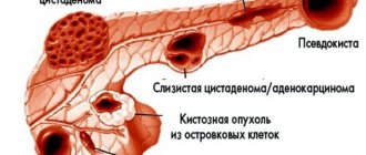
What is a neuroendocrine tumor of the pancreas?
Pancreatic neuroendocrine neoplasms (PNEN) are a group of neoplasias (tumors) of different origin, structure and clinical manifestations.
Depending on whether these tumors can secrete hormones and amines, causing carcinoid and other clinical syndromes, they can be classified as functional (F-NEN) or nonfunctional (NF-NEN). Neuroendocrine neoplasms of the pancreas are the most common neuroendocrine tumors.
Most PNENs are low grade but have malignant potential. They grow slowly and remain stable for many years, at least when they are small. However, without treatment, most of these tumors grow and eventually metastasize to the liver.
Pancreatic neoplasms are rare overall: <2% of all pancreatic tumors. Their diagnosis and localization often require significant technology, and their successful treatment requires a thorough multidisciplinary approach.
For these reasons, a patient with a possible or confirmed pancreatic endocrine tumor is best treated in a large multidisciplinary center that includes endocrinologists, gastroenterologists, pancreatic surgeons, and medical oncologists.
Staging of the tumor process
Determining the stage is of key importance, as it completely determines further treatment. As with most other tumors, NET is usually divided into 4 stages - from I to IV. Moreover, in addition to the stage, the diagnosis includes a special coding - TNM, where:
- T (tumor, tumor)
– describes the primary tumor,
- N (nodes, lymph nodes)
– describes the spread of the tumor to the lymph nodes,
- M (metastasis, metastases)
– describes the presence or absence of metastases.
Another parameter describing a neuroendocrine tumor is Grade. It is used to describe the ability of a tumor to grow rapidly and spread throughout the body and comes in 3 grades:
- G1 (low grade)
– low growth and spread rate,
- G2 (intermediate grade)
– intermediate,
- G3 (high grade)
– high rate of growth and spread.
Classification
The term neuroendocrine tumor of the pancreas usually refers to insulinoma, gastrinoma or glucagonoma. Sometimes more rare VIPomas and somatostaniomas are added to this trio.
PNENs are defined as functional or nonfunctional depending on whether they cause a syndrome of hormonal hypersecretion—excessive production of hormones.
Of all functional PNENs, gastrinomas and insulinomas are the most common. Other neoplasias are usually considered together as a group called "rare functional PNENs."
Several such rare clinical syndromes have been proposed as possible functional PNENs. These include the following:
- calcitoninoma;
- parathyrinoma;
- growth hormone-secreting tumor (GRFoma);
- adrenocorticotropin hormone-secreting neoplasia (ACTHoma);
- neurotensinoma.
According to European and American oncologists, non-functional PNEN are more common than functional ones. The former are usually diagnosed in the fourth or fifth decades of life and often have already metastasized to the liver at the time of diagnosis.
INCIDENCE
- The incidence of neuroendocrine tumors of the gastrointestinal tract and pancreas is approximately 3-5 cases per 100,000 people per year.
- NETs of the gastrointestinal tract and pancreas can develop at any age, but more often after 50 years.
- When detected with glucagon, gastrin, VIP, and somatostatin, metastases to regional lymph nodes are detected in 60-80% of patients.
- The five-year survival rate for carcinoid cancer averages 50-67%. The best median survival rate occurs when the tumor is localized in the rectum (88%), bronchus (74%) and appendix (71%).
Clinical and pathogenetic characteristics of the main NETs
| Tumor type | Secreted hormones or amines | Clinical symptoms |
| Carcinoid | Serotonin | Carcinoid syndrome: hot flashes, diarrhea, bronchospasm, hypertension, heart damage. |
| Gastrinoma | Gastrin | Zollinger-Ellison syndrome, severe peptic ulcers |
| VIPoma | Vasointestinal polypeptide | Severe diarrhea (“pancreatic cholera”, Werner-Morrison syndrome) |
| Insulinoma | Insulin | Hypoglycemia |
| Glucagonoma | Glucagon | Diabetes, erythema migrans, irritation and redness of the tongue |
| Somatostatinoma | Somatostatin | Gallbladder dysfunction, cholelithiasis, impaired glucose tolerance |
CARCINOID
One of the most common tumors of the diffuse neuroendocrine system! The tumor secretes:
- serotonin
- bradykinin
- 5-hydroxytryptophan
- prostaglandins
- VIP
- histamine
This leads to the development of carcinoid syndrome.
CARCINOID SYNDROME
Manifestations:
- hot flashes (63-94%). Sudden appearance of deep red or purple erythema of the upper torso, mainly the face and neck. Accompanied by an unpleasant sensation of warmth, lacrimation, itching, swelling of the face and conjunctivitis, salivation and sweating, a feeling of pulsation.
- diarrhea (68-84%)
- bronchospasm (3-19%)
- shortness of breath (3-19%)
- telangiectasia (25%)
- heart changes (11-53%)
- pellagra-like syndrome with hyperkeratosis and pigmentation (2-6%)
- heart damage - carcinoid cardiac syndrome (Hedinger syndrome) is detected in more than 50% of patients.
The syndrome is caused by the development of fibrous changes in the endo- and myocardium of the right heart with damage to the valves. Stenosis and insufficiency of the tricuspid valve and pulmonary valve are characteristic.
CARCINOID CRISIS
- It can occur spontaneously or be provoked by stress, alcohol, certain foods (for example, cheese), and injections of catecholamines.
- Initial attacks last 2-5 minutes, and later their duration can increase to several hours.
- Crises more often occur with daily excretion of 5-HIAA more than 200 mg and are provoked by stress, anesthesia, biopsy or surgery, chemotherapy, causing high mortality.
CLASSIFICATION OF NEO (WHO, 2002)
- Well-differentiated neuroendocrine tumor (benign).
- Well-differentiated neuroendocrine carcinoma (low-grade).
- Poorly differentiated neuroendocrine carcinoma (small cell).
- Mixed exocrine and endocrine carcinoma.
- Tumor-like lesion.
Characteristics of highly differentiated NETs
- low proliferative potential
- ability to secrete various biological substances
- low sensitivity to chemotherapy.
These include various carcinoids of the foregut, midgut and hindgut, pheochromocytoma, and medullary thyroid cancer.
Characteristics of low-grade NETs
- highly malignant tumors with high proliferative potential
- sensitive to chemotherapy and radiation therapy.
These include small cell cancer of the lung and other organs.
Characteristics of mixed exocrine and endocrine carcinomas
- these include various pancreatic tumors (except carcinoids)
- The sensitivity to chemotherapy of these tumors is moderate.
CLASSIFICATION OF CARCINOID TUMORS (WILLIAMS AND SANDLER, 1963)
- Upper (2-9%) (foregut): tumors of the respiratory tract, thymus, esophagus, stomach, duodenum, pancreas. Characterized by low secretion of serotonin, increased secretion of histamine and various hormones, an atypical course of carcinoid syndrome, frequent metastasis to the bones.
- Middle (75-87%) (midgut): tumors of the small intestine, appendix, cecum, ascending colon. Hypersecretion of serotonin and other vasoactive substances, carcinoid syndrome.
- Lower (1-8%) (hindgut): tumors of the transverse colon and descending colon, sigmoid and rectum. Carcinoid syndrome is not typical; liver metastasis is common.
Classification depending on the presence or absence of carcinoid syndrome
- Hormonally active tumors are characterized by hypersecretion of hormones. Clinical symptoms allow early diagnosis.
- Hormonally inactive tumors (no symptoms of hormone hypersecretion) – up to 50% of neuroendocrine tumors. Clinically manifested with a large tumor mass - at this stage the tumor metastasizes, and can also manifest itself as symptoms of intestinal compression.
LABORATORY DIAGNOSTICS
- Determination of the level of serotonin and its metabolites in urine. The most common test is to measure the excretion of 5-HIAA (5-hydroxyindoleacetic acid) in 24-hour urine. However, this analysis may give false positive results depending on the nature of the food consumed by patients (citrus fruits, bananas, pineapples, kiwi, nuts).
- Determination of the level of chromogranin A in the blood (an increase in the level of chromogranin A is observed in 87-99% of patients), the norm is less than 4.5 mmol/l. This plasma marker is the most reliable for NET.
- Determination of the level of excretion of neuron-specific enolase in the blood.
Reasons for appearance
10% of functional NENs become a manifestation of hereditary familial endocrine tumors:
- multiple endocrine neoplasia syndrome type 1 (MEN1);
- von Hippel-Lindau disease (VHL);
- neurofibromatosis type 1 (NF-1);
- tuberous sclerosis (TSC).
The reason for the development of these pancreatic neuroendocrine neoplasms is hereditary switching off of the corresponding tumor suppressor gene during fetal development.
Although the outdated term “islet cell tumor” is often used today to identify neoplasms of the endocrine pancreas, it is a misnomer. Many pancreatic neuroendocrine tumors do not develop directly from islet cells.
Most of these neoplasms arise from APUD stem cells. These are the precursor cells from which all types of pancreatic tissue are subsequently formed. They are located in the epithelium of the ducts of this organ.
The fact that many gastrinomas and somatostatinomas are located near the glandular tissue of the pancreas, but not within it, supports the view that these neoplasms may develop extrapancreatically.
Choosing a foreign clinic for NET treatment with Booking Health
Treatment of rare tumors of the neuroendocrine system requires a responsible approach to choosing a medical institution and doctor. When planning treatment abroad, Booking Health will help you make the right choice. Booking Health is an international medical tourism operator that has been organizing treatment for patients from 75 countries abroad for more than 10 years. The compliance of the quality of the company’s services with international standards is confirmed by the prestigious ISO 9001:2015 certificate.
Booking Health staff will help you:
- Select a clinic and doctor based on the annual qualification report
- Establish direct contact with your doctor
- Familiarize yourself with the preliminary examination and treatment program
- Receive clinic services at a favorable price, without surcharges for foreign patients (save up to 50%)
- Make an appointment for the desired date, without long waits
- Feel confident at all stages of the medical program thanks to independent external monitoring
- Purchase and send necessary medications to your home country
- Maintain contact with the clinic after completion of treatment, receive examination results and discharge statements
- Monitor bills from the clinic
- Organize control and additional examinations
- Book accommodation, plane tickets, transfer
- Translate medical documents into your native language, communicate with a doctor in one language
Fill out the “Send a request for treatment” form on the official Booking Health website and wait for a call from a medical consultant who will help you choose a medical program and prepare for your trip.
Symptoms of the disease
Functional tumors lead to hormonal hypersecretion syndromes.
| Neoplasia | Symptom(s) |
| Insulinoma | Hypoglycemia is a decrease in blood glucose levels. Sweating, weakness, tremors, hunger, etc. |
| Gastrinoma (ulcerogenic pancreatic adenoma) | It manifests itself as a protracted gastric ulcer that is difficult to treat - abdominal pain, nausea, vomiting, etc. |
| VIPoma | Uncontrollable diarrhea, decreased potassium levels in the blood, decreased acidity of gastric juice, dehydration, weight loss. |
| Glucagonoma | Glucose intolerance, dermatitis, stomatitis, glossitis, deep vein thrombosis, depression, heart failure. |
| Somatostatinoma | Symptoms of diabetes mellitus and cholelithiasis, steatorrhea, achlorhydria. |
Nonfunctional neoplasms cause nonspecific symptoms, such as vague abdominal pain, and are usually discovered incidentally.
The distinction between functional and nonfunctional PNEN is based on clinical presentation, and there is no absolute difference in hormone expression between functional and nonfunctional PNEN.
For example, tumor cells in a small PNEN may express glucagon, resulting in borderline elevated glucagon levels. Clinically, however, this PNEN is defined as nonfunctional because a small increase in glucagon is not sufficient to cause glucagonoma syndrome.
Treatment of neuroendocrine tumor in Germany
Each of the presented oncologies has its own signs of manifestation, but an accurate diagnosis can be made only after a thorough diagnosis of the patient. The Nordwest Clinic in Germany uses the latest modern equipment and effective research methods to detect diseases, as well as unique technologies to combat them. Our clinic is one of the few that uses 177-Lutetium dotatate therapy for neuroendocrine tumors. Please note that all neuroendocrine tumors cannot be treated according to the same regimen; the clinical recommendations of the attending physician are very important in each specific case. When you contact us, you can be sure that the therapy will be tailored individually, taking into account the characteristics of the development of oncology.
Diagnostics
Because recognition of hormonal hypersecretion syndrome requires considerable clinical experience and the symptoms of nonfunctional tumors are nonspecific, the diagnosis of PNEN often occurs quite late.
Endocrine testing, imaging, and histologic findings are essential for accurate diagnosis of PNEN.
A complete diagnosis must establish the nature of PNEN, assess tumor grade, identify primary and metastatic sites, and determine whether the tumor is functioning. If hormonal hypersecretion syndrome is suspected, appropriate biochemical testing is performed to determine it, followed by imaging, endoscopy and biopsy.
If a tumor in the pancreas or liver is incidentally identified by imaging, it is usually biopsied to confirm the presence of PNEN. Biochemical testing is ideally performed even if hormonal hypersecretion syndrome is not obvious, as it may be at a subclinical stage, and hypersecreted hormones can be used as tumor markers during subsequent evaluations.
Diagnosis of the disease
In most cases, neuroendocrine neoplasms are discovered incidentally when a person is being examined for another reason. A tumor can be detected using diagnostic methods such as:
- Ultrasonography.
- Endoscopic examinations: gastroscopy, colonoscopy, bronchoscopy.
- X-ray with contrast enhancement. Before the examination, the patient is given a solution to drink, which helps to clearly see the contours of the stomach and intestines.
The diagnosis is confirmed with a biopsy. Having discovered a pathological formation, the doctor removes a piece of tissue from it and sends it to the laboratory for examination under a microscope. If the tumor is suspected to be a pheochromocytoma, biopsy is resorted to only in extreme cases, as it carries serious risks.
Computed tomography and MRI help to detect a tumor, assess its size, location, degree of spread in the body, and detect metastatic foci. PET scanning is used to search for metastases. Your doctor may order tests that help detect hormones produced by the tumor, such as a urine test for 5-HIAA, a metabolic product of serotonin. Determining the level of chromogranin A in the blood helps monitor the effectiveness of treatment.
Treatment
Patients with pancreatic neuroendocrine neoplasms are treated individually to balance the effects of hormonal hypersecretion. Many pancreatic endocrine tumor syndromes are potentially life-threatening. Therefore, initial medical therapy is aimed at stabilizing the patient's condition to allow a complete preoperative assessment.
All resectable functional or nonfunctional PNEN are recommended to be treated surgically. Different types of surgical intervention are used:
- tumor enucleation (removal of the visible part of the tumor while preserving healthy organ tissue);
- enucleation in combination with pancreatic resection;
- distal pancreatectomy;
- pancreatoduodenectomy (removal of the pancreas and part of the intestine);
- etc.
The type of surgical intervention is determined individually, based on the characteristics of a particular disease in a particular patient. At the same time, neuroendocrine pancreatic cancer requires mandatory excision of lymph nodes.
All resectable insulinomas should be removed surgically, regardless of tumor size. 85–90% of patients with this type of PNEN can be completely cured in this way. Insulinomas account for approximately 35–40% of functional PNENs. Of these, malignant insulinomas account for only about 5–10% of the total.
Systemic therapy is necessary for patients with residual disease after surgery and for the treatment of inoperable tumors.
Synthetic analogues of the hormone somatostatin (growth hormone) are effective against functional PNEN. Their effect is especially pronounced in VIPomas and glucagonomas.
There is no clear evidence that somatostatin analogues treat nonfunctional PNEN, but they are likely to inhibit tumor growth.
Two somatostatin analogues, octreotide and lanreotide, and their long-acting formulations are currently available, and a new analogue, pasireotide, is still in the clinical trial phase with encouraging preliminary results.
Examination for suspected NET
- Collection of complaints and anamnesis (medical history),
- Blood and urine tests to identify biochemical markers of a neuroendocrine tumor,
- CT or MRI,
- PET-CT with gallium (68Ga-DOTATATE and 68Ga-DOTANOC),
- Glucose PET-CT (FDG)
- Scintigraphy with 111 In-Octreotide,
- FGDS (fibrogastroscopy),
- FBS (fiber bronchoscopy),
- FCS (fibrocolonoscopy),
- Endo-ultrasound,
- Capsule endoscopy.
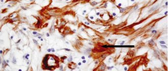
Biopsy plays a special role in diagnosis.
– sampling of a tumor fragment to accurately determine its type. A biopsy can be performed using various methods, for example, with FCS or FGS. After the biopsy, the tumor fragment is sent for histological and immunohistochemical examination (under a microscope) to determine the type and aggressiveness of the tumor.
Which examinations will be used in your situation will be determined by your attending physician. Often, when examining and planning treatment, it is necessary to consult a related specialist, for example, a cardiologist.
Treatment of neuroendocrine tumors
Given the rarity of NETs and the complexity of diagnosis, each patient is discussed by a multidisciplinary team before starting treatment. It includes:
- oncologist-chemotherapist,
- oncologist-surgeon,
- endocrinologist,
- morphologist,
- radiology specialist,
- if necessary, additional specialists: cardiologist, anesthesiologist, etc.
During the analysis of the clinical situation of each individual patient, a collegial individual treatment plan is developed, and, if necessary, additional examination.
Progressive treatments for neuroendocrine pancreatic neoplasia in Belgium
In clinics in Belgium, modern methods of minimally invasive surgery, radiation therapy, biological therapy and chemotherapy are used in the treatment of PNEN.
Microinvasive surgical and radiological methods include:
- radioactive polymer microspheres;
- chemoembolization;
- mild embolization;
- radiofrequency ablation (RFA);
- percutaneous alcohol ablation;
- microwave ablation.
Systemic interferon-α2A is used in clinics in Belgium for the treatment of PNEN, which are insensitive to somatostatin analogues.
The mTOR inhibitor everolimus (RAD001) and the tyrosine kinase inhibitor sunitinib are effectively used in Belgian clinics to slow the progression of neoplasia.
Chemotherapy is available for intermediate- and high-grade neoplasias. Temozolomide and capecitabine are the drugs of choice that can be used in patients with rapidly progressing PNEN.
The method of Peptide Receptor Radionuclide Therapy (PRRT) Lutetium Lu 177-dota-tate, developed by Belgian scientists in collaboration with colleagues from the Netherlands, is being successfully introduced into practice. It is effective against well-differentiated PNEN. But this method is not yet widely used, so it is best suited for neoplasias with high malignant potential that are resistant to other systemic treatments
Estimated cost of treatment for NET:
| Price | |
| Diagnosis of neuroendocrine tumor (NET) | from 1,010 euros |
| Chemotherapy for neuroendocrine tumor (NET) | from 2,383 euros |
| Treatment of neuroendocrine tumor (NET) by laparoscopic surgical resection | from 12,579 euros |
| Therapy with Lutetium-177-DOTATATE (Lu-177-DOTATATE) for neuroendocrine tumor (NET) | from 13,965 euros |
| Treatment of neuroendocrine tumor (NET) of the stomach using embolization or chemoembolization | from 23,676 euros |
| Treatment of neuroendocrine tumor (NET) of the small intestine using embolization or chemoembolization | from 23,808 euros |
Send a request for treatment
Complications
Patients with pancreatic endocrine neoplasia may experience morbidity associated with tumor growth.
- Patients with tumors in the head of the pancreas sometimes have a bile duct blockage, a pancreatic blockage, or both.
- Chronic abdominal pain may occur due to the compressive effect of a large intra-abdominal tumor or obstructive pancreatitis.
Morbidity caused by the effects of excess hormonal production by functional pancreatic neoplasms occurs earlier than the neoplastic effects in the course of the disease.
- If left untreated, patients with insulinoma may experience hypoglycemic seizures and even open coma.
- Before the development of effective antisecretory drugs (eg, H2 blockers and proton pump inhibitors), patients with gastrinoma often experienced life-threatening gastrointestinal bleeding from peptic ulcer disease.
- Patients with VIPomas are at risk of becoming dehydrated from diarrhea. They may also develop fatal cardiac arrhythmias due to associated hypokalemia.
- The morbidity associated with the hormonal effects of glucagonoma can be polymorphic. Patients may have diabetes, malnutrition, stomatitis, and other symptoms similar to severe nutritional deficiencies.
Survival prognosis
The prognosis depends on what type of tumor was diagnosed, how aggressive it is, and what stage it is at. Treatment has one of two goals:
- Completely remove tumor foci. If the examination results do not reveal signs of the presence of a tumor in the body, remission is stated.
- Relieve the patient of symptoms and contain the growth of the tumor. In this case, the patient can live quite a long time.
It is worth talking to your doctor before starting treatment, asking what the goal will be, what results can be expected, and what is planned to be done if the situation worsens and the tumor recurs.
Book a consultation 24 hours a day
+7+7+78
Forecast
The prognosis for most types of PNEN is favorable. But even that neuroendocrine tumor of the pancreas, for which the prognosis is considered good, requires constant monitoring. Because increased growth rate can become an alarming factor that worsens survival.
Because these tumors typically grow slowly and have a relatively low metastatic potential, and because no specific criteria have been defined to predict their behavior, the distinction between benign and malignant neoplasms is based on the presence of metastatic disease.
The five-year survival rate for PNEN without treatment is:
- 80% for all stages;
- 90–100% for localized disease;
- 40% for regional disease;
- 29% for distant metastases.
Long-term disease-specific survival (DSS) of surgically resected tumors exceeds 50% at 20 years.
Because even metastatic pancreatic endocrine neoplasms typically grow slowly, the prognosis of patients with these tumors is relatively good compared with that of patients with nonendocrine cancers.
More than 90% of patients with insulinomas have benign tumors without evidence of metastases, and up to 97% of these patients are completely cured by surgical resection.
Patients with gastrinomas have a worse prognosis. 60% of these tumors are malignant. However, survival rates differ strikingly between patients whose gastrinoma metastases are limited to lymph nodes and those with liver metastases, as follows:
- patients whose gastrinoma metastases involve only lymph nodes may live 25 years or longer, and their life expectancy is indistinguishable from that of people without gastrinoma.
- patients with liver metastases have a 5-year survival rate of 20-30%, compared to 90% for patients without liver metastases.
Approximately half of all VIPomas have metastases at the time of diagnosis or surgery, and approximately one third of patients are completely cured by surgery.
Most somatostatinomas (84%) have already metastasized at the time of presentation, but a number of patients survive 5 years after combined surgery and chemotherapy.
Among endocrine neoplasias, glucagonomas tend to be relatively large (5–10 cm) at diagnosis. In addition, up to 80% of such neoplasias show invasive or metastatic growth. Glucagonoma syndrome is recognized relatively late in most patients, and surgical treatment is possible in less than 20% of all patients. In this regard, such a pancreatic neuroendocrine tumor has the most serious prognosis.
Special diagnostic tests
According to statistical information from the SEER (Surveillance, Epidemiology, and End Results) registry, up to 50% of patients already have single or multiple metastases at the time of diagnosis of NET. The reason for this is the asymptomatic or low-symptomatic course of the early stages of the pathology. Clinicians often talk about neuroendocrine tumors as a “diagnosis of exclusion,” which is established after a comprehensive examination and exclusion of more common nosologies.
Diagnosis of neuroendocrine tumors begins with a history and clinical examination, including assessment of the condition of all groups of lymph nodes. The next step is a general laboratory examination:
- Clinical and biochemical blood test
- Coagulogram, blood pH assessment
- Blood test for electrolytes (potassium, magnesium, calcium, chlorine)
- Infectious markers (antibodies to HIV and hepatitis viruses)
- Clinical urine analysis
As a rule, it is more informative to determine specific markers of NETs, namely:
- Assessment of chromogranin A level in blood plasma
- Assessment of the level of pancreatic polypeptide in blood plasma
- Analysis of 24-hour urine for 5-hydroxyindoleacetic acid
- Blood test for serotonin
- Blood tests for tumor-specific markers, including gastrin, insulin, calcitonin, ACTH, epinephrine and norepinephrine, somatostatin, glucagon, VIP and others
Instrumental diagnostic methods include imaging examinations, if possible, with the collection of tumor tissue samples for further immunohistochemical research:
- X-ray, X-ray studies of the gastrointestinal tract
- CT and/or MRI, with or without intravenous contrast
- Endoscopic examination of the respiratory system
- Endoscopic examination of the digestive system
- Skeletal bone scintigraphy
A comprehensive examination allows us to establish the causes of development and localization of the tumor, the degree of its invasion into surrounding tissues and histological structure. Only after receiving complete information, an interdisciplinary team of doctors begins to develop a treatment regimen.
Find a specialized clinic