Internal bleeding of the stomach is not an independent disease, but acts as a symptom and complication of other diseases. It is important to understand that this symptom occurs not only when the stomach is damaged, but can also indicate problems not related to the gastrointestinal tract: blood clotting disorders, vascular diseases, metastatic growth of a tumor from a neighboring organ.
General symptoms, the severity of the patient’s condition, and diagnostic studies allow the doctor to correctly determine treatment tactics.
Causes of stomach bleeding
Often the cause of stomach bleeding is an untreated ulcer.
The main causes of hemorrhages can be divided into frequent, infrequent and rare.
Frequent sources of the problem:
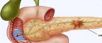
- An ulcer is a serious disease that is often life-threatening. Bleeding can occur either from a new “young” ulcer or from a chronic one. Long-existing ulcers are dangerous due to more massive bleeding, especially when it is located on the lesser curvature of the stomach. This is due to anatomical features - large branches of the left gastric artery are located here.
- Tumors cause acute bleeding only in 10% of cases; more often, bleeding is detected in the same way as with erosion - during FGS or stool examination.
- Erosion is superficial destruction of the gastric mucosa. In most cases, they are multiple, located in the fundus of the stomach, less often in the antrum. Heavy (profuse) bleeding with erosive gastritis is rare; more often it is drip leakage of blood, which is detected by chance, during FGS or stool examination for occult blood.
- Mallory-Weiss syndrome - occurs when the cardiac part of the stomach is ruptured in the longitudinal direction. This situation is possible when there is a sudden increase in pressure in a full stomach. It occurs in pregnant women with severe toxicosis (due to constant vomiting), athletes when lifting weights, and blunt abdominal trauma.
The following processes are less common:
- Henoch-Schönlein disease (hemorrhagic vasculitis) - after some kind of infection (ARVI, laryngitis, tonsillitis), toxic damage to the capillaries occurs and multiple hemorrhages occur throughout the gastrointestinal tract.
- Postoperative gastric ulcer.
- Acute leukemia.
- Gastroesophageal reflux disease.
- Iatrogenic damage (taking non-steroidal anti-inflammatory drugs, corticosteroids, antihypertensive drugs)
The rarest causes are: scurvy, hemophilia, thrombocytopenic purpura, aplastic anemia, sarcoidosis.
The main diseases that lead to stomach bleeding are listed here, but in general there are many more of them.
Types
The stomach is well supplied with blood, all its structures are penetrated by a capillary network. The venous system of the organ is also developed, which, for example, forms a pronounced venous plexus at the junction of the esophagus and the stomach. Capillaries, veins, and even small arteries can be injured or destroyed by various pathological processes, as a result of which blood begins to leak into the stomach cavity.
It can occur quickly or slowly, with loss of a small or significant amount of blood. Also, bleeding in the stomach may be accompanied by other pronounced signs of pathology or proceed almost unnoticed.
Based on the various characteristics of this process, the following classification of gastric bleeding has been created:
- acute (requiring emergency medical attention) or chronic (occurring over a long period of time);
- obvious (having a characteristic clinical picture) or hidden, that is, occurring with virtually no change in the patient’s condition;
- mild, moderate, severe (each degree of severity is characterized by certain clinical and laboratory parameters);
- ulcerative (complicating gastric ulcer) or non-ulcerative, which are formed against the background of other diseases.
The type of hemorrhage is determined when the patient seeks medical help and is reflected in the formulation of the diagnosis. This is extremely important for the development of therapeutic tactics, as well as for further prognosis.
Symptoms of acute gastric bleeding
Internal bleeding of the stomach can be hidden or obvious
Symptoms of acute bleeding depend on a group of factors: the rate of blood loss, the volume of fluid lost, the person’s age, and the presence of additional diseases.
In the clinic of acute bleeding, two periods are distinguished: latent and obvious.
The first occurs from the moment blood enters the intestines from the stomach. The second is from the moment blood is detected in vomit, feces or gastric contents (with FGS).
The severity of the clinical manifestations directly depends on the rate of blood loss. If it is more than 750 ml per day, the patient feels weakness, sweating, and severe headache. As blood pressure drops, a person notices a noise in the head, “flickering of spots,” and fainting occurs. In some cases, the only sign of bleeding is sudden loss of consciousness.
Vomiting indicates the presence of bleeding; its color can indicate the massiveness of blood loss. “Coffee grounds” indicates slight bleeding, since hemoglobin has time to turn into hydrochloric acid hematin under the influence of hydrochloric acid.
With heavy bleeding, cherry-colored vomit. If the blood is bright red, the bleeding most likely comes from the esophagus or is very massive in the stomach. Vomiting is often one-time, its repetition within two hours indicates continuous bleeding.
Diagnostic features
When a patient is admitted to the hospital, if he is conscious, the doctor clarifies the complaints, the presence of provoking factors, and examines the patient. Clinical data may suggest gastric or intestinal bleeding, but its exact source can only be determined with further examination. To do this, gastroscopy is performed using endoscopic equipment, during which esophageal varices, stomach ulcers, polyps or diverticula, and ruptures of the mucous membrane may be detected.
A clinical and biochemical blood test, determination of coagulability and platelet count are required, and stool is examined for the Gregersen reaction. If necessary, an ultrasound of the abdominal organs, diaphragm, chest, radiography of the stomach, angiography, radioisotope scanning, and MRI are performed.
The information obtained during the examination helps to carry out a differential diagnosis of bleeding in the stomach, that is, to exclude intestinal or pulmonary foci of blood loss. In addition, it is necessary to accurately identify pathologies leading to blood loss.
Symptoms of chronic bleeding
Internal stomach bleeding leads to severe anemia
Symptoms of chronic blood loss are vague and depend on the degree of iron deficiency anemia. Often the body compensates for losses so well that the patient walks independently and lives a normal life with a hemoglobin level of 30-40 g/l. The main symptoms of chronic gastric bleeding are signs of anemic syndrome. Severe weakness and fatigue appear, which increases even from slight physical activity.
The condition of the skin changes: the skin is pale, dry, flaky, and “stubs” appear in the corners of the mouth. Women immediately notice brittle nails and cracks in the skin of their hands.
With severe anemia, patients feel tachycardia even at rest, and shortness of breath appears. The condition of the oral cavity changes - there is a burning sensation in the tongue, difficulty swallowing, redness of the tip of the tongue. Sometimes there is hoarseness, coughing, redness and itching in the palate.
In especially severe cases, olfactory tastes change - patients may fall in love with the musty air of the basement, the smell of shoe polish or car tires. Sometimes there is a desire to eat sand or earth.
Symptoms: how to recognize the presence of pathology
Any first aid - pre-medical or medical - cannot be provided to a patient until the presence of specific symptoms of pathology has been established, the degree of damage and the level of threat to life have not been determined.
General symptoms of gastrointestinal bleeding are represented by the following manifestations:
- weakness, severe dizziness;
- increased sweating;
- darkening of the eyes;
- cooling the extremities;
- blanching of the skin and mucous membranes.
However, a characteristic sign that allows you to identify a specific type of bleeding is the admixture of blood in the stool (black stool, or melena), as well as vomiting of fresh blood and “coffee grounds.”
Treatment
In the acute period, the following manipulations are performed to stop bleeding:
- Drug hemostasis: the patient is injected intramuscularly with hemostatic drugs (Vikasol, Etamzilat, Aminocaproic acid).
- Endoscopic hemostasis (vessel clipping, photocoagulation, sclerotherapy).
- Arterial embolization.
- Surgical intervention (vascular ligation).
At the same time, the volume of circulating blood is replenished: saline solutions are administered (Disol, Trisol, Ringer's solution), microcirculation in the vessels is improved (Reopoliglyukin).
In case of severe blood loss, blood is transfused and iron-containing drugs are administered. In the future, iron supplements are prescribed for 3-6 months.
After stopping the bleeding, treatment tactics will depend on the disease that caused the disease.
First aid
For stomach bleeding, there is little that can be done at home. If you suspect a pathology, you must:
- urgently call an ambulance;
- the patient should be placed with the leg end raised; if he loses consciousness, the head must be turned to the side to prevent aspiration of vomit;
- Ice should be placed on the patient’s stomach;
- You cannot give food or drink, or take medications on your own.
Folk remedies
Photo: oprostatite.info
If gastric bleeding develops, traditional methods cannot be used. In the absence of timely specialized care, this condition can lead to the development of hemorrhagic shock and death of the patient. It is necessary to immediately call an ambulance and not self-medicate. During the period of remission, the following remedies can be used:
- 1 tbsp. l. horsetail is steamed for an hour in 250 ml of boiling water. The broth is filtered and consumed half a glass each time after meals.
- A tablespoon of pink immortelle flowers is infused in 250 ml of boiling water. Then the infusion is filtered and given to the patient 1 tbsp. l. every 2 hours.
- A decoction of blueberries is brewed and drunk like tea three times a day.
Parsley helps prevent stomach bleeding. In addition, decoctions of chamomile, bearberry, and yarrow are used. All these herbs have hemostatic properties. Any folk remedies are allowed to be used only after the approval of a specialist.
The information is for reference only and is not a guide to action. Do not self-medicate. At the first symptoms of the disease, consult a doctor.
What is capsule endoscopy?
In 2000, a group of doctors in England reported using a new tool to determine the causes of intestinal bleeding in the small intestine. This device was called an endoscopic capsule and measures 26 mm x 11 mm - the size of a large pill.
It consists of a battery with an 8-hour lifespan, a strong light source, a camera and a small transmitter. Once swallowed, the capsule begins to transmit images of the inside of the esophagus, stomach and small intestine to a receiver worn by the patient. The capsule takes two pictures per second, for a total of about 55,000 images per session. After 8 hours, the patient returns the receiver to the doctor, who downloads the information onto a computer and then reviews each bowel image taken in detail, looking for abnormalities that are possible sources of bleeding. The capsule continues its path through the patient’s colon and is excreted naturally.
The given technical parameters may differ for different capsule endoscopy systems. There are video capsules that work longer than 8 hours and shoot more than 2 frames per second.
Swallowing the capsule is fairly easy and safe; however, the capsule may become stuck in the small intestine. Video capsule delay may occur in the following cases:
- if the patient has previously undergone abdominal surgery that caused scarring in the intestines
- patients have a pathology in the intestine that has led to a severe narrowing of its lumen.
If the capsule gets stuck, it will need to be removed endoscopically or surgically. The positive point is that capsule retention almost never leads to acute obstruction, and the site of retention is directly adjacent to the pathological focus. In this case, along with the extraction of the capsule, therapeutic measures are taken to remove the pathology found.
In 15% of cases, the capsule does not have time to pass through the entire small intestine and photograph its mucous membrane because its battery runs out, in which case the study may have to be repeated. This phenomenon is more typical for video capsules with an operating time of 8 hours. For video capsules with 12 hours of operation, the likelihood of incomplete examination of the small intestine is significantly lower.
Like X-rays, the video capsule is a purely diagnostic method and cannot be used to take a biopsy, carry out treatment, or perform tattooing for subsequent surgical treatment. In addition, the capsule cannot be controlled. Therefore, the doctor does not have the opportunity to return to the suspicious area and examine it in more detail. Despite these limitations, capsule endoscopy is the gold standard for searching for occult intestinal bleeding in the small intestine (occult is intestinal bleeding that cannot be detected by standard endoscopic methods, but all symptoms of bleeding are present).
What is intraoperative enteroscopy?
In some cases, enteroscopy may require surgery. Intraoperative enteroscopy is performed in the operating room under general anesthesia. The surgeon, often working with a gastroenterologist or endoscopist, inserts the endoscope through the patient's mouth, or through a small incision in the small intestine (enterotomy). Next, the surgeon helps the endoscope move through the intestine, as if stringing loops of intestine onto it. This allows a complete examination of the small intestine. The advantage of intraoperative enteroscopy is that it allows the doctor to treat the cause of bleeding at the time of detection (for AVMs), or to remove pathologies or polyps found.
Because it is an invasive surgical procedure, intraoperative enteroscopy is used in extreme cases when other methods have failed to find or treat the source of the bleeding. Overall, this method is effective in treating the source of intestinal bleeding in approximately 70% of cases.
Popular publications:
- Video capsule endoscopy: frequently asked questions - 11/12/2013 17:02 - Read 48366 times
- Black feces Why? Causes? — 01/07/2015 05:06 — Read 48124 times
- Capsule endoscopy. Description, reviews, advantages and disadvantages - 03/24/2013 17:15 - Read 42869 times
- Abdominal syndrome. Abdominal pain - 01/10/2015 05:02 - Read 24133 times
- Capsule colonoscopy - indications, contraindications, price - 10/30/2015 07:20 - Read 20247 times
Key words: Small bowel bleeding, small bowel bleeding, capsule endoscopy, endoscopy, enteroscopy
- < Back
- Forward >
Infusion therapy
Begin infusion therapy with the introduction of balanced salt solutions.
Important! If there are signs of ongoing bleeding or unstable hemostasis has been achieved, blood pressure should be maintained at the minimum acceptable level (SBP 80-100 mm Hg), i.e. infusion therapy should not be too aggressive. Blood transfusions are carried out if adequate infusion therapy fails to stabilize the patient’s hemodynamics (blood pressure, heart rate). Consider the need for blood transfusion:
– when the hemoglobin level decreases below 70 g/l. when bleeding has stopped;
– with ongoing bleeding, when hemoglobin is below 90-110 g/l.
In case of massive blood loss (more than 50-100% of the blood volume), transfusion treatment is carried out in accordance with the principles of “Hemostatic resuscitation”. It is believed that each dose of packed red blood cells (250-300 ml) increases hemoglobin levels by 10 g/l. Fresh frozen plasma is prescribed for clinically significant coagulopathy, including drug-induced coagulopathy (for example, the patient is receiving warfarin). And in case of massive blood loss (>50% of blood volume). If reliable hemostasis is achieved, there is no need to administer FFP even with significant blood loss (more than 30% of the blood volume). Dextrans (polyglucin, rheopolyglucin), hydroxyethyl starch (HES) solutions may increase bleeding and their use is not recommended.
What is deep enteroscopy of the small intestine?
In cases where the lesion was found deep in the small intestine, beyond the reach of a standard endoscope, other techniques may be necessary. One option for further evaluation or treatment of the lesion is double-balloon enteroscopy. In 2004, the FDA (Food & Drug Administration) approved a new type of endoscope known as a double-balloon enteroscope (this name is given to this endoscope because it uses a system of two cylinders). This endoscope allows you to reach very distant parts of the small intestine (in some cases, all the way to the ileum - this is the last segment of the small intestine). This enteroscope can also be inserted through the anus, making it easier to examine the distal (farthest from the stomach) small intestine (to do this, the endoscope must first pass through the colon). In some cases, examination through the mouth and through the anus can examine the entire length of the small intestine, although this is not always possible. Because the double balloon enteroscope examination is much more complex than standard endoscopy (the procedure often takes several hours, as opposed to standard endoscopy, which usually does not exceed 20 minutes), it is usually performed only in cases where the source of small intestinal bleeding is inaccessible to standard endoscope and was found using one of the radiation diagnostic methods or using capsule endoscopy.
In one study, a double-balloon enteroscope was able to detect the source of bleeding in 74% of patients. Other studies have shown that when the source of intestinal bleeding is found, treatment success is achieved in 63 to 71% (in these studies, treatment is considered successful if the patient does not require further blood transfusions).
In addition to double-balloon enteroscopy, two other methods of examining the small intestine have become available. One is single-balloon enteroscopy, which is a similar procedure to double-balloon enteroscopy, although in this case the enteroscope only has one balloon attached to it and is designed to facilitate the procedure. The second option for deep enteroscopy of the small intestine is using a special spiral tube, which is placed on the endoscope and allows it to be advanced to distant areas of the small intestine. This device works differently than balloon enteroscopies and allows for shorter procedure times. Currently, it is only used for pathologies that can be reached by inserting an endoscope through the mouth.