First - 3 real stories of our patients
Story 1: weight loss, expensive creams and gall bladder
Patient P., 36 years old. The new workplace places high demands on appearance. The patient went on a strict diet, went in for sports and lost 8 kg in a month. However, skin rashes have intensified - areas of redness, peeling, and acne have appeared. Treatment by a cosmetologist did not help, and even expensive skin products did not produce results.
On the advice of another dermatologist, the patient came for examination to a gastroenterologist-hepatologist. At the appointment, the patient recalled that while playing sports she sometimes felt a tingling sensation in her right side, but did not pay attention. The doctor ordered an ultrasound, which revealed changes in the gallbladder: due to irregular nutrition, it was spasmodic, thick bile began to accumulate, and this inhibited intestinal motility. The doctor prescribed treatment and recommended eating 4-5 times a day in small portions. Gradually the condition of the skin improved, the pain stopped bothering me.
Conclusion: competent cosmetologists know how the condition of the skin is connected with the functioning of internal organs. Therefore, it is often advised to consult a gastroenterologist if you have problem skin. Listen up, beautiful skin starts with health on the inside.
Story 2: Skin rashes and hepatitis
Patient K, 34 years old. I have never suffered from allergies. Suddenly, hives-like rashes appeared, which began to recur regularly. The patient repeatedly consulted an allergist and hematologist. Antiallergic drugs did not help at all. It got to the point where rashes began to appear for no apparent reason at all. The condition worsened, with weakness, drowsiness, and fatigue.
Noticing this, the patient came for examination to a gastroenterologist. The curator prescribed tests, ultrasound with elastography. Diagnosis: chronic hepatitis C, genotype 1a, stage 2 fibrosis. Upon further examination, Giardia was discovered - it was they that caused the rash on the skin. The patient completed the appropriate course of treatment and antiviral therapy, and the rash disappeared.
Conclusion: if you have rashes, do not rush to buy expensive medications and go on a strict diet. Often the reason is different. Check your liver and gallbladder.
Story 3: Itchy skin and cirrhosis of the liver
Patient O., 54 years old, contacted the EXPERT Polyclinic on the recommendation of a dermatologist with complaints of itchy skin. Long-term treatment by a dermatologist turned out to be ineffective, and a consultation with a gastroenterologist-hepatologist was scheduled.
The doctor found out that several years before the appearance of itchy skin, changes were detected in the biochemical blood test due to problems with the production and removal of bile from the liver. The patient worked with chemical fertilizers for many years. In addition, the gynecologist prescribed hormonal contraceptives, which she took for a long time, to normalize the menstrual cycle.
The patient was fully examined according to one of the comprehensive diagnostic programs, including a liver biopsy. Toxic and drug-induced hepatitis were excluded. All this helped establish the final diagnosis: primary biliary cirrhosis of the liver. The patient was prescribed bile acid preparations and treatment for skin itching. Now the patient feels well, the skin itching has disappeared, and the biochemical parameters of the liver are normal.
Conclusion: if your liver has been overloaded - you have been in contact with toxic substances, or have been taking medications for a long time - get examined periodically. This way you can catch a potentially dangerous disease at an early stage.
Dermatological symptoms of liver cirrhosis: the significance of an interdisciplinary problem
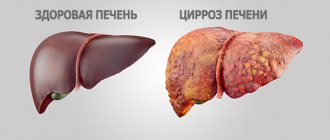
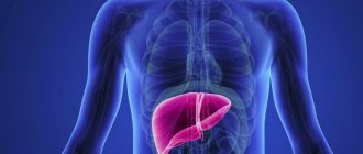
The liver of an adult weighs about 1.5 kg. It is covered with a thin, strong connective tissue membrane – Glisson’s capsule [1]. Most of the liver is located on the right side of the body. The liver is projected onto the anterior abdominal wall of the epigastric region. The upper border of the liver normally begins in the 10th intercostal space on the right in the mid-axillary line. From here it rises steeply upward and medially. Along the right nipple line, the liver border can normally reach the 4th intercostal space. Further, the border of the liver descends to the left, crosses the sternum slightly above the base of the xiphoid process, the upper border of the liver reaches the middle of the distance between the left sternum and the left nipple line. The lower border of the liver also begins in the 10th intercostal space on the right, but goes obliquely and medially, crosses the 9th and 10th costal cartilages on the right, runs along the area above the womb obliquely to the left and up, crosses the costal arch at the level of the 7th left costal cartilage and in the 5th intercostal space it connects with the upper border [2]. The location of the inferior border of the liver is one of the most important clinical characteristics of its size. Normally, it is determined below the edge of the costal arch on the right; the protrusion should be no more than 2 cm. The liver consists of 2 main lobes, the right lobe is much larger than the left. The lower surface of the liver is called visceral and is in contact with some parts of the gastrointestinal tract and the right kidney. The upper surface of the liver is smooth, directly adjacent to the diaphragm. On the lower surface of the liver there is a short, deep transverse groove - the porta hepatis. The relative mass fraction of the liver is not the same at different periods of a person’s life: in a newborn, the liver occupies most of the abdominal cavity, and the mass is 1:20 of body weight; in an adult, the weight of the liver is 1:50 of body weight, and its skeletotopy corresponds to that described above [2]. The liver consists of parenchyma formed by hepatocytes and connective tissue stroma. Hepatocytes are functional liver cells that perform at least 500 different functions, from storage (glycogen) to detoxifying (glucuronide). The uniqueness of the liver as an organ also lies in the fact that it is both an exocrine and endocrine gland. The endocrine secretion comes from the liver directly into the bloodstream, and the exocrine secretion is bile. The latter enters the hepatic duct, gall bladder, and duodenum. The common bile duct (ductus choledochus) opens into the duodenum, forming the nipple of Vater. In 1 day, from 0.5 to 1 liter of bile enters the intestine, the dynamics of its intake is determined by digestive need. If there is no such need, bile is deposited in the gallbladder [2]. Bile contains bile pigments (bilirubin), bile salts, proteins, cholesterol, and tissue fluid crystalloids. The main function of bile is to emulsify dietary fats, which is a preparation for enzymatic action. Normally, the amount of bile pigment - bilirubin in the blood is small: its total amount should not exceed 20 µmol/l, and the amount of bilirubin determined by the direct diazoreaction (Jendraszek method) should not exceed 3.4 µmol/l [3]. The latter is a bilirubin glucuronide. The so-called “indirect” bilirubin is a yellow pigment bound to blood proteins that has not been detoxified in the liver. A small amount of toxic “indirect” bilirubin in the blood does not lead to disruption of overall homeostasis. Liver diseases inevitably affect the condition of the skin and its appendages. In this case, symptoms arise, the assessment of which, even in the absence of special studies, makes it possible to make an accurate diagnosis and direct the treatment process in the right direction. One of these serious diseases is cirrhosis of the liver. It is necessary to distinguish between 2 processes, as a result of which the organ loses most of its specific functions. The loss of these functions occurs as a result of the death of specialized organ elements, in this case hepatocytes, and their replacement with connective tissue. These 2 processes are cirrhosis and fibrosis.
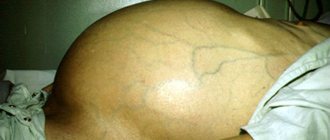
Fibrosis is an inactive process, representing scar changes in any organ; once arising, for example, as a result of an abscess, fibrosis fills the missing structure of the organ, although it is functionally untenable. This process does not pose any danger, because it is not active and does not progress. Cirrhosis is an active process of replacement of functionally specialized tissue with connective tissue that no longer performs specialized functions. The main danger of cirrhosis is its steady progression, accompanied by the death of the functional elements of the organ - hepatocytes. The uniqueness of the liver as an organ also lies in the fact that it has pronounced regenerative capabilities and is capable of restoring some of the lost lobules. It is this ability to regenerate that is lost in cirrhosis. Damaged hepatocytes regenerate more slowly than they are replaced by connective tissue. The causes of cirrhosis come down to the action of a number of agents, the activity of which exceeds the adaptive capabilities of the organ [4–6]. The pathogenetic classification of liver cirrhosis is based on the principle of taking into account portal hypertension, the development of cirrhosis as a result of previous necrosis of hepatocytes, and prolonged stagnation of bile. In accordance with this clinical and morphological classification, liver cirrhosis is distinguished: portal, postnecrotic, biliary, mixed. Portal cirrhosis is the most common type of liver cirrhosis (up to 40% of all cases). This species received its name due to the often developing hypertension in the portal vein system of the liver. According to the WHO clinical classification, this is micronodular cirrhosis, and its causes are often alcohol intoxication, fatty hepatosis, pathological conditions with protein-vitamin deficiency; Infectious lesions, for example, Botkin’s disease, may also be important [7]. Another synonym for portal cirrhosis is septal, since it is characterized by the formation of connective tissue septa that fragment the liver lobules. General clinical symptoms of portal cirrhosis include weakness, loss of appetite, pain in the right hypochondrium, alternating constipation and diarrhea, and bloating. The liver is palpable in 85% of cases, the spleen in 40% of cases. Jaundice develops at the beginning of the process in only 12% of patients. An enlarged liver in the early stages of cirrhosis may not manifest itself clinically, and only an increase in the size of the organ indicates trouble. Gradually, the consistency of the liver becomes denser, its surface becomes lumpy, and an increase in size may be replaced by a decrease. Splenomegaly appears later than hepatomegaly [7]. Very quickly, with portal cirrhosis, congestion develops in various venous pools: esophagogastroduodenoscopy reveals varicose veins of the esophagus (from which bleeding is sometimes possible), veins of the anterior abdominal wall (“Medusa’s head”, Fig. 1), and overflow of hemorrhoids [7]. Due to stagnation, the liquid part of the blood plasma leaks into the abdominal cavity, and ascites is formed, sometimes reaching significant sizes. Massive edema develops in the lower extremities. The appearance of ascites always indicates the presence of hepatocellular failure. Indeed, in addition to stagnation, the release of fluid into the tissue is facilitated by disruption of albumin synthesis. This point, combined with sodium retention, leads to a decrease in intravascular colloid osmotic pressure. Sodium retention is due to increased synthesis of aldosterone and decreased inactivation in cirrhosis.
Postnecrotic cirrhosis accounts for up to 30% of all cirrhosis and, according to the WHO classification, corresponds to macronodular cirrhosis. In most cases, this form of cirrhosis occurs as a result of viral hepatitis, as a result of which it is also called “post-hepatitis”. Other causes of this form of cirrhosis are hepatotoxic poisons. Under the influence of all these factors, necrosis of the liver parenchyma occurs, and massive necrosis is followed by collapse of the remaining stroma. The collapsed stroma turns into scars, between which areas of liver tissue are preserved. Since the regenerative ability of the liver is preserved for some time, large nodes of functionally still healthy liver parenchyma are formed between the layers of connective tissue [7]. But, unfortunately, this form of cirrhosis is characterized by rapid progression of the disease, and signs of hepatic cellular failure quickly come to the fore: pain in the right precostal area, dyspeptic disorders. Jaundice develops in most patients and occurs in waves. Astheno-vegetative disorders are often associated. If portal cirrhosis is characterized by slow progression of ascites, then in postnecrotic cirrhosis ascites has a wave-like course and in the early stages can even resolve on its own for some time [7, 8].
Biliary cirrhosis accounts for 5–10% of all liver cirrhosis. There are primary and secondary biliary cirrhosis. Primary biliary cirrhosis is an inflammatory autoimmune disease of the interlobular and septal bile ducts. The bile ducts are gradually destroyed under the influence of viruses, drugs, and other intoxicants; this condition leads to ductopenia, persistent cholestasis, and progressive liver failure. Thus, the basis of primary biliary cirrhosis is intrahepatic cholestasis. This form of cirrhosis most often affects women aged 40–60 years, the intensive rate is 4–15 cases per 105 people. The average life expectancy of patients with clinical manifestations is 7–10 years [8]. Secondary biliary cirrhosis occurs with cholangitis, congenital defects in the structure of the bile ducts, in the presence of various long-term obstacles to the outflow of bile (stone, scar, neoplasm). Connective tissue develops around the bile canaliculi and along the periphery of the hepatic lobules, resulting in the formation of so-called “false lobules.” Secondary biliary cirrhosis is based on extrahepatic cholestasis. General clinical symptoms of biliary cirrhosis are determined by cholestasis. Characterized by jaundice, itching, steatorrhea, osteoporosis, and bleeding. In clinically pronounced cases, portosystemic encephalopathy develops [8].
Although itching can occur with any liver damage, it is especially painful with biliary cirrhosis. Sometimes its intensity can be compared to the itching of skin lymphoma or severe atopic dermatitis. Itching drives the patient into a frenzy, sometimes to suicide [8]. A distinctive feature of skin itching in liver failure is the presence of only secondary elements - excoriations (scratching), while the primary elements of all kinds of rashes, characteristic of most itchy dermatoses, are absent (Fig. 2). There are no papules, vesicles, tubercles, only sometimes urticaria are found. The functional characteristics of liver cirrhosis of any origin include the following parameters [4, 9]: 1. Hepatocellular failure: – compensated (only changes in stress test parameters, bilirubin level – <34 µmol/l, prothrombin time (PTT) – 1–4 s , albumin level – >35 g/l); – subcompensated (bilirubin level – 35–50 µmol/l, albumin level – 28–35 g/l, PTT – 4–6 s); – decompensated (albumin level – <28 g/l, bilirubin level – > 51 µmol/l, PTT – > 6 s). 2. Portal hypertension: – moderate; – sharply expressed. Cirrhosis is characterized by an intrahepatic form of portal hypertension. 3. Activity of cirrhosis: – inactive; – active (moderate, pronounced). 4. Ascites: – no; - soft; – tense. 5. Encephalopathy: – no; – mild (grade 1–2); – severe (grade 3–4). The death of cells and tissues with such a huge functional set (as already noted, at least 500) that the liver tissue has, determines the whole variety of skin symptoms manifested in a patient with cirrhosis of the liver. Jaundice and itchy skin are essential symptoms of liver cirrhosis. Shades of yellowness can vary widely - from pale yellow to yellow-red. There is even the concept of “subictericity,” when jaundice is subjectively barely discernible; the sign is well defined on the sclera. Jaundice becomes clinically noticeable when bilirubinemia is at least 34–36 μmol/L (2–3 mg%) [1, 9]. The distribution of yellowness over the surface of the skin may be uneven; it is often more pronounced on the trunk and sclera, and to a lesser extent on the extremities. Acute pathology of the hepatobiliary system is often projected in the peri-umbilical zone: with acute cholecystopancreatitis, hemorrhages may appear here, and with a rupture of the common bile duct, a sharp yellow coloration may occur. Mild jaundice occurs in atrophic forms of cirrhosis.
The cardinal symptoms of cirrhosis - jaundice and skin itching - are often accompanied by others that are important not only for verification of the diagnosis, but also for the clinical assessment of the functional state of the liver, the degree of compensation of the pathological process, and therefore for the prognosis. In the therapeutic medical history, the general characteristics of the patient’s skin, as a rule, are not included in a separate status localis. This is one of the initial stages of examining a patient. Portal cirrhosis is characterized by dark pigmentation of the skin, mostly in its exposed areas, which is associated with the deposition of melanin in the dermis; in turn, these melanin deposits are caused by an increased content of estrogens and steroid hormones. There is a so-called sallow complexion. With any form of liver cirrhosis, changes in the hairline occur. Of all the skin appendages, hair is perhaps the most sensitive to intoxication. In patients with liver cirrhosis, a lack of hair in the armpit area can often be found. An increased content of estrogen leads to the fact that even men lose hair in the beard and mustache area and develop gynecomastia [5]. The development of erythema of the palms is a symptom characteristic not only of cirrhosis, but also of any chronic liver disease. However, in cirrhosis, “liver palms” are an integral part of the clinical picture. In addition to erythema, smoothness of thenar and hypothenar is noted. This symptom can be observed not only in patients with liver cirrhosis, but also during pregnancy, rheumatoid polyarthritis, and also in healthy adolescents [5].
The clinical picture of liver cirrhosis is often complemented by various neoplasms. All neoplasms arising against this background are benign. They can be roughly divided into vascular, “storage neoplasms” and hyperkeratotic. Vascular neoplasms include all kinds of hemangiomas, primarily stellate spider-shaped hemangiomas, which are almost as characteristic a sign of liver damage as “liver palms”. They are also called "spiders". They are considered a prognostically unfavorable sign. These “spiders” are localized almost exclusively in the area of the outflow of the superior vena cava: on the forehead, back of the head, shoulders, and the anterior wall of the chest. Morphologically, stellate hemangiomas are punctate, 1–3 mm, ectasia of dark red, cherry-colored vessels. With vitropression, weak pressure with a glass slide, especially when using a dermatoscope, pulsation of the central vessel can be detected. The mechanism of their formation is apparently due to a number of mediators and hormones released during the period of death of hepatocytes. Heparin is one such hormone. In hepatopathy, both arterioles and venules are affected [10]. Spider veins should be differentiated from angioma-like elements in Rendu-Osler-Weber disease (hereditary hemorrhagic telangiectasia), Fabry-Anderson angiokeratoma, Fordyce, and spider nevi. The latter are vascular nevi, exist from early childhood and are not accompanied by pathology of internal organs. Equally harmless is Fordyce angiokeratoma, which is a congenital malformation of the vascular wall. But Fabry–Anderson angiokeratoma requires close attention, because it is a fatal disease [11]. A characteristic but relatively rare symptom of liver cirrhosis is a bluish, crimson or red tongue with severe atrophy of the mucosa and papillae. The lips also become red, as if varnished. This sign is observed in cirrhosis, but is often difficult to interpret and distinguish from other pathological conditions. For example, this sign may be the only manifestation of incipient lichen planus. In addition, atrophy and “varnished” language is often accompanied by candidiasis of the oral mucosa, its atrophic form. Cyanotic tongue, although without pronounced atrophy, can be observed with mitral and mixed heart defects, in which cirrhosis of the liver is not uncommon.
In general, the vascular system is greatly affected by liver cirrhosis. Such patients develop various variants of livedo (Fig. 3) - a kind of dilatation of skin vessels: according to the type of mesh - reticular livedo (livedo reticularis), rings - annular livedo (livedo annillaris), tree crown - tree-shaped livedo (livedo racemosa) [12] . In severe cases, especially at the stage of gradual transition of chronic active hepatitis to cirrhosis, in such patients multiple hemorrhages appear on the skin of the trunk and limbs, and although this condition is called “hepatic purpura,” the nature of the hemorrhages varies from small petechiae to ecchymosis and vibice. If even small-point hemorrhages or hemorrhages in the sclera are detected, it is recommended to perform well-known clinical techniques for assessing the condition of the vascular wall: “tourniquet”, “pinch”, and check for the presence of the Rumpel-Leede symptom. Sometimes the vascular wall suffers so much that hemorrhagic dermographism is detected [12]. Patients may complain of frequent nosebleeds. “Accumulation neoplasms” are represented by xanthomas and xanthelasmas. They are manifestations of functional disorders of lipid metabolism. These neoplasms are not specific to liver cirrhosis and rather indicate a predisposition to lipid deposition in tissues. But since lipid metabolism disorders are an indispensable component of the pathogenesis of liver cirrhosis, the appearance of xanthomas and xanthelasmas is already a clinical component of the clinical and laboratory syndrome of dyslipidemia. Peculiar lipid deposits can form both against the background of hyperlipidemia and in a normolipidemic state. The most common types of cholesterol and lipid deposits in the skin are flat xanthomas and xanthelasmas [13]. They are yellowish-whitish nodules measuring 1 to 5 mm and round or ovoid in shape. Lipid deposition occurs in the upper layers of the dermis, where there is a cluster of foam cells. The cytoplasms of these cells are filled with lipids. Typically, when diagnosing persistent dyslipidemia, multiple flat xanthomas are important.
In addition to flat ones, there are also multiple eruptive xanthomas (Fig. 4), which are larger formations - nodes 5–8 mm in size, painless, scattered throughout the skin; the largest number of them is found on the extensor surfaces of the limbs, back, and buttocks. Often these nodes have an inflammatory rim around the main element [14]. When carrying out differential diagnosis for multiple flat and eruptive xanthomas, one should take into account not only hyperlipidemic and dyslipidemic conditions, but also nosologies that are not directly related to lipid metabolism: these are various hematological diseases, often severe; life-threatening conditions (histiocytosis X, leukemia, myeloma and other systemic diseases). Rarer types of xanthoma are tuberous and tendinous, they do not correlate with liver cirrhosis. Hyperkeratotic neoplasms on the skin in liver cirrhosis can be represented by senile keratomas. These are hyperkeratotic neoplasms of brownish color, 1–2 cm in size, rising above the surface of the skin, dense and rough to the touch. They are localized almost exclusively on the skin of the torso, sometimes located along Langer's tension lines. If there are few of these neoplasms, then they represent an independent nosology and reflect only the tendency of the skin to the appearance of such neoplasms. If, over the course of a year or even a shorter period, a massive burst-like eruption of these keratomas in huge numbers suddenly occurs, then this condition can be regarded as Leser-Trelat syndrome [15], a paraneoplasia caused by gastrointestinal carcinoma. If the number of keratomas slowly increases over 3-4 years, and eventually other symptoms and skin itching join them, then a liver examination is necessary. A specialist can judge a lot by changes in the condition of the patient’s nails. The fact is that another component of the pathogenesis of liver cirrhosis is a violation of protein metabolism, including a violation of keratin synthesis. Often in such patients we find changes in the structure and color of the nails. The endogenous nature of these changes is indicated by their symmetry and multiplicity. For example, single dotted whitish spots on one of the nails of the hands, gradually moving towards the free edge, not only do not indicate the presence of a serious disease, but are not even considered as a pathological condition. To consider the symptom of leukonychia (Fig. 5) as a significant sign, symmetry, multiplicity of changes, as well as a large affected area are required. The most typical for toxic (alcoholic) cirrhosis is total leukonychia - a symptom, although rare, at the same time clearly correlates with the severity of the process [5]. Nonspecific changes in the structure of the nails are much more common: - onychorrhexis - cracks in the free edge of the nail; − onychoschisis – separation of the nail parallel to the horizontal surface; - pinpoint depressions (Rosenau's symptom) similar to psoriatic ones, but in smaller quantities; − longitudinal ridges of nails (like “roof slats”) – found normally in old people, and in young people – with corresponding severe diseases; − hapalonychia (onychomalacia) – a kind of softening of the nail plate due to disturbances in its mineralization; − koilonychia – concave, “spoon-shaped” nails are a variant of hapalonychia, but in combination with other symptoms (subicteric sclera, sallowness of the face, “varnished tongue”) can serve as a criterion for severe intoxication and, accordingly, an unfavorable prognostic sign. Pathology of the vessels of the nail bed is a common occurrence in toxic hepatitis with the outcome in cirrhosis. A splinter symptom is often observed - an expansion of the capillaries of the nail bed and an increase in their permeability, manifested in the form of a thin short “thread” visible through the nail plate (Fig. 6). The appearance of this symptom is a relatively early indication of liver dysfunction, and with cirrhosis this symptom appears regularly.
Hypertrophic lesions of the nails, their excessive compaction (scleronychia), and Hippocratic nails are not typical for cirrhosis of the liver. The classic sign of a long-term drinker has always been facial erythema - a symptom described in many works of fiction. Meanwhile, facial erythema itself, or rosacea, does not directly correlate with the severity of liver damage, but only indicates the fact of alcohol abuse. It is under the influence of alcohol that the blood vessels in the face dilate, which later becomes persistent. However, it is believed that the extreme degree of rosacea - rhinophyma - still correlates in a certain way with the severity of liver damage and can serve as an indicative symptom of established cirrhosis, giving rise to a targeted search. Many of the described skin changes are such indicative symptoms. It is believed that rhinophyma represents the final infiltrative-productive stage of rosacea development. The process can be localized not only in the area of the nose (rhinophyma), but also in the chin (gnathophyma), on the ears (otophyma), and eyelids (blepharophimosis) [15]. Of the 4 types of rhinophyma (glandular, fibrous, fibroangiomatous, actinic), liver cirrhosis is most characterized by the form associated with insolation - actinic. But all of the listed forms of rhinophyma can be observed. Urticaria is a nonspecific syndrome and, in general, is not characteristic of cirrhosis. But it may be one of the signs of active viral hepatitis. In advanced cases of cirrhotic lesions, the appearance of urticaria is caused rather by toxic influences [1, 16].
Thus, all the described symptoms of cirrhotic liver damage can be divided into 2 groups: unconditional and indicative. The unconditional symptoms include the complex “jaundice - skin itching - signs of portal hypertension.” Individually, none of the symptoms is unconditional, but the above complex allows one to suspect cirrhosis. All other symptoms should be considered indicative, in the presence of which an in-depth and targeted study of the liver is indicated. It should also be mentioned about the possibility of the appearance of the 3rd group of symptoms, which are usually referred to as “paraneoplastic dermatosis,” during the transformation of cirrhosis into hepatocellular carcinoma. With the initial appearance of jaundice, patients usually first turn to a therapist and an infectious disease specialist, with severe skin itching - to a dermatologist, and with the appearance of vascular and other neoplasms on the skin - to a cosmetologist. And if in the first 2 cases the patient’s path, as a rule, ends with the establishment of an accurate diagnosis, then for a cosmetologist this path often ends at the stage of removing the cosmetic effect. Thus, all specialists, including cosmetologists, play an important role in the initial tentative diagnosis of liver disease.
Literature 1. Tsirkunov L.P. Skin symptoms in the diagnosis of somatogenic diseases. M.: Znanie-M, 2001. 368 p. 2. Prives M.G., Lysenkov N.K., Bushkovich V.I. Human anatomy. 10th ed. St. Petersburg: Hippocrates, 1997. 704 p. 3. Clinical assessment of laboratory tests / trans. from English / ed. WELL. Titsa. M.: Medicine, 1986. 480 p. 4. Sadovnikova I.I. Liver cirrhosis. Questions of etiology, pathogenesis, clinical picture, diagnosis, treatment // Breast cancer. 2003. T. 5. No. 2. 5. Tischendorf F.V. Diagnosis by external signs. Atlas-handbook of clinical and differential diagnostics / trans. with him. M.: Med. lit., 2008. 320 p. 6. Nam YH et al. Liver cirrhosis as a delayed complication of Stevens-Johnson syndrome // Intern Med. 2011. Vol. 50 (16). R. 1761–1763. Epub 2011 Aug 15. 7. Therapy / trans. from English, revised and additional / ed. acad. RAMS A.G. Chuchalina. M.: GEOTAR-Medicine, 1999. 1026 p. 8. Pinheiro NC et al. Refractory pruritus in primary biliary cirrhosis // BMJ Case Rep. 2013 Nov. 14. 2 013. pii: bcr2013200634. doi: 10.1136/bcr-2013-200634. 9. Lippert G. International system of units (SI) in medicine / trans. with him. M.: Medicine, 1980. 208 p. 10. Silverio AO et al. Are the spider angiomas skin markers of hepatopulmonary syndrome? // Arq Gastroenterol. 2013 Jul-Sep. Vol. 50 (3). R. 175–179. doi: 10.1590/S0004-28032013000200031. 11. Elkin V.D., Mitryukovsky L.S. Selected Dermatology. Rare dermatoses and dermatological syndromes. Handbook of diagnosis and treatment of dermatoses. Perm, 2000. 699 p. 12. Skin pathology. In 2 volumes. Volume 1. General pathology of the skin. / V.G. Akimov, V.I. Albanova, I.I. Bogatyreva and others / ed. V.N. Mordovtseva, G.M. Tsvetkova. M.: Medicine. 1993. 336 p. 13. Rational pharmacotherapy of diseases of the digestive system. Hand. for practicing doctors / V.T. Ivashkin, T.L. Lapina and others / edited by. ed. V.T. Ivashkina. M.: Litterra, 2003. pp. 389–464. 14. Therapist's Handbook / ed. F.I. Komarova. M.: Medicine. 1979. 656 p. 15. Potekaev N.N. Rosacea. M. – St. Petersburg: ZAO “BINOM Publishing House”, “Nevsky Dialect”, 2000. 144 p. 16. Therapeutic Handbook of the University of Washington (series “Foreign Practical Guides to Medicine” No. 1) / ed. M. Wooddy, A. Whelan / trans. from English M.: Praktika, 1995. 832 p.
What does the skin have in common with the liver?
One of the many tasks of the liver is to remove waste products from the body. If for some reason the liver cells stop doing this job, the secretion increases through the skin. This manifests itself as rashes, changes in skin color, etc.
Check your liver if you see:
- early age-related changes - fatty hepatosis, hepatitis of various origins
- blood dew (Tuzhilin syndrome) is a sign of a number of diseases of the liver, gallbladder and pancreas (photo No. 1)
- vesicles and papules - often appear during an autoimmune process in the liver and viral hepatitis (photo No. 2)
- acne – fatty liver degeneration and cell inflammation – non-alcoholic steatohepatitis
- spider veins (so-called spiders) - frequent signs of hepatitis, cirrhosis, fatty hepatosis, alcoholic liver disease (photo No. 3)
- jaundice is a sign of the release of bile pigments into the blood due to a mechanical obstruction to the outflow of bile or damage to liver cells (photo No. 4)
- liver palms - appears due to the accumulation of estrogen in cirrhosis
- excoriation - scratching due to itching of the skin in hepatitis and cirrhosis
- urticaria - although not directly related to the liver, it may indirectly indicate increased allergization caused by a disease in this organ
- bruises without visible injury are a sign of bleeding disorders, pancreatic diseases
- white spots on the nails, cracks in the corners of the mouth - a sign of vitamin deficiency, anemia, intestinal diseases
- stretch marks (striae) - often appear with the accumulation of fluid and an increase in the volume of the abdominal cavity in cirrhosis
- enlarged abdomen and “jellyfish head” - ascites and dilation of the subcutaneous veins of the abdominal cavity - signs of liver cirrhosis and portal hypertension, also found in cancer of internal organs (photo No. 5).
Signs of disease on the face
When examining a patient who has liver disease, the doctor immediately sees signs on the face. Due to metabolic disorders in the body, the skin of the face most often becomes oily, rashes, pimples, and age spots of different sizes and shades (from light brown to black) appear. The face becomes pale, the skin ages sharply, becoming covered with wrinkles.
A typical manifestation of the disease is general severe weight loss. External signs that appear with liver diseases include enlargement and asymmetry of the abdomen, which are observed with hepatitis, cirrhosis due to an increase in liver volume or due to the appearance of fluid inside the abdominal cavity.
External signs of liver disease help not only the doctor determine the diagnosis and prescribe further examination, but also serve as a signal to the patient himself that he should immediately seek help from professionals. The face and skin of the body, reacting to liver problems, “scream” about the need for urgent treatment.
It is important to know!
- What is liver elastography?
- Which doctor treats hepatitis C?
- What is liver fibroscanning?
- Which doctor should I contact if I have hepatitis B?
What to do if you notice changes in your skin?
Step 1. Consult your doctor and get an individual examination plan - biochemical tests - ALT, AST, bilirubin, alkaline phosphatase, GGTP, markers of viral hepatitis, immunological tests, ultrasound, and others, according to the doctor’s recommendations. Step 2. Find out the test results, diagnosis and prognosis of the disease. Step 3: Obtain a treatment plan that takes into account comorbidities.
Don't wait until your skin condition gets worse. Check your liver. In the early stages, recovery can be achieved. And the specialists of the EXPERT Polyclinic - hepatologists and dermatologists with extensive experience - will be happy to help. Health and beauty to your skin!