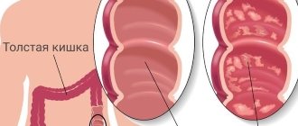
Causes of esophageal varicose veins
The disease develops against the background of increased pressure in the portal vein (also known as the portal vein), but other pathologies (hypertension, congenital anomalies, and so on) can contribute to its occurrence. Esophageal varicose veins, the causes of which lie in pathological changes in the portal vein, often begin to progress with previously diagnosed severe diseases of the liver or pancreas - cirrhosis, tumors, insufficiency. The disease often develops against the background of severe diseases of the thyroid gland, in which case we will be talking about pathological disorders of the vena cava.
According to statistics, esophageal varicose veins are more often diagnosed in men aged 50 years and older.
List of used literature:
- Angiography and transcatheter endovascular interventions for bleeding from esophageal varices. All-Union Conference “Radiation diagnostics and X-ray diagnostics of liver and kidney diseases”, Leningrad, 1984, pp. 22-24 (co-authors V.I. Prokubovsky, M.N. Ovchininsky, S.A. Kapranov).
- Possibilities and prospects for the development of X-ray endovascular interventions for bleeding from esophageal varices. VII All-Union Symposium “X-ray Endovascular Surgery”, 1985, pp. 133-134 (co-authors M.N. Ovchininsky, V.A. Cherkasov, S.A. Kapranov, S.B. Bakhvalov).
- Methods and tactics of endovascular hemostasis for profuse bleeding from esophageal varices. Republican conference of surgeons “Surgical treatment of portal hypertension, diseases and injuries of the liver”, Kharkov, 1986, pp. 60-62 (co-authors V.I. Prokubovsky, M.N. Ovchininsky, V.A. Cherkasov, S.A. Kapranov ).
- Ways to increase the effectiveness of endovascular interventions for bleeding from esophageal varices. Republican scientific and practical conference “X-ray contrast research methods and endovascular surgery”, Alma-Ata, 1986, pp. 167-169 (co-authors V.A. Cherkasov, M.N. Ovchininsky, S.A. Kapranov).
- A method for complex embolization of varicose veins of the esophagus and stomach. Rationalization proposal of industry significance 0-2976 dated October 2, 1987 (co-authors M.N. Ovchininsky, S.A. Kapranov, A.S. Belenky).
Karmanov
Author: Kapranov S.A.
Doctor of Medical Sciences, Professor, twice Laureate of State Prizes of the Government of the Russian Federation in the field of science and technology, Laureate of the Lenin Komsomol Prize, author of more than 350 scientific papers on medicine, 7 monographs, and 10 patents for inventions in medicine
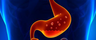
Esophageal varicose veins: symptoms
Depending on the degree of development, the following signs are distinguished:
- First degree. The diameter of the veins is increased by no more than 3 mm, the formation of single nodes is possible. Diagnosis of the disease is possible only by the endoscopic method; there are no clinical signs.
- Second degree. A rather dangerous period of varicose veins of the esophagus - there are no symptoms (even the mucous membrane of the organ still remains unchanged), but at the same time the contour of the veins already becomes unclear, and X-ray diagnostics reveal thickenings on the veins (harbingers of nodules). Already at this stage, bleeding is possible, which in the absence of immediate medical attention often leads to death.
- The third and fourth degrees of varicose veins of the esophagus are characterized by severe symptoms - the patient is often bothered by heartburn, pain occurs in the sternum, shortness of breath is possible, not only during physical activity, but also at rest. Often the disease is diagnosed only in the later stages of its development, when the first bleeding appears; blood is released directly from the esophagus or found in the vomit.
Diagnostic methods
The reason to consult a doctor if you suspect varicose veins of the esophagus are the clinical symptoms listed above. This is especially true for patients at risk - those suffering from arterial hypertension, liver cirrhosis, cholelithiasis).
To confirm the diagnosis, the doctor prescribes the patient to undergo a series of tests:
- blood tests - general and biochemical;
- fibroesophagoscopy;
- Ultrasound of the gastrointestinal tract;
- X-ray examination.
The most informative method for diagnosing pathology is fibroesophagoscopy. This diagnostic method involves inserting a tube equipped at the end with an optical system into the esophagus. The image is displayed on the monitor screen.
Thanks to this study, the doctor can evaluate:
- condition of the venous wall;
- degree of vein expansion;
- the risk of developing an aneurysm (thinning of the venous wall and its subsequent rupture);
- presence of bleeding and its cause.
Attention! If bleeding from the veins of the esophagus has already begun, then during esophagoscopy it becomes difficult to determine the cause of this complication; moreover, the study may provoke even greater progression of complications.
Less informative, but safer for a patient with suspected bleeding, is radiography with contrast. This study allows you to assess the condition of the esophagus as a whole and determine the degree of neglect of the disease.
Since in most cases, varicose veins of the esophageal vessels most often occur along with diseases of the liver and other gastrointestinal tract organs, during diagnosis attention should be paid to differentiating the following pathologies:
- polyposis of the stomach;
- hernia;
- peptic ulcer;
- oncological neoplasms in the gastrointestinal tract;
- hemorrhagic diathesis.
Treatment
Treatment of varicose veins of the esophagus begins with finding out the true causes of the development of the disease. If a pathology of the liver, thyroid or pancreas is diagnosed, then it must be treated - the provoking factor must be eliminated or its influence must be weakened. Medications include vitamin complexes, antacids and astringents. If bleeding is diagnosed, there is always a risk of its reoccurrence. In this case, the following is prescribed:
- blood transfusion;
- hemostatic drugs;
- introduction of a special probe that can compress the vessels.
Varicose veins of the esophagus, bleeding in which occurs too often, require surgical treatment.
Surgery is also performed if there is no effect from therapeutic treatment. Can esophageal varicose veins be stopped in this case? The prognosis is more favorable - the survival rate of patients increases 3 times. After any treatment, you must follow the dietary recommendations of doctors, undergo regular examinations and avoid excessive physical activity.
You can find out more about what esophageal varicose veins are and which doctor you should contact on the pages of our website Dobrobut.com.
2.What are the symptoms of bleeding from varicose veins?
As we have already said, varicose veins of the esophagus themselves do not cause any discomfort. But if a vein ruptures and starts bleeding, it is dangerous and requires emergency medical attention. Symptoms of varicose veins of the esophagus or stomach and the onset of bleeding from them may include:
- Vomiting blood;
- Black or bloody stools;
- Low blood pressure;
- Increased heart rate;
- Shock (in particularly serious cases).
Visit our Gastroenterology page
3.Indications
Today, almost every gastroenterological patient undergoes (including more than once, if necessary) the procedure of FEGDS, or fibroesophagogastroduodenoscopy. It is no coincidence that a flexible thin endoscope probe, equipped with high-tech manipulators, a video camera, and lighting, has become the gold standard for diagnosis and minimally invasive surgery for gastrointestinal diseases.
Esophageal vein ligation is also an endoscopic procedure.
After a thorough clinical, laboratory and instrumental examination (using the same FEGDS), the condition of the esophageal veins, the risk of bleeding and the feasibility of intervention are assessed. It is extremely important to inform your doctor about taking any medications for other diseases (some medications will need to be temporarily eliminated or replaced). Eating should also be excluded 8-12 hours before.
The term “ligation” involves the constriction of blood vessels with a ligature - a thin and strong surgical thread. Indeed, ligature loops (rings) are applied to the veins of the esophagus bulging with nodes, as a result of which the varicose vessels collapse within a few minutes, become sclerotic, “leave” the lumen of the esophagus and are switched off from the blood supply circuit. The number of ligating rings is determined by the number, size, condition of varicose nodes and other features of a particular case: in different situations it can be from 2-3 loops to 20 or more.
Local anesthesia with sedative premedication is used. This is absolutely enough for patients to tolerate the procedure without any problems: some heaviness and pressure are felt, but these sensations are not pain as such. Only in a few special cases is endoscopic ligation performed in medicated sleep or under general anesthesia.
The total duration of the procedure usually does not exceed one hour.
The patient then spends approximately the same amount of time in the observation room, from where, in the absence of obvious postoperative complications, he is sent home (but not driving his own car: driving vehicles and other high-risk mechanisms is contraindicated for the next 24 hours).
The ligatures come out naturally in about a week.
About our clinic Chistye Prudy metro station Medintercom page!
Diet for patients with varicose veins
When esophageal varicose veins are diagnosed, the patient is immediately introduced to dietary habits. After all, diet is the key to a carefree life for a patient with varicose veins.
If you adhere to it, not only human health will be preserved, but also the digestive system. In this case, proper nutrition means 4-6 meals a day.
The patient also needs to remember that dinner should take place no later than 3 hours before bedtime.
Particular preference should be given to the following products:
- rich in vitamin E. These include green onions, vegetable oil, chicken yolk;
- containing a lot of vitamin C;
- bioflavonodes are beneficial for body cells;
- routinely containing, currant nuts;
- stimulating elastin production, seafood;
- rich in plant fiber.
All food should be at normal temperature, it should not be too cold or hot.
It is impossible for acid from the stomach to enter the esophagus. To do this, you need to lie in the correct position, so the head of the bed for a patient with VRVP is raised by 10 cm or even more.
You cannot do extraneous things, for example, watch your favorite movie or read books while eating. This negatively affects the process of food absorption, which is undesirable in this case.
Healthy nutrition plays an important role in the treatment of varicose veins; a person can help his body and strengthen it. Nutrition has a beneficial effect on the condition of varicose vessels and their walls, and on the amount of fluid in the body.
If you add a few simple exercises to your diet to train your blood vessels, this will have a positive effect on the condition of the body; the risk of blood clots or the accumulation of excess fat in the veins will be much lower.
When a patient has varicose veins of the esophagus, he needs to drink a lot of fluid throughout the day, about two liters, water should be at least a liter.
It is important to avoid harmful foods so as not to worsen your condition. These include:
- strong brewed tea and coffee;
- alcoholic drinks;
- spices and various additives;
- sugar-containing products.
There is a special colored diet for patients who have esophageal varicose veins. It involves the consumption and preparation of products selected according to color scheme. You can eat orange, red, blue, green, yellow vegetables and fruits.
You cannot eat dairy products or salty foods. It is better to refuse meat. Also helpful would be therapeutic fasting and a fasting day about once a week.
Varicose veins of the esophagus with cirrhosis of the liver
Liver cirrhosis becomes a factor in the development of varicose veins in 70% of cases. With this pathology, healthy organ cells are replaced by scar tissue, which interferes with normal blood flow. Stagnation forms in the portal vein, which leads to varicose veins in the lower part of the esophagus.
Factors for the development of liver cirrhosis are:
- drinking alcohol;
- viral hepatitis;
- taking medications;
- genetic predisposition;
- In newborns, cirrhosis of the liver occurs due to diseases suffered by the mother during gestation: herpes, hepatitis, rubella.
4.Advantages and disadvantages
In comparison with alternative methods of treating varicose veins of the esophagus (sclerotherapy, open surgery), endoscopic ligation has a number of undeniable advantages. This is a minimally invasive, usually outpatient procedure with minimal trauma and, accordingly, minimal risk of postoperative complications, infection, hepatotropic undesirable effects, harm from general anesthesia, etc. At the same time, the effectiveness of the method is very high.
Possible complications include painful swallowing, mechanical damage to the esophagus, and bleeding. In practice, such effects are very rare, but they are not completely excluded. Therefore, any clearly pathological phenomena in the postoperative period (severe pain, nausea and vomiting mixed with blood, abnormally dark stools, etc.) should be reported to the doctor immediately.
Bleeding of portal origin accounts for 10-30% of all bleeding from the upper gastrointestinal tract.
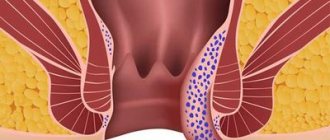
Varicose veins of the esophagus and stomach are formed with portal hypertension - a persistent increase in portal pressure above 12 mmHg. They are localized in the submucosal layer of the lower esophagus and represent portosystemic collaterals connecting the portal and systemic venous circulation. Bleeding as a result of rupture of varicose veins of the esophagus and stomach is one of the most severe complications of portal hypertension, accompanied by high mortality rates.
I. Endoscopic classifications of esophageal varices of the esophagus.
In 1983, K.-J.Paquet identified 4 degrees of varicose veins (VV) of the esophagus:
1 tbsp. Single venous ectasia (verified endoscopically, but not determined radiographically). 2 tbsp. Single well-demarcated vein trunks, mainly in the lower third of the esophagus, which are clearly expressed during air insufflation. The lumen of the esophagus is not narrowed, the esophageal mucosa above the dilated veins is not thinned. 3 tbsp. The lumen of the esophagus is narrowed due to bulging of the esophagus in the lower and middle thirds of the esophagus, which partially collapse during air insufflation. Single red markers or angioectasias are detected at the apices of the varicose veins.
4 tbsp. In the lumen of the esophagus there are multiple varicose nodes that do not collapse with strong air insufflation. The mucous membrane over the veins is thinned. At the tops of the varix, multiple erosions and/or angioectasia are detected.
In 1997, N. Soehendra, K. Binmoeller proposed a three-grade classification of varicose veins separately for the esophagus and stomach.
VVV of the esophagus: grade 1 - the diameter of the veins does not exceed 5 mm, elongated, located only in the lower third of the esophagus. 2nd degree - the diameter of the veins is from 5 to 10 mm, tortuous, located in the middle third of the esophagus. Grade 3 - diameter more than 10 mm, tense, with a thin wall, located close to each other, “red markers” on the surface of the veins.
Gastric varicose veins: grade 1 - the diameter of the veins does not exceed 5 mm, poorly visible above the gastric mucosa. 2nd degree - diameter from 5 to 10 mm, single, polypoidal appearance. Grade 3 - diameter more than 10 mm, in the form of extensive conglomerates of polypoid-type nodes with thinning of the mucosa.
The Japanese Scientific Society for the Study of Portal Hypertension in 1991 developed rules for recording endoscopic signs of varicose veins of the esophagus and stomach, consisting of 6 main positions:
I. Determination of the prevalence of varicose veins in the esophagus and varicose veins of the stomach relative to the cardia.
II. Shape (appearance and size).
III. Color as an indirect sign of the wall thickness of the air valve.
IV. “Red markers” - telangiectasias, red cherry spots, hematocyst spots.
V. Signs of bleeding - in case of acute bleeding, its intensity is established; in the case of spontaneous hemostasis, the nature of the thrombus is assessed.
VI. Changes in the mucous membrane of the esophagus.
II. Methods for diagnosing PG and bleeding from varicose veins
1. X-ray examination of the esophagus with a barium suspension is a simple and accessible method. First degree of VRVP: changes, as a rule, are not detected, but sometimes the relief of the mucous membrane is formed by thickened and convoluted folds, similar to gastric folds. The second degree of ERVP is manifested by single and group accumulation defects on the relief or contour of the esophagus of a round, oval or convoluted shape. The third degree of varicose veins - the folds of the mucous membrane of the esophagus are constantly expanded, in the c/3 and n/3 of the esophagus large nodes, grape-shaped and polyp-shaped conglomerates are clearly visible, sharply bulging and narrowing the lumen of the esophagus.
2. Esophagogastroduodenoscopy (EGD) - provides important information about the condition of the esophagus, stomach and duodenum during the development of PG syndrome, the source of esophagogastric bleeding, its location, intensity, degree of varicose veins, and the presence of concomitant pathology. Allows for objective monitoring of the quality and effectiveness of the treatment.
3. Ultrasound diagnostics - makes it possible to assess a number of objective signs of PG: expansion and the appearance of tortuosity of the portal, splenic and superior mesenteric veins; varicose veins of the upper stomach with thickening of its walls; increase in the size of the liver and spleen; the appearance of portocaval anastomoses; ascites; slowing of blood flow in the portal vein according to the results of a Doppler study; decrease in volumetric blood flow in the portal vein and its branches according to the results of duplex angiography.
4. The magnetic resonance imaging method allows you to: obtain an image of the parenchymal organs of the abdominal cavity, large vessels, and retroperitoneal space; diagnose diffuse and focal diseases of other abdominal organs and retroperitoneal space; determine the exact size of the liver, spleen, the presence of ascites, the condition and image of large vessels.
5. Endoscopic ultrasonography is a non-invasive method for assessing the condition of the great vessels of the portal vein system and collaterals. Endosonographic monitoring of sclerotherapy or vein ligation allows early identification of up to 30% of cases of inadequate vein obstruction and objectification of the results of therapy.
The leading role in the diagnosis of varicose veins of the esophagus and stomach and bleeding of portal origin, determining the risk of the first bleeding or its recurrence, as well as the effectiveness of conservative and surgical treatment, belongs to endoscopy. Often, endoscopic examinations are the first and, at this stage, the main method for diagnosing PG, which first manifested itself as acute esophageal-gastric bleeding from the esophageal esophagus or stomach, which occurs in 50% of such patients with a mortality rate of up to 60% (K.-J. Paquet, 1983; AKBurraughs, 1993).
The main sources of bleeding from the upper digestive tract are varicose veins, predominantly from the distal esophagus. Gastric varices are less common and are usually less well diagnosed due to the structural features of the mucous membrane and the difficulty of examining the cardia in a retroflexed position, especially against the background of ongoing bleeding. Dilation of the venules and capillaries of the mucous membrane and submucosal layer of the stomach leads to portal hypertensive gastropathy, which, during endoscopic examination, is characterized by the presence of foci of red spots on the mucous membrane, hyperemia, mosaic pattern of the mucous membrane, and in more severe cases, diffuse dark red spots or intramucosal hemorrhages . It is believed that up to 25% of bleeding can be caused (photo 4.) by gastropathy (T. McCormack, 1993).
III. Modern approaches to the treatment of acute bleeding from varicose veins
Treatment of variceal bleeding includes three main areas: treatment of active bleeding (current bleeding); prevention of recurrent bleeding; prevention of first bleeding.
Studies have shown (Groszmann RJ, 1990, Franshis R.De, 1990, Borisov A.E., 2001) that non-selective β-blockers (propranolol, nadolol, timolol, etc.) are used for this purpose and are currently recommended as drugs choice in patients with varicose veins before the first bleeding with dosage selection in order to reduce portal pressure below 12 mmHg. (Pagliaro L. et al., 1994). At the same time, the combination of β-blockers and ISMN (isosorbide mononitrate) has a synergistic effect on reducing portal pressure and may be more effective in preventing variceal bleeding (Feu F. et al., 1995) than the use of β-blockers alone. However, this combination has a large number of side effects and its use is possible only under strict control of portal pressure. The role of drug therapy in acute variceal bleeding is to enhance the effect of endoscopic treatment. Vasoactive drugs can be used once the diagnosis of variceal bleeding is confirmed. The administration of drugs such as somatostatin or its analogues can be extended for the period after sclerotherapy (Besson I. et al., 1995; Amico GD et al., 1999).
If, despite urgent sclerosing, drug or combination therapy, bleeding does not stop or early relapse occurs (in approximately 10-20% of patients), then in these cases a number of authors (Sanyal AJ et al., 1996; Cormick PAMc et al ., 1994) use a transjugular portosystemic shunt (TIPS), which causes an immediate decrease in portal pressure.
Short-term antibiotic prophylaxis is currently considered the standard for patients with variceal bleeding (Rimola A. et al., 1999) in cirrhosis, because they always have a high risk of developing severe bacterial infections (three oral, two intravenous), and antibiotics are an effective means of preventing infection (Bernard B. et al., 1999).
Methodology of endoscopic methods of hemostasis for varicose veins
Today, among the endoscopic methods of stopping bleeding and preventing its recurrence, the most widely used method is endoscopic injection sclerotherapy (ES).
The technical features of the method differ according to the method of administration of the sclerosing agent: a) intravasal sclerotherapy - if the drug is injected directly into the varicose vein; b) paravasal sclerotherapy - when the drug is injected into the submucosal layer from several points near the varicose vein; c) combined method - involves the use of a combination of the first two methods.
Endoscopic ligation of VRVP (EL)
The last decade has been marked by an active search for an alternative endoscopic method that would be as effective as injection sclerotherapy, but would be simpler to perform and lead to a lower percentage of complications. There are three different clinical tasks of EL:
I. EL - as a diagnostic method for stopping bleeding from varicose veins.
II. EL is used as a method to prevent recurrent bleeding from the SVC.
III. Preventive treatment - prevention of the first bleeding.
Since March 1998, we have performed endoscopic interventions on 140 patients with varicose veins of the stomach. The age of the patients ranged from 14 to 68 years, 126 patients were Child-Pugh class B.
Depending on the level of blood flow disturbance in the portal system, patients were divided into two groups: with extrahepatic and intrahepatic block. The first group included 12 patients with umbilical vein thrombosis and post-traumatic compression of the portal vein trunk. The second group consisted of patients with liver cirrhosis of various etiologies.
To determine the severity of liver cirrhosis and assess the life prognosis of patients, the Child-Pugh criteria system was used. The forecast of the risk of occurrence or recurrence of bleeding was based on endoscopic criteria for assessing varicose veins: the degree of dilatation of the veins, their distribution in parts of the esophagus, the presence of markers, taking into account the functional state of the liver according to Child-Pugh.
A fundamental point in choosing treatment tactics for patients with portal hypertension is the presence of a history of hemorrhage. Based on this, we identified groups of patients in whom endoscopic interventions were performed at the height of bleeding, for the purpose of primary prevention, as well as its relapses, and developed a treatment algorithm for each group of patients.
- The group of patients with a history of bleeding from varicose veins included 60 patients. The main task in such patients is to prevent recurrent bleeding. For this purpose, ligation and/or sclerosis of VRVP was used.
- The group of patients without a history of bleeding from varicose veins consisted of 68 people. Treatment of esophageal varices in patients at high risk of bleeding was aimed at primary prevention of bleeding. In these patients, endoscopic ligation (EL) was considered the treatment of choice. The motive for active actions aimed at eliminating varicose veins, in addition to endoscopic and clinical criteria, was the fact of the territorial remoteness of settlements from specialized medical institutions.
- Treatment of acute variceal bleeding was carried out in 12 patients:
Endoscopic interventions for ongoing bleeding from varicose veins included two aspects: stopping bleeding and preventing early relapses. For this purpose, sclerotherapy (ES) of VRVP was performed in the bleeding area and along the contoured varicose veins. If conditions allowed, namely: a clearly visible area of venous damage, technical equipment and trained personnel, EL was performed. At the height of bleeding, ES was performed in 4 and EL in 8 patients. Endoscopic hemostasis was effective in 9 patients. In 2 patients, recurrent bleeding from the urinary tract was the cause of death. Another patient with class C cirrhosis died with increasing liver failure and severe posthemorrhagic anemia due to bleeding from acute ulcers of the gastric outlet.
ligation of VRVP was performed in 138 patients (157 interventions). We used: HX-21L-1 ligation device, caps with straight and beveled ends (MN-595), NM-1K injectors (Olympus, Japan). Patients underwent from 2 to 6 ligation sessions using from 3 to 17 ligatures in one stage.
The maximum observation period for patients is 9.5. Bleeding in the postoperative period was observed in 4 patients, two with a fatal outcome.
The rapid and successful introduction of the method of ligation of esophageal and gastric varices into the program for the prevention and treatment of portal bleeding is due to a number of its advantages compared to sclerotherapy (Table 1).
Table 1
Comparative assessment of complications
with endoscopic methods of eradication of varicose veins (T. Marek et al., 2003)
| Possible complications | EL | ES |
| Total number | 2% | 22% |
| Bleeding | no data | 1-35% |
| Ineffective hemostasis | 14% | 5-23% |
| Deep ulcers | 2,6% | 12,5-18% |
| Effusion into the pleural cavity | no data | 48-60% |
| Perforation of the esophagus | no data | 1-6% |
| Scar strictures | 0-2 % | 22-33% |
| Transient bacteremia | 5,7 | 5-17,2% |
| Fever | no data | 11-55% |
| Recurrent bleeding | 26-37% | 44-48% |
| Mortality from ongoing bleeding | 28% | 45% |
EL achieves the desired effect faster, has fewer complications and is easier to tolerate by patients (Table 2).
table 2
Advantages of the endoscopic ligation method (Marek T. et al., 2003)
| Criteria | EL | ES |
| Efficiency of hemostasis | 86% | 77% |
| Mortality | 28% | 45% |
| Recurrent bleeding | 37% | 48% |
| Complication rate | 2% | 22% |
results
The immediate and long-term results of using the method of endoscopic ligation of varicose veins are encouraging. A higher efficiency of the endoscopic ligation method was noted in comparison with the injection sclerotherapy method, which was manifested in a decrease in the number of recurrent bleedings by an average of 20% and a decrease in mortality by an average of 10-15%. It should be emphasized that to achieve complete elimination of varicose veins using this method requires 2-3 sessions less than with injection sclerotherapy, the percentage of eradication of varicose veins with ligation (70-80%) is higher than with injection sclerotherapy (40-60% ).
Analyzing the complications associated with the use of EL and ES and comparing them with literature data, we were convinced that the EL method is not highly aggressive. The eradication effect of this method is high in the first months after the intervention. We consider the development of relapse of esophageal varicose veins in a short time after their eradication to be an indication for the use of ES.
Conclusions:
1. Endoscopic ligation of URVP is:
- an effective method of primary (preventive) prevention of bleeding;
- an effective means of hemostasis at the height of bleeding from VRVP;
- an effective way to prevent bleeding in patients with a history of bleeding from varicose veins;
- a minimally invasive and widely used method with a minimum number of complications;
2.Endoscopic methods for correcting PG should be carried out in combination with drug therapy for the underlying disease.
3. It is advisable to carry out endoscopic methods for the correction of PG in specialized medical and diagnostic institutions with a modern diagnostic base, where complex (conservative, endoscopic, angiographic and surgical) treatment is possible.
A.V. Filin, L.M. Myaukina, E.V. Kim, A.A. Owl
Leningrad Regional Clinical Hospital, St. Petersburg
Literature:
1. Andreev G.N., Apsatarov E.A., Ibadildin A.S. Diagnosis and treatment of complications of portal hypertension. - Almaty: Kazakhstan, 1994. - 317 P.
2. Borisov A.E., Kuzmin-Krutetsky M.I., V.A. Kashchenko V.A. // Bleeding of portal origin. - St. Petersburg, 2001. - 126 S.
3. Lytkin M.I., Eryukhin I.A., Didenko V.M. Long-term results of treatment of patients with portal hypertension complicated by gastroduodenal bleeding // Vestn. hir. - 1984. - No. 12. - P. 11-15.
4. Patsiora M.D., Scherzinger A.G. Surgical treatment of intrahepatic portal hypertension // Surgery of portal hypertension (mistakes and dangers). - M. - 1984. - P. 6-15.
5. Sherlock Sh, Dooley J. // Diseases of the liver and biliary tract: Prkatic manual: Translated from English. / Ed. Z.G. Aprosina, N.A. Mukhina. - M.: Geotar Medicine, 1999. - 187s.
6. Bernard B, Grange JD, Khac EN: Antibiotic prophylaxis for the prevention of bacterial infections in cirrhotic patients with gastrointestinal bleeding: a meta-analysis. Hepatology - 1999, 29: 1655-1661.
7. Besson I, Ingrand P, Person B: Sclerotherapy with or without octreotide for acute variceal bleeding. N Engl J Med - 1995, 333:555-560.
8. Casado M., Bosch J., Garcia-Pagan JC Clinical events after transjugular intrahepatic portosystemic shunt: correlation with hemodynamic findings // Gastroenterology. - 1998. - No. 114. - R. 1296-1303.
9. D'Amico G, Politi F, Morabito A, and the Liver Study Group of V Cervello Hospital: Octreotide compared with placebo in a treatment strategy for early rebleeding in cirrhosis. A double-blind, randomized pragmatic trial. Hepatology -1999, 28: 1206-1214.
10. De Franchis R., Pascal JP, Burroughs AK Definitions, methodology and therapeutic strategies in portal hypertension. A Consensus Development Workshop // J. Hepatol. - 1990. - No. 15. - R. 256-261.
11. Escorsell A, Ferayorni L, Bosch J: The portal pressure response to B-blockade is greater in cirrhotic patients without varices than in those with varices. Gastroenterology - 1997, 112: 2012-2016.
12. Feu F, Garcia-Pagan JC, Bosch J: Relation between portal pressure response to pharmacotherapy and risk of recurrent variceal haemorrhage in patients with cirrhosis. Lancet - 1995, 346: 1056-1059.
13. Garcia-Tsao G. Portal Hypertension and Bleeding Gastroesophageal Varices // Best Practice of Medicine. — January. - 2000.
14. Garcia-Tsao G., Groszmann RJ, Fisher RL: Portal pressure, presence of gastroesophageal varices and variceal bleeding // Hepatology. - 1985. - No. 5. - P. 419-424.
15. Groszmann RJ, Bosch J., Grace N.: Hemodynamic events in a prospective randomized trial of propranolol vs placebo in the prevention of the first variceal hemorrhage // Gastroenterology. - 1990. - No. 99. - R.1401-1407.
16. Lo GH, Lai KH, Cheng JS: Emergency banding ligation versus sclerotherapy for the control of active bleeding from esophageal varices. Hepatology 1997, 25: 1101-1104.
17. Marek T., Kohut M., Rey JF ESGE CD-ROM ON ENDOSCOPY COMPLICATIONS, 2003.
18. McCormick PA, Dick R, Panagou EB: Emergency transjugular intrahepatic portosystemic stent shunting as a salvage treatment for uncontrolled variceal hemorrhage. Br. J. Surg. - 1994, 81: 1324-1327.
19. Moitinho E., Escorsell A., Bandi JC. Prognostic value of early measurements of portal pressure in acute variceal bleeding // Gastroenterology. - 1999 - No. 117. - R.626-631.
20. Monthly Vital Statistics Report: Advance report of final mortality statistics // Monthly Vital Stat Report. - 1995. - No. 43. - R. 24-33.
21. Nolte W., Hartman H., Ramadori G. Portal Hypertension-Pathophysiologie und Therapieansaetze // Ztsch. Fuer Gastroenterologie. - 1994. - Bd.32.- S. 447-459.
22. North Italian Endoscopic Club for the Study and Treatment of Esophageal Varices: Prediction of the first variceal hemorrhage in patients with cirrhosis of the liver and esophageal varices. A prospective multicenter study. N Engl J Med. 1988, 319: 983-989.
23. Pagliaro L., D'Amico G., Pasta L.: Portal Hypertension in Cirrhosis: Natural history // Portal Hypertension. Pathophysiology and Treatment. - Oxford: Blackwell Scientific. - 1994. - P. 72-92.
24. Paquet KJ. Endoscopic paravariceal injection sclerotherapy of the esophagus - indications, technique, complications, results of a period of nearly 14 years // Gastrointestinal Endoscopy. - 1993. - No. 29. - P. 317.
25. Rimola A, Garcia-Tsao G, Navasa M: Diagnosis, treatment and prophylaxis of spontaneous bacterial peritonitis. J Hepatol 1999, In press.
26. Sanyal AJ, Freedman AM, Luketic VA: Transjugular intrahepatic portosystemic shunts for patients with active variceal hemorrhage unresponsive to sclerotherapy. Gastroenterology. - 1996, 111: 138-146.
27. Sarin SK, Lamba G, Kumar M: Comparison of endoscopic ligation and propranolol for the primary prevention of variceal bleeding. New Engl J Med 1999, 340:988-993.
28. Sauerbruch T., Wotzka R., Kopcke W. Prophylactic sclerotherapy before the first episode of variceal hemorrage in patients with cirrhosis // New Engl. J. Med. - 1988.-319.-P.8-15.
29. Schlichting P., Christensen E., Faurholdt L. Main causes of death in cirrhosis // Scand. J. Gastroenterol. - 1983. - No. 18. - R. 881-888.
30. Shah V., Garcia-Cardena G., Sessa WE, Groszmann RJ The hepatic circulation in health and disease: report of a single topic symposium // Hepatology. - 1998. - No. 27. - R. 279-288.
31. Viallet A., Marleau D., Huet M.: Hemodynamic evaluation of patients with intrahepatic portal hypertension. Relationship between bleeding varices and the portohepatic gradient // Gastroenterology. - 1975. - No. 69. - R. 1297-1300.