One day hospital 3rd KO
Kravchenko
Olga
12 years of experience
Oncologist
Make an appointment
Colon cancer is a pathological process of development of a malignant tumor formed in the epithelial tissue of the small or large intestine. During its formation, normal epithelial cells are transformed into atypical ones, prone to rapid, uncontrolled division. In 99% of cases, the disease develops in the large intestine and is called colorectal cancer. It ranks second in prevalence in the age group of 45 years and older.
Symptoms
With bowel cancer, the first symptoms are usually minor, and many patients do not pay attention to them. You should definitely visit a gastroenterologist or proctologist if you experience:
- alternating constipation and diarrhea without significant changes in diet;
- constant discomfort - bloating, periodically felt pain;
- during defecation – a feeling of incomplete completion of the process;
- frequent urge to defecate, often painful;
- decreased performance, constant fatigue;
- anemia;
- causeless weight loss.
Over time, these manifestations intensify and are joined by more pronounced symptoms of colon cancer:
- difficult defecation, accompanied by cramping pain;
- ribbon-shaped or too thin stools, often mixed with blood;
- prolonged heartburn, sour belching, bitter taste in the mouth;
- periodic nausea and vomiting;
- acute pain in the abdominal area, accompanied by fever;
- painful urination, blood in the urine.
Symptoms
Symptoms of a neoplasm depend on its type, size and location. Small neoplasms not exceeding 2-3 cm do not manifest themselves clinically. And only when they increase do signs appear:
- Abdominal pain of unknown localization.
- Intestinal obstruction. It can develop against the background of obstruction or intussusception of the intestinal wall due to impaired peristalsis.
- Polyps, gamangiomas and colon cancer may bleed. With profuse bleeding, scarlet blood or melena will be released from the anus. With minor bleeding, there may be streaks of blood in the stool; with hidden bleeding, iron deficiency anemia develops.
- Discharge of mucus from the anus – villous polyps, cancer.
- Dyspeptic disorders: constipation, diarrhea, bloating.
- Feeling of a foreign body in the rectum.
Causes and risk factors
The mechanism of development of intestinal cancer has not yet been precisely established. However, the factors that increase the likelihood of the disease have been studied quite well. Among them:
- inherited predisposition;
- the presence of polyps in the intestines;
- chronic inflammatory diseases - Crohn's disease, ulcerative colitis, diverticulosis, chronic obstruction;
- unbalanced diet with excessive amounts of red meat, smoked meats, spicy seasonings, fatty and fried foods;
- exposure to ionized radiation, carcinogenic compounds;
- obesity, sedentary lifestyle;
- age over 45 years.
Causes of intestinal neoplasia
At the moment, it is generally accepted that intestinal neoplasia is a polyetiological disease. Several reasons can lead to their development:
- Dietary features: overeating, eating large amounts of meat, spicy, pickled, smoked and salty foods, alcohol abuse, lack of dietary fiber, plant fiber and vitamins.
- Inflammatory bowel diseases - ulcerative colitis, Crohn's disease.
- Hereditary predisposition - several syndromes have been described in which multiple intestinal neoplasms develop, which are prone to malignancy.
- Age. The older a person is, the higher the likelihood of developing neoplasia.
Stages
When signs of bowel cancer appear, it is important to determine the stage of the disease, since the choice of treatment strategy directly depends on this. There are four main ones, to which the so-called zero stage is often added - the stage at which benign neoplasms (polyps) or chronic inflammation with erosion of the mucous membrane appear in the intestine. These conditions often become precancerous.
- The tumor does not exceed 2 cm in size and is located within the mucous membrane. Lymph nodes are not affected.
- The size of the tumor increases to 5 cm, the malignant tissue spreads into the submucosal layer. Two or three regional lymph nodes are affected, there are no metastases.
- The neoplasm grows up to 10 cm, penetrates the muscle layer of the intestinal wall, but does not extend beyond the organ. Partial or complete occlusion of the intestinal lumen is possible. Almost all regional lymph nodes are affected, and metastases are found in nearby organs.
- The tumor grows more than 10 cm, grows through all layers of the intestinal wall and penetrates into neighboring tissues. The breakdown of cancer tissue begins with the spread of cancer cells throughout the body - to distant lymph nodes and organs. The presence of distant metastases is the main sign of the fourth stage, regardless of the size of the cancer tumor.
Benign intestinal tumors
Polyps . These are relatively benign neoplasms growing from the intestinal mucosa. They look like spherical, mushroom-shaped or branched growths. Some polyps are located on a thin stalk, others on a broad base. The main danger of these neoplasms is their possible malignancy (malignant degeneration), therefore they are recommended to be removed in a timely manner.
Intestinal lipoma . This is a very rare neoplasm, occurring in 0.035-0.4% of cases of all benign intestinal neoplasms. Typically, lipoma is represented by a single neoplasm, but there may be variants of multifocal lesions.
Diagnosis of these neoplasms at the preoperative stage is a difficult task. As a rule, it is discovered during a morphological examination of material removed during surgery for other intestinal diseases.
Morphologically, the lipoma is represented by well-differentiated adipose tissue with fibrous stroma. Usually the surface is smooth and covered with unchanged mucous membrane. If the tumor is large, there may be erosions and ulcers on the mucosa (they are associated with ischemic lesions and traumatization by feces).
Intestinal hemangiomas . Strictly speaking, hemangioma is not a tumor formation. It is essentially the result of changes in the blood vessels (malformation). Conventionally, it can be divided into two categories:
- Capillary, represented by a network of blood vessels of the smallest caliber - capillaries, lined with hyperplastic epithelium.
- Cavernous - formed by larger blood vessels that are located between the connective tissue stroma.
The main symptom of hemangioma is bleeding. With large neoplasms, obstruction of the intestinal lumen with the development of intestinal obstruction is possible.
Diagnostics
Laboratory and instrumental diagnosis of intestinal cancer includes the following procedures:
- biochemical blood test to detect the level of bilirubin, ALT, AST;
- analysis for tumor markers;
- colonoscopy - examination of the inner walls of the large intestine using an endoscope with a tissue biopsy of detected tumors;
- irrigoscopy - an X-ray examination of the intestine, performed with a radiopaque substance, to identify deformed areas and narrowings;
- Ultrasound of the abdominal cavity to detect metastases in other organs;
- CT scan of the abdominal cavity, necessary to determine the size of the tumor, its extent and exact location.
In addition, there may be a need for additional studies of the chest, liver, brain and other organs if metastasis to them is suspected.
Attention!
You can receive free medical care at JSC “Medicine” (clinic of Academician Roitberg) under the program of State guarantees of compulsory medical insurance (Compulsory health insurance) and high-tech medical care.
To find out more, please call +7, or you can read more details here...
Treatment
The treatment tactics for an intestinal tumor, the symptoms of which are described above, are determined individually for each patient depending on the stage of the disease, the age and general condition of the patient, the location of the pathological formation and other factors. As a rule, oncologists use a combination of several methods to achieve results in the most gentle way.
- Surgery. In the early stages, the surgeon removes the area of the intestine affected by the disease, and samples of the removed tissue are sent for histological analysis. The edge of the removed tissue should not contain malignant cells, otherwise the intervention must be repeated. For an inoperable tumor that blocks the intestinal lumen, palliative intervention is performed - a colostomy is formed (an opening for the discharge of feces) or a stent is installed to expand the lumen.
- Chemotherapy. Potent drugs are prescribed before surgery to reduce the size of malignant tissue, and after surgery to prevent relapses. For inoperable tumors, chemotherapy drugs increase the patient's life expectancy and reduce adverse symptoms.
- Radiation therapy. Ionized radiation is often combined with chemotherapy to achieve the most effective procedures.
- Targeted therapy. Drugs that target specific types of protein are prescribed to block cell growth and division. They are prescribed in later stages to reduce tumor growth in order to prolong the patient's life.
- Immunotherapy. A new trend in oncology uses the body's own immune system to fight tumors. The drugs unblock immune cells, thereby stimulating them to attack malignant cells.
Diagnosis and treatment of intestinal cancer in Moscow
If you have symptoms of intestinal cancer, contact the Meditsina clinic for diagnosis and subsequent treatment of cancer. Here you will find:
- highly qualified specialists - oncologists, gastroenterologists, surgeons, radiologists;
- modern diagnostic equipment of the latest generation;
- an excellently equipped laboratory that performs all types of tests;
- comfortable inpatient department.
Call to find out more about the problem you are interested in and make an appointment.
Prevention of intestinal neoplasia
The following recommendations will help reduce the risk of developing intestinal neoplasia:
- Normalization of nutrition. It is necessary to eat enough vitamins, fiber and dietary fiber. Do not overeat, limit the consumption of carcinogenic foods: smoked meats, marinades, easily digestible carbohydrates, fatty meats, fried foods, spices, alcohol.
- Maintain a physical activity regime.
- Avoid chronic constipation and diarrhea and promptly treat gastrointestinal diseases.
Considering that intestinal cancer develops from polyps, their timely removal is necessary. For this purpose, all people over 50 years of age are recommended to have a colonoscopy at least once every decade. This procedure will allow you to examine the intestinal mucosa and simultaneously remove detected tumors.
| More information about treatment at Euroonco: | |
| Oncologist-gastroenterologist | RUB 5,100 |
| Chemotherapy appointment | RUB 6,900 |
| Emergency oncology care | from 12,100 rub. |
| Palliative care in Moscow | from 44,300 rubles per day |
| Radiologist consultation | RUB 11,500 |
Book a consultation 24 hours a day
+7+7+78
Questions and answers
How to recognize bowel cancer?
The initial symptoms of bowel cancer are similar to a number of other, less dangerous diseases, so patients often do not pay attention to them. If for no apparent reason:
- loss of appetite, aversion to meat dishes;
- stomach ache periodically;
- there is constant discomfort in the abdomen, gas formation has increased;
- prolonged constipation began, alternating with bouts of diarrhea;
this means that you should contact a gastroenterologist or proctologist for examination as soon as possible.
Is there a cure for bowel cancer?
Patients with intestinal tumors can be treated at any stage of cancer. The chances of complete relief from malignant pathology remain even in the most advanced cases.
Colon cancer with metastases: how long do patients live?
Without medical care, the life of a patient with metastases to other organs is 6-11 months, depending on the rate of development of secondary tumors. When completing a course of treatment, the five-year survival rate in patients with metastases is 30-45%, i.e. at least thirty people out of a hundred live more than five years after surgery.
Attention! You can cure this disease for free and receive medical care at JSC "Medicine" (clinic of Academician Roitberg) under the State Guarantees program of Compulsory Medical Insurance (Compulsory Medical Insurance) and High-Tech Medical Care. To find out more, please call +7(495) 775-73-60, or on the VMP page for compulsory medical insurance
IV. Benign and malignant swellings of the colon, diagnosis, treatment.
In the remaining 20 years, a significant increase in the size of the fatty tissues of both the small and large intestines is expected.
To the intestines, benign swellings are brought: polyps, leiomyomas - swellings that develop from smooth intestinal ulcers, rhabdomyomas - swellings from transverse dark intestinal ulcers, angiomas, neuromas , etc. Stinks can be single or multiple.
Clinic. The good plumpness of both the small and large intestines may not show itself for a long time. If you may experience pain in the abdomen without clear localization, diarrhea, seeing blood and mucus in the stool, weight loss.
Likuvannya. The method of selecting the removal of benign tumors of the small intestine is an operation that involves resection of the affected section of the intestine with obstructive cytological findings of the removed tumor.
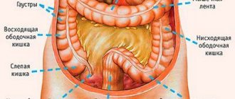
Anatomical appearance of the large intestine
Intestinal polyps
Likuvannya. Apply electrocoagulation to the polyp through the rectoromanos cop. In case of malignant transformation of the nolip, carry out prompt delivery.
Fair rectal swellings constitute 50-70% of all rectal swellings. They occur in 2.5-7.5% of all patients with rectal pathology. Separate the epithelial
swellings (polyps),
villi
non-epithelial
swellings (leiomyomas, lipomas, fibroids, vessel swellings, etc.).
Epithelial cupric tracts
In the 2nd-3rd month of intrauterine development, it is due to the gradual reduction of the supra-spine group of tail ulcers.
Enter the Cupric canal and you will get to the spinal cord. It is important to appear in people aged 18-30 years. In cupric's passages there may often be growths, hairs, and the appearance of sebaceous and sweat deposits. The morphological warehouse is divided into:
1) epithelial cupric tracts;
2) presacral puffiness;
3) tail-like appendages.
Clinical signs.
The formed abscess after its independent opening usually develops into a purulent nocia, which is formed along the middle line of the interstitial recess, above the cupric, near the anus, 3-4 cm above the anal ring.
The diagnosis is made visually and confirmed with additional festulography.
Likuvannya.
We want to carry out sanitation of the noritsa, for which we use antiseptics, sprinkling with medicinal solution KMpO4, 5% iodine.
Operationally
Colon cancer. The onset of illness for colon cancer takes 3rd place after colon cancer. People aged 40-60 years old and older suffer from illness. The localization of cancer varies - the cecum, the hepatic, splenic and rectal - sigmoidal kuta. Clinically, cancer of the right and left half of the colon is divided. Cancer of the right half grows by exophytic growth and does not cause disruption of the passage of stool through the intestine.
Cancer of the left half of the colon is characterized by infiltrative growth with the development of intestinal obstruction. Ailments include abdominal pain, weight loss, underlying weakness, seeing mucus and blood in stool, constipation, flatulence.
The diagnosis is confirmed after an hour of radiological obstruction of the colon with administration of a contrast enema; The only remaining time to clarify the diagnosis is to use colonoscopy
.
Likuvannya is more surgical . Conduct resection of the colon between healthy tissues. In case of inoperable swellings, a bypass anastomosis is performed between the colon and small intestine, or a piece of anti-natural outlet is formed to drain feces and gas. Feces are collected from a special stool bag, which is fixed with a gum belt to the patient’s stomach. It is necessary to systematically disinfect colostomy bags with an antiseptic solution. Feces are not to blame for being wounded on the skin
Surgery – removal of colon swelling
Colon cancer
It affects people between 50 and 60 years of age.
Dissociate exophytic fluff
colon, as growth near the lumen of the intestine, in the form of a polyp, node, or common
endophytic swelling
(infiltrative cancer), as growth at the thickness of the intestinal wall.
Clinic. The main signs of colon cancer lie in the localization of the swelling, the type of growth, size, and the appearance of deformation. It should be noted that the clinical symptoms of cancer of the right and left half of the colon are different. When swelling is evident in the right half of the colon in patients, there is no pain in the right respiratory lobe or right hypochondrium, progressive anemia, caused by chronic hemorrhage or intoxication with decomposition products , obstructive and puffy (the only manifestation of which is puffiness) forms of colon cancer. The most common complications of colon cancer are due to intestinal obstruction, perforation, and bleeding.
Likuvannya. The main treatment method for colon cancer is surgery.
Before symptomatic operations, bypass intestinal anastomoses and colostomies should be performed. After radical operations, it is possible to carry out courses of chemotherapy
List of sources
- Tsukanov A.S., Shelygin Yu.A., Achkasov S.I., Frolov S.A., Kashnikov V.N., Kuzminov A.M., Pikunov D.Yu., Shubin V.P. Principles of diagnosis and personalized treatment of hereditary forms of colorectal cancer. Bulletin of the Russian Academy of Medical Sciences. — 2019
- Association of Oncologists of Russia. Practical recommendations for drug treatment of patients with colon cancer. — 2014
- V.V. Martynyuk. Colon cancer (morbidity, mortality, risk factors, screening). St. Petersburg Medical Academy of Postgraduate Education. — 2000
Malignant intestinal tumors
Malignant neoplasms of the intestine are divided into tumors of epithelial and non-epithelial origin. Epithelial tumors develop from the epithelium of the intestinal mucosa (colorectal cancer). Most often, these are adenocarcinomas, squamous cell carcinoma, less common are small cell, signet ring cell, medullary and undifferentiated cancer. Cancer is always preceded by benign neoplasia or chronic inflammatory processes - polyps, Crohn's disease, ulcerative colitis.
Non-epithelial tumors include leiomyosarcoma, angiosarcoma and Kaposi's sarcoma.
Based on the growth pattern of a malignant neoplasm, the following are distinguished:
- Exophytic tumors grow into the intestinal lumen.
- Diffuse infiltrative tumors - spread inside the intestinal wall.
- Annular neoplasms cover the intestinal wall along its circumference.
Book a consultation 24 hours a day
+7+7+78


