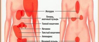
Causes of pain in the epigastric region
Among the pathological causes of pain in the epigastrium are:
- gastritis;
- ulcer;
- gastrointestinal obstruction;
- gurgle;
- esophageal stenosis;
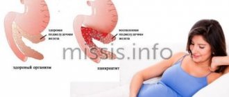
- pancreatitis;
- appendicitis;
- cryptosporidiosis.
Various inflammatory diseases at the initial stage may manifest themselves as mild pain. The occurrence of deep damage to the gastric or intestinal mucosa leads to increased pain. The nature of the pain can determine the severity and danger of the disease.
Gastritis
Gastritis is an inflammatory disease of the gastric mucosa. Pathology occurs due to poor nutrition, increased or decreased gastric acidity, or infection by a bacterium called Helicobacter Pylori. To treat gastritis, it is necessary to eliminate all possible provoking factors and adjust the diet.
Symptoms include epigastric pain after eating, heartburn, fatigue, and nausea. Gastritis is considered the most common gastrointestinal disease. If a pathology of the digestive system is suspected, gastritis is diagnosed in 8 out of 10 cases. If left untreated, the disease becomes more complicated (erosions and ulcers occur).
Stomach ulcer
A stomach ulcer is a deep inflammatory lesion of the mucous membrane. Among the causes are:
- the presence of sluggish bacterial inflammation of the mucous membrane (gastritis);
- long-term use of non-steroidal anti-inflammatory drugs;
- alcohol abuse;
- increased stomach acidity.
An ulcer is characterized by the occurrence of severe pain in the epigastrium 1-2 hours after eating. The pathology is also accompanied by sour vomiting, after which the pain becomes less noticeable.
Diagnostics
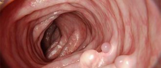
Based on the localization of pain, its nature, intensity, as well as its connection with food intake, the doctor determines their possible cause. To reliably diagnose the disease, an objective examination is carried out, which includes ultrasound examination of the digestive system, fibroesophagogastroduodenoscopy (examination of the mucous membrane of the upper digestive tract using a special endoscope), radiography of the hollow organs of the digestive system with a contrast agent.
If necessary, the doctor may additionally prescribe computed tomography or magnetic resonance imaging, which can visualize even small structural changes in organs and tissues.
Diagnosis of pain in the epigastric region
Epigastric pain is a non-unique symptom, the presence of which makes it impossible to make a diagnosis. An in-depth examination is required using the following methods:
- gastroscopy (FGDS) of the stomach and intestines;
- general and biochemical blood test;
- Ultrasound or CT scan of the abdominal cavity;
- analysis of stool and urine.
With the help of FGDS, it is possible to carefully examine the stomach and duodenum using an endoscope. A blood test gives a general idea of the condition of the body. If gastritis or peptic ulcer is detected, a breath test is additionally prescribed to determine the presence of Helicobacter bacteria.
To make an accurate diagnosis, it is important to use instrumental, hardware and laboratory diagnostic methods. If a person is admitted to the hospital with acute pain, differential diagnosis is used. First of all, the most dangerous pathologies are excluded.
Recommended video:
Pain in the epigastric region
Alexei
May 21, 2020
Hello! Periodic pain in the epigastric region, sometimes the area of pain is not entirely clear and radiates to the right and left. The nature of the pain is tolerable, there is no severe pain, it looks like a burning sensation and as if something is stabbing, sometimes it feels like something is stuck in the esophagus. There is belching after eating, sometimes heartburn (it doesn’t hurt much). Pain and discomfort do not depend on food intake, the only concern is the feeling of fullness in the stomach after eating, and as if there is a lump closer to the esophagus. There is no bitterness in the mouth or nausea. Appetite is good. I passed the following tests - blood, urine, feces, FGDS. Did an ultrasound of the abdominal cavity. Here are the results: BLOOD - ALT - 23.7 (normal 0.0-50.0); AST-18.7 (norm 0.0-50.0); alkaline phosphatase - 101 (normal 30-120); Gamma-GT - 16.5 (normal 0.0-55.0); Alpha amylase - 59.0 (normal 28.0-100.0); Lipase - 14 (normal 3 -67); Total protein - 74.7 (normal 66.0-83.0); Albumin 46.8 (normal 35.0-52.0); Total bilirubin - 24.9 (normal 5.0-21.0); Direct bilirubin - 4.3 (normal 0.0-3.4). M O C A - urine alpha amylase - 131 (normal 0-490). A N T I T E L A to Helicobacter pylori, IgG - 0.4 IU/ml. K A L general analysis - consistency - soft; Brown color; pH - neutral; stercobilin - present; bilirubin - absent; muscle fibers with striations - absent; muscle fibers without striations - single; connective tissue - absent; neutral fat - absent; fatty acids - absent; soap - a small amount; intracellular starch - absent; extracellular starch - absent; iodophilic flora - absent; digestible fiber - absent; indigestible fiber - present; mucus - absent; red blood cells - absent; leukocytes - absent; helminth eggs - not found; protozoa - not detected; yeast fungi - not detected. Ultrasound of the Abdominal cavity: LIVER. Location: typical. Contours: smooth. Sound conductivity: unchanged. Parenchyma structure: homogeneous. Parenchyma echogenicity: average. Dimensions: thickness of the right lobe 97 mm (normal up to 125 mm), CVR 119 mm (normal up to 150 mm), thickness of the left lobe 97 (normal up to 60 mm), CVR 78 mm (normal up to 100 mm) Presence of space-occupying formations and their characteristics: not identified. Vascular pattern of the parenchyma: preserved. Intrahepatic ducts: not dilated. Hepatic veins: not dilated. Portal vein: 9.5 mm (normal up to 14 mm). Inferior vena cava: 18mm (normal 25mm) Hepatic artery in the area of the porta hepatis: not dilated. GALL BLADDER. Location: typical. The presence of kinks and constrictions: in the middle third, when changing the position of the body, they are partially straightened. Dimensions: 74*19mm. Walls: 2mm (norm up to 3mm). Clearance: free. Common bile duct: 4 mm, not dilated, the lumen in the area accessible to visualization is free. PANCREAS. head 24 mm (normal 11-30mm), body 11 mm (normal 4-21), tail 20 mm (7-28). Contours: unclear. Echogenicity of the parenchyma: isoechoic of the liver. Parenchyma structure: homogeneous. Pancreatic (Wirsung) duct: not dilated. Presence of space-occupying formations and their characteristics: not identified. SPLEEN. Location: typical. Shape: typical. Contour: smooth. Structure: homogeneous. Echogenicity is not changed. Dimensions 96*36. Presence of space-occupying formations and their characteristics: not identified. Splenic vein at the level of the hilum of the spleen: not wide. Free fluid in the abdominal cavity is not detected. CONCLUSION: No organic pathologies were identified. F G D C. pH = 4.0 - against the background of bile reflux. The ESOPHAGUS is freely passable along its entire length, its walls are elastic, the mucous membrane of the abdominal segment is hyperemic, with fibrin deposits. The rosette of the cardia is elastic, does not close completely, retrograde prolapse of the gastric mucosa is determined against the background of reflux. The STOMACH is of normal shape and size, the walls are elastic, they expand freely with air. In the stomach on an empty stomach there is a lot of foamy mucous content mixed with a moderate amount of cloudy, flaky bile. The mucous membrane is normal, with areas of hyperemia. The folds of the mucous membrane are of the correct orientation, soft, mobile, and can be expanded freely with air. Peristalsis can be traced in all departments. The GOILKEEPER is of the correct shape, gapes, we pass freely for the videoscope. The duodenal bulb is normal, cone-shaped, the mucous membrane is hyperemic. HORSESHOE duodenum with normal lumen, contains bile. The mucous membrane is hyperemic, with multiple lymphangiectasia (the “semolina” phenomenon). The relief has been preserved. The BDS is not visually changed. CONCLUSION: distal catarrhal reflux - esophagitis. Indirect signs of hiatal hernia. Superficial gastritis. Duodenitis. Duodeno-gastric reflux. Indirect signs of the pancreatobiliary zone.
The question is closed
Pain in the epigastric region
Serious and life-threatening situations
Some diseases can lead to a serious threat to human life. The most dangerous pathologies of the gastrointestinal tract are appendicitis and gastric or intestinal ulcers. Appendicitis in the absence of surgical treatment leads to rupture of the appendix and infection in the abdominal cavity.
The ulcer may be complicated by bleeding. With prolonged progression, the risk of perforation (the appearance of a through hole in the intestines or stomach) increases significantly. When perforation occurs, secondary peritonitis develops. The mortality rate for perforated ulcers, even with timely treatment, is about 7%.
Medical assistance, which doctor to contact
If a person begins to experience discomfort and mild pain in the epigastrium, then he needs to make an appointment with a doctor. First you will need to see a therapist. After this, visit a gastroenterologist. After the diagnosis, it will be possible to determine the cause of the ailment.
If the pain is unbearable, then you need to call an ambulance. Acute pain in the epigastric region is a reason for hospitalization of a person in the hospital. In a hospital setting, the necessary treatment regimen is selected. If possible, conservative treatment is carried out with medications.
Epigastric pain - causes
There are several main causative factors that provoke the development of pain, these include:
- Gastritis is an inflammation of the gastric mucosa, which can be triggered by various factors (poor nutrition, an infectious process caused by the specific bacterium Helicobacter pylori). In this case, pain in the epigastric region is often accompanied by belching of sour or unpleasant-smelling air, bloating and unstable stools (diarrhea is replaced by constipation and vice versa).
- Peptic ulcer is the formation of a defect in the mucous membrane (ulcer) of the duodenum or stomach. Accompanied by point pain, which intensifies on an empty stomach.
- Enteritis is an inflammation of the small intestine, which leads to diffuse pain and dyspeptic symptoms (bloating, unstable stool, loss of appetite).
- Colitis is an inflammation of the colon, the appearance of signs from the epigastrium most often provokes a pathological process of the transverse colon. As with enteritis, dyspepsia syndrome can develop.
- Pancreatitis is a pathology of the pancreas, accompanied by intense pain that spreads to the back (girdles).
Somewhat less frequently, acute pain in the epigastrium can be a manifestation of pathology of the heart (unstable angina, myocardial infarction) or lungs (pneumonia, pleurisy, atelectasis).
What medications can be used
Specific medications can be prescribed only after determining the type and nature of the disease. Among the classes of drugs that are most often used in the treatment of gastrointestinal pathologies are:
- gastroprotectors (accelerate the recovery of the affected gastric mucosa);
- antibiotics (prescribed when a bacterial infection is detected);
- food enzymes (prescribed for indigestion and inflammation of the pancreas);
- painkillers (used for severe discomfort at the initial stage of treatment);
- enterosorbents (used for intoxication and various poisonings);
- proton pump inhibitors (used for high acidity).
The dosage and duration of use of the drug is determined by the doctor. Before making a diagnosis, it is prohibited to use painkillers and antispasmodics, as this may complicate the diagnosis.
Treatment
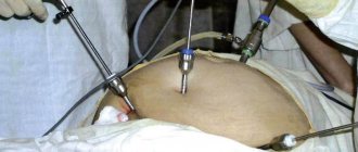
After a reliable objective determination of what causes the epigastric pain, the doctor prescribes appropriate treatment. It may include conservative therapy (the use of drugs from various pharmacological groups) or surgical intervention. The first surgical department of the hospital performs surgical interventions with the aim of radical treatment of the causes of discomfort in the epigastric region. The most often performed is excision of an ulcerative defect of the stomach or duodenum, removal of dead tissue in acute pancreatitis. To reduce the morbidity of the operation, laparoscopy is used.
Painful sensations require contacting a specialist doctor, since their independent treatment can cause complications, the most dangerous of which are intense bleeding from the ulcer and death of pancreatic tissue.
Traditional medicine
If, after examination, uncomplicated gastritis, bulbitis or duodenitis is diagnosed, then treatment with folk remedies is possible. Herbal remedies that relieve inflammation and help fight infection are preferred. You can use the following recipes:
- St. John's wort infusion. This herb has analgesic, anti-inflammatory and antimicrobial effects. To prepare the infusion, you will need to pour 1 tablespoon of dry St. John's wort into 250 ml of boiling water. Then let the liquid brew for 2-3 hours. Take 75 ml before meals 2-3 times a day for a week.
- Chamomile decoction. Chamomile has an antiseptic, anti-inflammatory, choleretic and astringent effect. It is often used for gastrointestinal pathologies to normalize digestion and eliminate pain. To prepare the medicine, you will need to take 3 tablespoons of chamomile, add 300 ml of liquid, bring to a boil and boil for 30 minutes. The decoction is consumed 3-4 times a day, 50 ml.
Before using traditional medicine recipes, you should consult your doctor. When treating diseases of the gastrointestinal tract, it is necessary to correctly combine folk and pharmacy remedies.
Diet for gastrointestinal diseases
If epigastric pain is associated with gastrointestinal pathology, you will need to use a gentle diet. The main principles of proper nutrition are:
- fractional meals in small portions 4-6 times a day;
- exclusion of too hot and cold dishes;
- exclusion of hard-to-digest foods;
- exclusion of fried, salted, spicy and smoked.
It is recommended to give preference to liquid dishes. The basis of the diet can be various cereals and soups. It is also very important not to drink alcohol throughout the entire therapy.