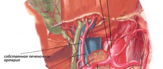
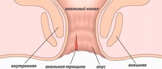
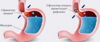
Many organs are susceptible to prolapse: stomach, intestines, kidneys, liver, uterus, rectum. However, prolapse does not occur in all people and not always.
“Omission is not a rare diagnosis, but it’s not widely known,” says general practitioner Olga Alexandrova . “Many people live for years with this pathology, not knowing about its existence, but find out about it by chance, during an ultrasound examination. Others, on the contrary, spend years being treated for non-existent diseases and do not even know the true cause of their suffering.
Question and answer Is prolapse of the liver and kidney dangerous? Physiologically, people who are tall, thin, underweight (fat tissue in moderation helps maintain internal organs in the correct position), who often have congenital weakness of muscles and ligaments, are predisposed to prolapse.
Prolapse can occur due to sudden weight loss. Organs, suddenly left without “support”, can “slide.”
Another reason is hard physical work, carrying heavy weights, and excessive fitness activities (especially exercises with weights). Lack of physical activity, in turn, can also lead to prolapse. Physical inactivity weakens the muscular corset and ligamentous apparatus, which provides “support”.
In men, prolapse often occurs due to a chronic debilitating cough, in women - after pregnancy and childbirth, or more precisely, due to the lack of proper preparation for them.
Question answer
How to improve digestion using folk remedies?
Symptoms
Symptoms of the disease directly depend on the organ that was involved in the pathological process. Taking this fact into account, the following symptom complexes can be identified:
- Associated with bladder prolapse: Difficulty urinating, feeling of incomplete emptying of the bladder, frequent urination, urination in small portions, sudden (imperative) urge to urinate, loss of urine due to physical activity or urgency
- Associated with rectal prolapse: difficulty defecating, the need to help with your hand (press on the prolapsed vaginal walls) during defecation.
- Associated with uterine prolapse: nagging pain in the lower abdomen, discomfort during sexual intercourse.
At the same time, the main symptom characteristic of all patients and leading them to the doctor is the feeling of a foreign body in the vagina. Moreover, most patients have a violation of the support of several organs at once, and therefore the clinical picture may be more multifaceted than described above.
| anonymously to the doctor, through the feedback form, we will try to help you. | Ask a Question |
What can be done? How is prolapse of internal organs treated?
Traditional medicine offers 2 methods of treating prolapse of internal organs: the use of a bandage or surgery. An alternative method is osteopathic treatment, which allows you to return the internal organs to their places and thereby really solve the problem. Prolapse of organs is always associated with a violation of their ligamentous system and a decrease in smooth muscle tone. An osteopathic doctor relieves spasm and hypertonicity, normalizes blood circulation and mobility, returns the sagging organ to its original place, restoring connections between all body systems. As a result of the work of an osteopath, microcirculation of blood, lymph, metabolic processes and nerve conduction are normalized. When the organ returns to its place, the functioning of the entire organism is normalized.
Osteopathy is effective for prolapse of all internal organs - stomach, intestines, kidneys, spleen, reproductive organs. Soft and painless effects, the absence of side effects and contraindications, combined with high efficiency, make osteopathy one of the best methods for treating prolapse.
Causes
The main cause of pelvic organ prolapse is damage to the supporting apparatus of the pelvic floor. The main elements of the latter are ligaments and fascia, which intertwine the pelvic organs and fix (suspend) them to the walls of the pelvis. This can be compared to the lines of a parachute. The pelvic muscles and perineum act as support for the ligaments and fascia.
That is why at a young age, with preserved muscle tone, pelvic organ prolapse is quite rare, while at the same time, after 50 years, every second woman suffers from it. At the moment, there is no consensus on the cause of the development of this disease; most likely, like most pathologies, it has a multifactorial nature. However, the following main reasons can be identified:
- Pregnancy and childbirth. During this period, the qualitative composition of the pelvic floor tissues changes - they become more elastic and stretchable. In some patients, unfortunately, restoration of the previous properties of ligaments and fascia does not occur. On the other hand, during childbirth, some of the fascia, ligaments and muscles are damaged. This is especially true in the case of complicated births (large fetus, rapid labor, episiotomy (perineal incision), use of obstetric forceps, vacuum, etc. during childbirth).
- Hard physical work. Here a process similar to childbirth develops - the supporting structures of the pelvis simply cannot withstand the load and tear.
- Chronic constipation, respiratory diseases (accompanied by constant cough). This also includes obesity, which is also accompanied by increased loads on the ligamentous apparatus of the pelvic floor.
- Of course, hereditary weakness of connective tissue also contributes. Often such women have concomitant diseases such as hemorrhoids, varicose veins of the lower extremities, and pathology of the musculoskeletal system.
| Most patients receive care for free (without hidden additional payments for “network”, etc.) as part of compulsory health insurance ( under the compulsory medical insurance policy ). | Application for treatment under compulsory medical insurance |
Risk factors for developing prolapse
Risk factors for pelvic organ prolapse in women include vaginal births and number of births, age and obesity. Risk factors for recurrence of prolapse after surgical correction include damage to the levator ani muscles, treatment of advanced forms of prolapse, and familial forms of the disease.
Number of births
The risk of prolapse increases with the number of vaginal births in history.
The Oxford Family Planning study, which followed more than 17,000 women for 17 years, found that compared with nulliparous women, the risk of hospitalization for prolapse increased markedly after the first (4-fold) and second (8-fold) birth. and then it grew more slowly: after the third birth by 9 times, the fourth by 10 times, and so on.
Factors associated with labor and the development of prolapse include high fetal birth weight, prolonged second stage of labor, and maternal age less than 25 years at first birth. However, vaginal prolapse can also occur in nulliparous women.
Elderly age
The risk of developing prolapse increases with age.
In the Women's Health Initiative study (more than 27,000 women), there was a small but statistically significant progressive increase in the prevalence of rectocele with age (50 to 59 versus 60 to 69 and 70 to 79 years).
Obesity
Women who are overweight (BMI ≥25–29.9 kg/m2) and obese (BMI ≥30 kg/m2) have an increased risk of developing prolapse compared to their normal-weight counterparts. However, whether weight loss leads to regression of prolapse remains controversial, but there are reports of regression of prolapse in women after bariatric surgery.
Hysterectomy
The role of hysterectomy in the development of subsequent pelvic organ prolapse is controversial. The risk may depend on age, presence of prolapse before hysterectomy, and surgical approach.
Retrosymphyseal urethropexy or "suspension" procedure
(fixation of the uterus to the posterior surface of the symphysis pubis) can lead to a greater anterior deviation of the anterior vaginal wall, which changes the distribution of force along all vaginal walls. As a result, the apex and posterior wall of the vagina may become prone to the development of support defects, including enterocele or rectocele.
Increased intra-abdominal pressure
Chronic constipation and other conditions that cause repeated increases in intra-abdominal pressure, such as chronic obstructive pulmonary disease, can stretch and damage the pudendal nerve.
There is currently no reliable data regarding whether the risk of prolapse is increased in women who lift weights. One study of more than 1,000 women found that women doing physically demanding jobs had significantly more severe prolapses than other categories of women.
Connective tissue pathology
Some connective tissue diseases (eg, Ehlers-Danlos syndrome) or congenital abnormalities (eg, bladder exstrophy) contribute to the development of prolapse. Women with hypermobile joints have a higher prevalence of prolapse than women with normal joint mobility.
Family history
Potential genes and patterns of inheritance are unknown. Most likely, we are talking about connective tissue dysplasia as the cause of prolapse.
Diagnostics
Diagnosis of pelvic organ prolapse does not cause difficulties for a specialist dealing with this issue. It is enough to conduct a standard gynecological examination and perform several provocative tests (pushing hard or coughing). During the examination, it will be possible to tell which part of the vagina/organ has suffered the most: the anterior wall/bladder, the upper part of the vagina/uterus and the posterior wall of the vagina/rectum.
The degree of prolapse is also determined: if the prolapsed organs do not extend beyond the vagina, then this is degree 1 or 2; if this happened, then grade 3 or 4 (with complete prolapse of the vaginal walls). Unfortunately, doctors who are not involved in the treatment of these patients often incorrectly assess the degree of the disease and incorrectly determine those parts of the pelvic floor that are involved in it. For the most part, problems in the treatment of this category of patients are associated with errors at the diagnostic stage.
Vaginal prolapse - symptoms and treatment
Conservative treatment
Conservative treatment is aimed at maintaining and strengthening the pelvic floor muscles, as well as preventing deterioration. It is recommended:
- when the vaginal walls prolapse without the patient’s complaints, when only the doctor sees the prolapse;
- in the presence of contraindications: severe diseases of the cardiovascular system with heart rhythm disturbances and periods of increased blood pressure; old age; diseases of the coagulation system due to the risk of bleeding or complications of anesthesia;
- if the patient refuses surgical treatment.
Exercises for the pelvic floor muscles
There are many variations of Kegel exercises. They are performed by the woman herself in a standing, sitting or lying position [21][23]. To find the “right” muscles, a woman needs to try to stop the flow of urine when emptying her bladder. At the same time, exactly those muscles of the pelvic day that need to be trained are tensed.
To achieve the best results, it is important to train in stages. The first stage is for patients with weakened pelvic floor muscles. One of the exercise options:
- Lie on your back, spread your legs slightly, bending them at the knees. The buttocks should lie firmly on the floor during the entire exercise.
- Tighten the pelvic floor muscles (the movement is the same as when stopping urination or retaining gas in the intestines). Do not hold your breath, make sure that when the pelvic floor muscles are tense, the muscles of the abdomen, back and legs remain relaxed.
- Keep the muscles tense for 2-3 seconds, then relax completely.
You can make sure that the pelvic floor muscles are really working using a mirror: the tendons in this area should be stretched. At first, it is recommended to perform 5 repetitions of the exercise 3 times a day. In the future, a set of exercises for prolapse of the vaginal walls is selected by the attending physician.
Exercises can be performed using various devices, for example, safe silicone cones of different weights. Inserting them into the vagina helps a woman understand and feel those same pelvic floor muscles. When the cone is in the vagina, the pelvic floor muscles contract reflexively.
Electrical stimulators and vibration stimulators . These devices are used to develop the strength of the pelvic floor muscles and vaginal walls, which are not amenable to controlled contraction. You can record your training results and track changes using several parameters using an application on your smartphone. Success can only be achieved with regular practice.
Gynecological pessary. This polymer medical product, installed in the vagina both by the woman herself and by a gynecologist to support the walls of the vagina and uterus from prolapse, comes in various shapes and sizes [24]. The pessary eliminates prolapse only while the woman is using it. Some pessaries for long-term continuous wear (2 weeks), cubic and mushroom-shaped pessaries (Arabin) are used during the day, and the woman removes them at night.
First of all, during the inspection and/or with the help of adaptation rings (Arabin), the shape and size of the product are selected. A properly sized pessary should not cause discomfort. In addition, the doctor should explain to the woman how to use and store the pessary. It is important to know that the product cannot be boiled. If dryness is present, estrogen-containing creams are often prescribed.
Prescribing hormone therapy
Hormonal therapy is prescribed taking into account age, concomitant diseases, the presence of menstruation, the presence/absence of contraindications in the medical history (thrombosis - blockage of blood vessels; diseases of the coagulation system; severe decompensated diseases of the liver, kidneys, cardiovascular system). Depending on these data, both combined estrogen-progestogen drugs or combined menopausal replacement therapy, as well as local vaginal hormonal drugs, may be recommended [11].
Bandage underwear
Used as a temporary stage of treatment (preparation before surgery or before selecting a pessary). It is a special elastic swimming trunks with a high waist. The device is hygienic, convenient to use, and prevents prolapse of the vaginal walls. The bandage can be purchased at a pharmacy.
Diet
Proper nutrition during vaginal prolapse - prevention of constipation and increased gas formation. Diet principles:
- eating small portions 5 times a day;
- avoiding overeating;
- enriching the diet with products containing plant fibers, vegetables, fruits, soups, cereals;
- refusal to eat legumes, fatty and fried foods.
Surgery
If conservative treatment is ineffective or there are direct indications, surgical treatment is used. Indications for surgery: 3-4 degree of pathology, disruption of neighboring organs, complete prolapse of the uterus or rapid progression of the disease.
The goals of surgical treatment are to achieve the best result using a minimally invasive technique:
- resolve complaints;
- restore the integrity and relationship of all organs involved in the prolapse, restore their functions.
When choosing a method of surgical treatment, the doctor evaluates several points: the degree and type of prolapse of the vaginal walls, the general condition and age of the woman, the desire to bear and give birth to a child in the future, the nature of sexual activity, the condition of the cervix, the presence of other diseases of the pelvic organs.
Before surgical treatment, the doctor discusses with the woman the scope of the operation, the risks of possible complications and rules of behavior after the operation.
The operations are performed under general anesthesia, epidural or intravenous anesthesia. Previously, the woman undergoes a complete preoperative examination: she takes blood and urine tests, does an ECG, fluorography of the chest organs, visits a therapist, anesthesiologist and other specialists in order to exclude contraindications to surgery in the presence of concomitant diseases.
In surgery, both the patient’s own tissue and synthetic materials are used to strengthen the ligamentous apparatus and muscles.
Main types of operations
1. Strengthening the walls of the vagina and pelvic floor using tissue from the woman’s genital area (anterior colporrhaphy, colpoperineolevatoplasty).
Anterior colporrhaphy is indicated for prolapse of the anterior vaginal wall and/or cystocele (bulging of part of the bladder). It is performed in a hospital under general anesthesia. Methodology: cutting out and excision of a flap from excess tissue of the anterior vaginal wall. The size of the removed fragment should correspond to the excess stretched or drooping vaginal wall. After removing the outlined flap, the edges of the incisions are sutured [1][2][12].
Colpoperineolevatoplasty is indicated for prolapse of the posterior vaginal wall and rectocele (prolapse and prolapse of the anterior wall of the rectum). Methodology: removal of bulging excess tissue from the posterior wall of the vagina by cutting out and applying muscle-fascial sutures. This method helps restore the integrity of the pelvic floor muscles.
During these operations, scars in the vagina and perineal tissues obtained during previous births can be eliminated.
2. Correction of prolapse by suturing the cervix or vaginal dome to the uterosacral ligaments.
Indicated for prolapse of the vaginal walls and enterocele (prolapse of the posterior vaginal vault). Access - vaginal. It is performed in a hospital under general anesthesia. Condition - the vagina must be of sufficient length (at least 7 cm), which is determined during a two-handed examination by approaching the vagina to the ischium. Methodology: fixation of the cervix or vaginal dome (in the absence of a uterus) to the sacrospinous ligaments, while the body of the uterus or the dome is pulled up [26].
3. Manchester operation - a combination of strengthening the ligamentous apparatus and surgery on the mucous-muscular layers of the vaginal walls.
Indicated for prolapse of the walls of the vagina and uterus, in the presence of a long cervix (more than 4 cm) and bulging of part of the bladder into the vagina. Access - vaginal. The technique consists of several stages:
- shortening the long cervix and suturing the cardinal ligaments together (the function of these ligaments is to keep the uterus from lateral displacement);
- anterior colporrhaphy - cutting out and excision of a flap from excess tissue of the anterior vaginal wall;
- colpoperineolevatoplasty - removal of bulging excess tissue from the posterior wall of the vagina by cutting out and applying muscle-fascial sutures.
After the above operations (points 1, 2 and 3), future pregnancy is not contraindicated, but vaginal birth is excluded.
4. Vaginal extirpation (removal) of the uterus in combination with anterior colporrhaphy and colpoperineolevatoroplasty.
It is performed on elderly patients who have completed their reproductive function, with all types of vaginal and uterine prolapse. After this operation, menstruation stops and pregnancy is no longer possible.
Access - vaginal. Methodology: removal of the body of the uterus and cervix with careful formation of the vaginal dome, which implies:
- cutting and excision of a flap from excess tissue of the anterior vaginal wall;
- removal of bulging excess tissue from the posterior wall of the vagina by cutting out and applying muscle-fascial sutures.
The patient is warned that complications are possible during vaginal operations:
- formation of hematomas and abscesses, which are eliminated during subsequent rehabilitation;
- the occurrence of periodic pain in the perineum as a result of entrapment of the pudendal nerve during surgery;
- ishalgia (pain along the sciatic nerve);
- pain during sexual intercourse.
It is important to keep in mind that after all the operations described above (points 1, 2, 3 and 4), complaints may return if the patient does not follow the doctor’s recommendations.
5. Operations aimed at partial obliteration (gluing) of the vagina.
These operations are offered very rarely, since after them it is impossible to be sexually active. They are performed only in old age with complete prolapse of the uterus (in the absence of problems with the cervix and endometrium), if there are no other alternatives.
6. Operations using synthetic materials to strengthen the ligamentous apparatus of the uterus and fix it.
Vaginal extraperitoneal colpopexy (TVM operation - transvaginal mesh). During this operation, non-absorbable synthetic mesh prostheses are used. Indications: repeated prolapses and prolapses in previously operated patients, the presence of pathologies of organs of other systems. Access - vaginal. Methodology:
- One mesh implant is placed under the bladder as a frame for the anterior vaginal wall.
- Another implant is fixed above the rectal wall to strengthen the posterior vaginal wall.
- By passing a prosthetic flap through the obturator foramen of the pelvic bones in the area of the inguinal folds in the buttock area, a complete reconstruction of the pelvic floor is performed [2].
Thus, being under the muscular layer of the vagina, the mesh prosthesis duplicates and strengthens the contour of the vaginal tube. This technique can be combined with removal of a long part of the cervix and with removal of the uterus with a intact cervix. The advantages of the operation are that the uterus can be preserved, there are practically no relapses of prolapse, and it does not interfere with sexual activity. Disadvantages: high cost of the operation.
Sacrocolpopexy. Today, the most effective technique is superior to operations through vaginal access [27]. Indications: in the presence of prolapse of the upper part of the vagina (anterior and/or posterior vaginal fornix). Access - laparoscopic or laparotomy During laparotomy on the abdominal wall along the suprapubic skin fold, a 15 cm long incision is made layer by layer with a scalpel until the doctor can visually see the pelvic organs. During laparoscopy, an incision is made with a scalpel, only 1 cm long. A special instrument, a trocar, is inserted, through which carbon dioxide enters the abdominal cavity, which inflates the abdomen. After this, a laparoscope is inserted into the patient's body. A video camera and a light source are connected to the laparoscope, which allows you to transfer the image from the video camera to the monitor, where the doctor sees the condition of the pelvic organs.
Technique: attaching the upper 2/3 of the posterior wall of the vagina (if the uterus is absent) or the body of the uterus (if the uterus is preserved) to the anterior longitudinal ligament of the sacrum using a synthetic non-absorbable mesh implant. Next, the mesh is fixed to the anterior wall of the vagina. Often, the uterosacral ligaments are fixed parallel to the mesh on both sides. Pros: there are practically no relapses, the uterus as a reproductive organ is preserved. Disadvantages: longer operation time (2.5-3 hours), high cost and long rehabilitation time.
Prolapse of the vaginal walls is very often accompanied by a symptom such as stress urinary incontinence; today there are options for solving this problem:
- Introduction of a hyaluronic acid preparation into the submucosal layer of the urethra of the anterior vaginal wall. As a result of the filling of tissue cells, their volume increases, pressure appears on the lumen of the urethra, which narrows it and prevents incontinence. It is performed under local anesthesia using a specific technique on an outpatient basis. The effect can last up to 1.5 years. This technique is very popular because it is easy to perform, minimally invasive and hypoallergenic. After administration, rehabilitation is not required, but in some cases, for a longer and more complete effect, local administration of estrogen-containing drugs may be recommended.
- The surgical method for stress urinary incontinence is sling (loop) surgery. Access - vaginal. Methodology: placing a synthetic non-absorbable loop around the bladder neck. There are several options; currently a common technique is to insert a loop from an incision in the anterior vaginal wall in the area of the middle part of the urethra through the obturator foramen of the pelvic bones, bringing the ends of the loop to the bend of the thigh and perineum (operation TVT-O) [23]. Anesthesia - intravenous/epidural or general anesthesia. It can be performed as a surgical operation within one day or in a gynecological hospital under general anesthesia. The duration of the operation is 15-20 minutes. The patient is able to sit and stand on the first day. This operation provides for correction of the degree of tension of the loop after the operation.
Rehabilitation after surgical treatment of vaginal prolapse
Recommendations for recovery after surgery may vary depending on the type of intervention. After all operations:
- For two months, sexual activity, lifting weights of more than 7 kg, running, abdominal exercises, jumping, visiting the steam room, swimming pool are excluded. Preventing constipation is important. All this can lead to divergence of the seams on the “tender” walls of the vagina and cause bleeding. Within two months the sutures dissolve.
- for three weeks it is forbidden to sit at a right angle.
- Physiotherapeutic procedures are excluded.
After strengthening the pelvic floor using tissue from the woman’s genital area (anterior colporrhaphy or colpoperineolevatoplasty), strict bed rest for three days is recommended.
After surgery via vaginal access, antiseptic or antibacterial suppositories are prescribed in the vagina in order to prevent inflammation and divergence (melting with pus) of the threads after suturing.
After operations using the laparoscopic or laparotomy method, antibiotic therapy is prescribed to prevent inflammatory processes in the pelvic organs and speed up recovery after surgery.
Long-term recommendations for preventing relapses: avoid carrying weights of more than 7 kg, prevent constipation, control body weight.
If a woman plans to become pregnant after surgery, she should be warned that vaginal birth may pose a risk of rupture of the vaginal and perineal walls in the future. Therefore, the labor management plan must be agreed upon with the obstetrician-gynecologist observing the pregnant woman. In each case, everything is decided individually. Thus, rehabilitation strongly depends on the discipline of the woman herself, her age and the characteristics of her body.
Treatment
Treatment of pelvic organ prolapse is perhaps the most difficult issue. First, it is necessary to treat the disease only if there are complaints. Secondly, and most importantly, the management of each patient must be strictly individual. All types of treatment for pelvic organ prolapse can be divided into conservative and surgical.
The first treatment option can be useful in the initial stages (1-2) of prolapse, for young patients, or when surgical treatment for some reason (the patient’s serious condition or the patient’s reluctance to undergo surgery) cannot be performed. Among non-surgical methods, the most relevant are the following: pelvic floor muscle training (Kegel exercises or biofeedback therapy) and pessaries. Unfortunately, these methods most often only allow to slow down the progression of the disease.
Our doctors
Perepechay Dmitry Leonidovich
Urologist, Candidate of Medical Sciences, doctor of the highest category
41 years of experience
Make an appointment
Kochetov Sergey Anatolievich
Urologist, Candidate of Medical Sciences, doctor of the highest category
35 years of experience
Make an appointment
Khromov Danil Vladimirovich
Urologist, Candidate of Medical Sciences, doctor of the highest category
Experience 36 years
Make an appointment
Operation
Of course, they will not replace damaged ligaments and fascia. Surgical treatment for severe forms of prolapse is the only effective treatment method. The most popular, studied and natural operations are those performed through the vagina. In this case, it is possible to restore all parts of the pelvic floor and achieve excellent cosmetic results with minimal risks for the patient.
hybrid techniques have become widespread.
, which allow you to restore the natural anatomy of the pelvic floor through a combination of organ-preserving techniques, maximum use of the patient’s own tissues and targeted/targeted use of synthetic materials in the busiest areas.
Symptoms of pelvic organ prolapse in women
- sensation of a foreign body and heaviness in the perineum, discomfort in the vagina
- urination disorders (cystocele and urethrocele): frequent urination, lack of satisfaction with the act of urination, urge to urinate, urinary incontinence when coughing and any exertion, etc.
- disorders of defecation and stool (rectocele, enterocele, sigmoidocele, perineocele, rectal prolapse): fecal incontinence, rectal bleeding.
- sexual dysfunction.
Low back or pelvic pain is often associated with prolapse, but studies have not confirmed this association.
Symptoms of prolapse are often related to body position: they are less noticeable in the morning or when lying on the back and worsen during the day when women are active and upright.
Many women with prolapse have no symptoms and treatment is usually not indicated. Some women themselves see the protrusion of organs beyond the genital opening.
Diagnostic procedures
If timely treatment was carried out and the degree was not advanced, doctors predict a favorable recovery. The walls of the vagina and the body of the uterus return to their normal position and continue to function. The opportunity to become pregnant and bear a child is not lost. In the third degree, complications and a more severe course of treatment and rehabilitation are possible. The functioning of the genital organs may be partially or completely lost. Observation by a gynecologist and muscle strengthening are constantly required to avoid relapse.
Pathology can be diagnosed during a routine examination in a gynecological chair. Also, discomfort can lead a woman to a forced appointment. The gynecologist determines the anatomical structure of the organs and assesses the level of development of cystocele or rectocele. The condition of the pelvic floor muscle tissue is checked.
The woman is sent for an ultrasound. Transvaginal ultrasound involves directing sound waves using a transducer to internal organs. It is inserted through the vaginal opening and illuminates the uterus, cervix and ovaries. The specialist assesses their condition based on the image on the screen.
In addition to the gynecological examination, the patient is sent to the urologist's office. The functioning of the bladder is checked using a urodynamic procedure.
After conducting all the necessary studies, the doctor establishes the diagnosis and its degree. Depending on the severity of the disease, urgent treatment is prescribed.
Prevention methods
Let's look at some recommendations that can reduce the risk of pelvic organ prolapse:
- Performing preventive exercises aimed at strengthening the pelvic floor. Experts recommend paying special attention to this point after 40 years, as well as after childbirth.
- Try to eat healthy and drink enough water to minimize the risk of constipation.
- Regular physical activity is recommended to maintain the normal position of the pelvic organs.
- Maintain a normal weight and try to avoid obesity.
- Be careful when lifting objects that are too heavy.
- During pregnancy, see a gynecologist and undergo timely diagnosis to exclude complications.
Get more detailed information about the treatment of pelvic organ prolapse during an in-person appointment with a specialist. Be healthy!
Our services
The administration of CELT JSC regularly updates the price list posted on the clinic’s website. However, in order to avoid possible misunderstandings, we ask you to clarify the cost of services by phone: +7
| Service name | Price in rubles |
| Ultrasound of the kidneys and adrenal glands | 2 700 |
| Urography intravenous | 6 000 |
| Laparoscopic nephropexy | 100 000 — 135 000 |
All services
Make an appointment through the application or by calling +7 +7 We work every day:
- Monday—Friday: 8.00—20.00
- Saturday: 8.00–18.00
- Sunday is a day off
The nearest metro and MCC stations to the clinic:
- Highway of Enthusiasts or Perovo
- Partisan
- Enthusiast Highway
Driving directions
