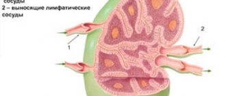
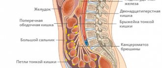
In recent years, there has been an increase in the number of patients with extrapulmonary and generalized forms of tuberculosis, the number of cases of abdominal tuberculosis (ABTB), which is primarily due to an increase in the number of patients with HIV infection. Particularly often, damage to the TB is observed in patients with HIV infection in the stage of secondary diseases. In TOBP, the retroperitoneal and mesenteric lymph nodes, intestines, peritoneum, and spleen are most often affected [1–4].
Diagnosis of TOBP due to the similarity of clinical manifestations with other nonspecific diseases of the abdominal organs is difficult, the clinical picture does not have pathognomonic symptoms, most often this category of patients is examined in general medical institutions for chronic pain syndrome in the abdomen, colitis, mesadenitis, ascites of unknown etiology. A highly effective diagnostic method that makes it possible to establish the diagnosis of TOBP, namely intestinal tuberculosis, is colonoscopy [5–8].
We set the goal of the study - to evaluate the effectiveness of colonoscopy in the diagnosis and effectiveness of treatment of patients with TOBP.
Material and methods
In the surgical department of the Moscow Scientific and Practical Center for BT, from 2010 to 2012, 63 patients with suspected TOBP aged from 22 to 77 years were examined and treated - 40 (63.5%) men and 23 (36.5%) women. Pulmonary tuberculosis was diagnosed in 49 (77.7%) patients, 5 (7.9%) were examined with suspected pulmonary tuberculosis without pulmonary tuberculosis. There were 31 (49.2%) HIV-infected patients. A total of 76 colonoscopies were performed, the study was performed both for diagnosis and to evaluate the effectiveness of treatment.
All patients in the comprehensive examination program for the diagnosis of TOBP underwent standard clinical and instrumental examinations: ultrasound of the abdominal organs, esophagogastroduodenoscopy, irrigoscopy, computed tomography of the abdominal organs and stool examination for Mycobacterium tuberculosis
(MBT) by fluorescent microscopy and inoculation on solid nutrient media.
Colonoscopy was performed according to the generally accepted method with the obligatory collection of material for histological examination, fluorescent microscopy and culture on solid nutrient media, and polymerase chain reaction (PCR).
When performing colonoscopy, we used a standard method of preparing the intestine for examination - using enemas or the drug Fortrans.
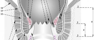
When assessing the colonoscopic picture, we focused on 3 types of tuberculous intestinal lesions: miliary, infiltrative and infiltrative-ulcerative form (see figure).
Miliary (a), infiltrative (b) and infiltrative-ulcerative (c) forms of intestinal tuberculosis.
During colonoscopy, erosions were most often found in the colon; tuberculous ulcers in all cases were localized in the right half of the colon, most often in the cecum, in the area of the bauhinine valve and the ileum. It was taken into account that the endoscopic picture of intestinal tuberculosis can be varied, although the changes are not specific. Oval or round ulcers, pseudopolyps, narrowing of the lumen, and rigidity of the intestinal walls may be observed. During intestinal endoscopy, swelling, hyperemia of the mucous membrane, the presence of ulcers with undermined edges, wall rigidity, and narrowing of the intestine are noted. Single, often multiple, ulcers are localized in the cecum, ileum, ascending, and colon. Tuberculous ulcers are often surrounded by a perifocal inflammatory shaft, often merge with each other, occupying a significant extent of the intestine, scarring, and produce ring-shaped narrowings of the intestine. Intestinal tuberculosis sometimes occurs in the form of an isolated hyperplastic pseudotumor process localized in the cecum, with a sharp thickening of the wall of the cecum, sometimes its ascending part, with an inflammatory process of specific tubercles, caseous decay, followed by scarring, narrowing of the intestinal lumen. To recognize intestinal tuberculosis and carry out differential diagnosis (with Crohn's disease, ulcerative colitis, intestinal tumors, amoebic dysentery, appendicitis), the results of histological examination are very important: with intestinal tuberculosis, along with sarcoid-like granulomas, foci of caseous necrosis appear. In some cases, the biopsy material reveals a picture of chronic nonspecific inflammation, but mycobacteria and epithelioid granulomas with Pirogov-Langhans cells, which form the basis of histological changes in tuberculosis, cannot be detected. This may be due to insufficient biopsy depth or predominantly submucosal localization of the pathological process. In such cases, the diagnosis is established based on a combination of clinical, laboratory and instrumental data.
Crohn's disease
- The diagnosis of Crohn's disease was most likely in the presence of the following characteristics: male gender, hematochezia, perianal lesions, intestinal obstruction, extraintestinal manifestation.
- Endoscopic signs were: longitudinal ulcers, identification of the mucous membrane in the form of a cobblestone pavement, intraluminal strictures, involvement of the rectum in the pathological process.
- Histological signs and findings according to computer enterography were: asymmetrical thickness of the intestinal wall, “comb” sign, fibrous-fatty proliferation.
Results and discussion
Visual signs of intestinal tuberculosis were detected in 47 (74.6%) patients; they were characterized by the presence of erosions, infiltrates and ulcers in the colon. All patients underwent a biopsy of erosions or ulcers for histological examination, fluorescence microscopy and culture on solid nutrient media, and PCR. In 24 (38%) cases, microscopy revealed the presence of epithelioid giant cell granulomas, Ziehl-Neelsen staining revealed acid-fast mycobacteria, in 2 (3.2%) cases colon adenocarcinoma was diagnosed, in 4 (6.4%) - cytomegalovirus ulcerative colitis, which is typical for patients with HIV infection; in the remaining 37 (58.7%) cases, nonspecific colitis was detected. It was mandatory to take a biopsy from ulcers and erosions to test for MBT using fluorescent microscopy - in 17 (27%) cases MBT was found. When cultured on solid nutrient media, a positive result was obtained in 21 (33.3%) patients, and in 4 patients in this group, fluorescence microscopy gave a negative result. A PCR study to detect MBT fragments gave a positive result in 28 (44.4%) cases. However, research using the PCR method, which has high sensitivity, cannot be classified as a strictly specific diagnostic method, since the detection of MBT fragments in a biopsy from tuberculous ulcers is possible not only in intestinal tuberculosis, but also when ingesting sputum in patients with pulmonary tuberculosis.
Based on the results of colonoscopies with biopsies, the diagnosis of tuberculosis of the abdominal organs was established in 54 (85.7%) patients.
All patients received chemotherapy for tuberculosis in accordance with standard anti-tuberculosis chemotherapy regimens. In 46 (85.2%) patients, against the background of anti-tuberculosis therapy, clear positive dynamics were noted in the form of an improvement in general well-being, a decrease in the severity of symptoms of tuberculosis intoxication, the disappearance of abdominal pain, and normalization of stool. In 9 (16.6%) patients, despite anti-tuberculosis chemotherapy, no clear positive dynamics were noted: a slight improvement in general well-being, moderate abdominal pain and diarrhea persisted. These patients underwent control colonoscopy, 6 - once, 3 - twice. During the control colonoscopy, in comparison with the previous one, no clear positive dynamics were noted: tuberculous ulcers persisted or even new ones appeared, infiltration of the intestinal wall. Histological examination revealed an active tuberculosis process. These patients had their anti-tuberculosis therapy regimen changed, and all of them experienced clinical improvement.
The results obtained allowed us to conclude that colonoscopy is a fast, inexpensive and diagnostically valuable method for diagnosing tuberculosis of the abdominal organs (intestines), allowing us to reliably establish this diagnosis in 80.6% of patients. In some cases, performing a control colonoscopy against the background of anti-tuberculosis therapy allows one to assess the dynamics of treatment and, if necessary, change the anti-tuberculosis chemotherapy regimen.
Diet for tuberculosis
Nutrition for tuberculosis should be aimed at strengthening the immune system.
- The patient should consume from 120 to 150 g of pure protein per day. It is needed for the production of antibodies. Sources of protein: fish, seafood, dairy products, lean poultry and fish, liver of cattle and fish.
- The amount of fat the patient needs is from 50 to 80 g per day. They are necessary to restore cell membranes that have been damaged by mycobacteria. To avoid a shortage of fats, you need to eat butter and vegetable oils, fish oil, lard, and animal fats in small quantities.
- Carbohydrates for tuberculosis should correspond to the age norm - about 400 g per day. They can be obtained from cereals and vegetables. Eating more than 80 g of confectionery products per day is not recommended.
- Mineral salts normalize metabolism and improve the functioning of the endocrine system, thereby increasing the body's defenses. Their sources can be: tomatoes, figs, cauliflower, herbs, cheeses, cottage cheese.
Prevention
Prevention of tuberculosis begins in infancy. On the 4th day of a child’s life, he is vaccinated with BCG, which is a weakened strain of mycobacteria. Since pathogens are very active against children, it is very important to develop immunity as early as possible. A weaker version of the vaccine, BCG-M, has been developed for premature infants. Normally, a bump appears at the injection site, and then a bubble with a yellowish liquid, which later bursts and becomes crusty. The vaccine does not completely protect against tuberculosis, but it does help prevent extrapulmonary infection in children.
It is also important to lead a healthy lifestyle, eat well, not smoke, and undergo regular examinations. Anyone can get tuberculosis, even from quite prosperous sections of society. The mechanism for the development of the disease is triggered by malfunctions in the immune system.
Symptoms
Tuberculosis can damage both the respiratory system and other organs of the human body. The lungs are affected in 85% of cases. The symptoms are very similar to any other diseases of the respiratory system, so you need to be extremely attentive to the body to diagnose the disease at an early stage.
Clinical symptoms of pulmonary tuberculosis:
- A dry cough, which, as the disease develops and progresses, becomes wet with the presence of blood in the sputum. Often prolonged coughing attacks occur at night. - The cough lasts for at least 3 weeks.
- Coughing up blood. The bacillus attacks the small arteries that supply the lungs with blood, which is why the blood is released with a cough.
- Sharp chest pain when coughing. It occurs due to irritation of the nerve endings of the pleura, which covers the lungs. In a healthy person, the pleural layers slide relative to each other, due to which inhalation and exhalation occur. The infected leaves are affected by Koch's bacillus, which leads to disruption of the functioning of the respiratory system.
- Increased shortness of breath with any physical activity. The fact is that the bacterium affects the lungs, which means they cannot supply the human body with the necessary amount of oxygen. Shortness of breath occurs as a result of a person’s attempts to inhale as much air as possible, of which he feels lacking.
- Fever (increased body temperature from 37.1 - 38 °C). But most cases are characterized by exceeding the norm by 1-2 degrees in the evening.
- Weakness and malaise as a result of increased temperature.
- Increased sweating at night.
- Weight loss.
A noticeable deterioration in the condition in most cases is observed in the evening and at night.
In children, tuberculosis is accompanied by additional symptoms:
- Enlarged lymph nodes and pain when touching them. This occurs because this is where a large number of bacteria are concentrated.
- Pale skin due to decreased blood supply.
- Sleep disturbances, problems concentrating, increased irritability and mood swings.
The non-pulmonary form of tuberculosis is much more difficult to diagnose:
- The only symptom is a slight but constant increase in body temperature, as a consequence of the body’s fight against the disease.
- Damage to the genitourinary system is characterized by the presence of blood in the urine and acute pain in the lumbar region. The consequence of the disease is infertility in women.
- The presence of blood in feces is also observed when the digestive tract is damaged. This also includes bloating, intestinal obstruction, and pain similar to those observed with pancreatitis or gastritis.
- If the infection affects the nervous system, symptoms similar to meningitis occur. Inflammation of the meninges occurs. The patient feels severe pain when turning and tilting the head, especially strong tension in the occipital muscles.
- If the disease affects the joints and bones, then the person becomes prone to frequent fractures, pathologies appear in the vertebral curves and postural disorders. Movements become constrained. Acute pain occurs in the back and joints.
Hospital treatment
Usually treatment occurs at home, but sometimes the complex course of the disease requires observation in a hospital. Once the diagnosis is confirmed, treatment in hospital may take approximately 3-4 months. During this period, to monitor the success of treatment, sputum is collected for analysis. If bacteria are not detected three times, the person is discharged and his condition is then monitored on an outpatient basis.
If the bacterium is particularly resistant to drugs, making their selection incredibly difficult, the patient may remain in the hospital for one to one and a half years.
The main indications for hospitalization are:
- non-pulmonary form. Treatment is done through chemotherapy;
- pulmonary form, not amenable to outpatient treatment due to the resistance of the bacterium;
- treatment of acute forms of infection;
- the need for surgical intervention.
Treatment
In countries with an increased number of cases of infection and spread of the disease, treatment is provided free of charge. This reduces the risk of infection, mortality and the likelihood of spreading infection during migration.
The main condition for treatment is complete cessation of smoking and drinking alcohol. Both the first and second greatly reduce the body’s immunity, making it unable to fight the disease. It is important to carefully follow all the necessary rules of personal hygiene, including for healthy people, to prevent the risk of disease.
Mycobacterium tuberculosis is able to quickly adapt and develop resistance to various drugs. Therefore, selecting a treatment regimen is a very long process.
Medicines are divided into several types:
Drugs that directly affect the causative agent of the disease and affect the resistance of the bacilli. They have bactericidal and bacteriostatic mechanisms of action.
- Isoniazid is the most effective drug, suppressing 90% of active and growing bacilli in a few days.
- Rifampicin is the best sterilizing agent.
- Pyrazinamide kills bacteria in the acidic environment of cells.
- "Ethambutol" is a chemotherapy drug that affects typical and atypical cells.
- The drug Streptomycin, which has an antimicrobial effect, is administered intravenously.
The second line of drugs to combat the disease includes tablets:
- "Protionamide" is a drug active in an acidic environment.
- "Cycloserine" is a bacteriostatic and bactericidal drug.
- “Ethionomide”, which has an effect on bacilli located outside and inside cells.
Intravenous drugs:
- Amikacin is a broad-spectrum antibiotic. Prescribed when bacilli are resistant to other drugs.
- "Kanamycin", active against acid-fast bacteria.
The dose of all medications must be calculated strictly by the attending physician, since it directly depends on the weight and age of the patient, as well as on the nature and stage of the disease.
Historical information
There is plenty of evidence that tuberculosis was familiar in ancient times. Direct evidence is the modified bones of the mummies of Egyptian pharaohs.
In the chronicle of the history of Ancient Greece, there are materials from Hippocrates, who identified the main symptoms of the acute form of the disease: sputum, weakness in the body, weight loss, cough and pain in the chest. The infected were evicted from cities to isolated areas. But both Hippocrates and the scientist Avicenna knew tuberculosis as a disease transmitted only by airborne droplets.
Throughout human history, the name of tuberculosis has varied. It was defined as the “white plague” and “flying death” because it affected a huge number of people. In 1200 BC, the Hindus called it consumption. Marriages with sick women were strictly prohibited. In 1819, the French doctors Laennec and Bailey called the disease “tubercle” (tuberculum). For a long time it was believed that tuberculosis was a non-infectious disease characteristic exclusively of the lower social strata of the population.
On March 24, 1882, the German scientist Robert Koch identified the causative agent of the disease - a bacterium named Koch's bacillus in honor of the researcher. He discovered it while studying the sputum of one of his patients. Robert then experimentally caused this disease in animals, which confirmed the correctness of his discovery. It was Koch who proved that tuberculosis is equally dangerous for representatives of both the upper and lower classes of the population. March 24 has officially received the status of World Tuberculosis Day.
Celebrities who suffered from the terrible disease were actress Vivien Leigh, sister writers (Charlotte, Emily and Anne) Bronte, playwright Anton Pavlovich Chekhov, seventh US President Andrew Jackson, writer George Orwell.