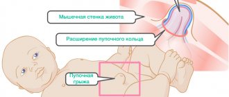
A hernia is an opening or “weak” spot in the muscles of the anterior abdominal wall – the so-called “hernial orifice” – through which organs or fatty tissue emerge under the skin. As a result, a noticeable subcutaneous bulge is formed, and the tissues extending into the hernial sac are compressed and cause pain and discomfort. Most often, intestinal loops fall out through the hernial orifice.
Ventral hernia of the anterior abdominal wall is a postoperative complication. In this case, the abdominal organs protrude under the skin in the area of the postoperative scar. Such a defect can reach large sizes, disrupt the functioning of the gastrointestinal tract, and be infringed.
How does a hernia of the anterior abdominal wall manifest?
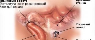
First of all, this is a cosmetic defect, that is, a noticeable visual protrusion. The “weakest” places of the anterior abdominal wall are the umbilical ring, inguinal canal, Spigelian line, and linea alba. The hernia consists of several parts:
- hernial orifice;
- hernial sac;
- contents of the bag;
- outer soft tissue shells of the bag.
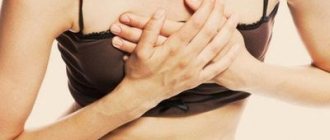
A hernia causes pain and discomfort, and some patients feel as if something has come out or experience a sensation of tearing. Other symptoms:
- feeling of pressure on the anterior abdominal wall;
- pain and burning;
- feeling of weakness;
- the bulge may appear when coughing, sneezing, straining and disappear when lying down;
- When straining, coughing, or lifting heavy objects, the pain intensifies.
What diagnostic methods may be needed?
In most cases, visual examination and medical history, including the date of surgery and the nature of rehabilitation, are sufficient to make a diagnosis. A general blood and urine test is required, and radiography and ultrasound of the abdomen may also be useful.
A hernia after abdominal surgery most often appears unexpectedly, and the patient’s task is to find his bearings in time and consult a doctor. “The First Family Clinic” is open every day at the address: Kolomyazhsky Prospekt, 36/2 (Udelnaya metro station, Pionerskaya).
What will happen if left untreated?
The bulge will gradually increase, will no longer disappear when lying down, and the pain will intensify. The most dangerous complication is strangulation, which can lead to organ necrosis followed by peritonitis, which is a direct threat to life. In case of strangulation, emergency removal of the abdominal hernia is performed.
Important! In this condition, you should not try to correct the defect yourself. It is urgent to call an ambulance.
Other possible complications of hernias:
- inflammation;
- impossibility of reduction;
- coprostasis.
Symptoms of postoperative ventral hernia
Depending on its location, size and contents, it manifests itself with various symptoms - from a slight feeling of discomfort to severe pain with minor physical exertion, nausea, constipation and vomiting. The most serious complication of a hernia is strangulation of the organs located in it.
There are several reasons for the occurrence of postoperative hernias. Most often, this is an infection in the wound after surgery, preventing normal tissue healing. Of no small importance are the presence of genetic defects in the formation of connective tissue and concomitant diseases, such as diabetes, obesity, chronic pulmonary disease (COPD), etc.
In addition, heavy physical activity in the early postoperative period can lead to the appearance of a hernia. Rarely, technical defects in wound suturing during the initial operation or poor-quality suture material can lead to the formation of a hernia.
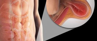
What is a strangulated hernia?
In case of strangulation, the internal organs located in the hernial sac are compressed by the ring of the hernial orifice. As a result, the vessels that feed the organs (most often the greater omentum and intestinal loops) are compressed.
Characteristic symptoms of infringement:
- traces of blood in the stool;
- pain in the area of formation that increases and does not go away with rest;
- inability to correct the defect while lying on your back, even by pressing on it (this is strictly not recommended!);
- the skin at the site of the protrusion changes color - from very pale to purple;
- the formation increases and thickens;
- the patient feels nauseous and vomits frequently.
- there is no transmission of the cough impulse.
It is known that acute appendicitis (AA) is the most common disease in emergency surgical practice, accounting for 20-40% of the reasons for hospitalization of patients in surgical hospitals [1, 2]. Postoperative ventral hernia (PVH) remains the most common complication in the long-term period after laparotomy, occurring in 3.8-20% of cases [16]. In external hernias of the anterior abdominal wall (inguinal, femoral, Spigelian line, postoperative), a vermiform appendix and omental processes of the colon are sometimes detected [3]. However, the detection rate of OA in the hernial sac is only 0.008% [6]. For the first time, the English surgeon Claudius Amyand described the observation of acute appendicitis in an inguinal hernia in 1735, and the Parisian surgeon Rene Jacques Croissant de Garengeot in 1731 - in a femoral hernia. We conducted an analysis of the literature, in which we summarized data on a rare localization of the disease and described our own observation. Unfortunately, the first two observations in the 20th century, described by M. de Andrade [8] in 1987 and P. Horgan [14] in 1991, were not studied by us due to the lack of texts of articles in electronic medical libraries.
In a literature review by C. Sugrue et al. [18] described a 78-year-old patient who was admitted with complaints of pain in the mesogastric region for 3 days, single vomiting, and the presence of a large tense and irreducible tumor-like protrusion in the area of the scar after upper midline laparotomy for cholecystectomy. Based on clinical and radiological examination methods, irreducible PIH was diagnosed. The contents of the hernial sac were the cecum and a macroscopically inflamed appendix. There were no signs of pathological changes in the small intestine or incomplete rotation of the large intestine. An appendectomy and in-lay plastic surgery of the abdominal wall were performed. Histological examination confirmed the presence of OA.
M. Gass et al. [11] described the observation of a 62-year-old patient with cramping abdominal pain, nausea and vomiting, and intermittent episodes of fever. Prescribed antibiotics alleviated these symptoms, but burning pain remained in the right iliac region, and palpation revealed signs of local peritonitis. Previously, the patient underwent cystojejunostomy with Roux-en-Y reconstruction for an extrapancreatic pseudocyst and drainage of the abdominal cavity with removal of a drainage tube in the area of the right lower quadrant of the abdomen. Computed tomography (CT) revealed a vermiform appendix under the skin with signs of acute inflammation (Fig. 1)
, which was confirmed by laparoscopy.
A laparoscopic appendectomy and suturing of a 3 cm long abdominal wall defect with a continuous suture with additional IPOM repair were performed (Fig. 2)
.
There are observations of OA in the hernial sac in PIH after operations for urological and gynecological diseases, as well as in transplantology practice. So,
Y. Dittmar et al. [9] described a 69-year-old patient with acute pain in the right lower quadrant of the abdomen. She underwent a right kidney transplant for chronic renal failure 4 years ago. The examination revealed PIH and an inflamed appendix in the hernial sac.
S. Bhagwandin et al. [5] described the observation of a 57-year-old patient with moderate obesity and PIH with a hernial orifice diameter of 12 cm. 5 years earlier, she underwent orthotopic liver and kidney transplantation with subsequent immunosuppression. She was admitted to the clinic with complaints of pain for 8 hours in the right lower quadrant of the abdomen, lack of appetite, fever, and nausea. CT scan without contrast revealed a blind-ending tubular structure with a thickened wall in the hernial sac (Fig. 3)
.
HER. Achkasov et al. [1] described a similar patient who had undergone radical nephrectomy for cancer of the right kidney 9 months earlier. During follow-up and CT scanning, an inflamed appendix in the lumbar hernia was detected. In all three cases, a herniolaparotomy was performed, the presence of OA was confirmed intraoperatively, an appendectomy and repair of the hernial orifice with a prolene mesh prosthesis were performed.
Gynecologists E. Galiñanes and A. Ramaswamy [10] reported a 27-year-old woman with PIH and complaints of abdominal pain associated with nausea, vomiting and anorexia for 24 hours. She had a history of hysterectomy with right oophorectomy for stage III cervical cancer. through lower transverse laparotomy according to Pfannenstiel. A diagnostic laparoscopy was performed, which revealed that the apex of the appendix was strangulated in the lateral edge of the hernial orifice. An appendectomy and herniorrhaphy were performed, and transfascial sutures were applied to close the defect. Morphological examination confirmed the diagnosis of OA.
At the end of the twentieth century, there was an active introduction of laparoscopic technologies in surgery, one of the expected advantages of which was a reduction in the likelihood of hernia formation. However, the problem of trocar hernia, the equivalent of PIH after laparotomy, has emerged. Ch. Menenakos et al. [15] described the observation of a 63-year-old man who was admitted with complaints of abdominal pain and an irreducible tumor formation in the right abdomen. One year before admission, the patient underwent laparoscopic anterior resection of the rectum for cancer with the formation of a preventive temporary end ileostomy, which was closed 3 months after the initial operation. Upon examination, two PIHs were identified: one in the area of the ileostomy and the other, painful and irreducible, in the area of insertion of a 12-mm trocar during laparoscopic surgery, in the right iliac region. During the operation, a strangulated and inflamed vermiform appendix was revealed. Appendectomy and hernia repair of both hernias were performed with sub-lay plastic surgery of the abdominal wall (Fig. 4)
.
In another observation by C. Sugrue et al. [18] reported a sick 81-year-old man who presented with abdominal pain and bile vomiting for 1 day. CT scan revealed free fluid in the abdominal cavity. For diagnostic laparoscopy, a trocar (10 mm) is installed above the navel and two ports (5 mm) in the right and left iliac fossa. It was revealed that the purulent effusion was caused by a covered perforation of the duodenal ulcer. After sanitation of the abdominal cavity, a drainage was installed in the right lateral canal and removed through a puncture from a 5-mm trocar in the right iliac region, which was removed on the 1st day after surgery. After 5 weeks, the patient began to complain of pain and swelling in the right iliac region. The examination revealed an irreducible tumor-like mass in the area where the 5-mm port was installed. On ultrasound, the contents of the hernial sac were represented by the apex of the small intestinal loop. During the operation, it was established that the hernial sac contained the distal part of the inflamed appendix (Fig. 5). M. Cerna` et al. [7] described the observation of a patient hospitalized with suspected abscess of the anterior abdominal wall caused by a strangulated PIH. The audit confirmed that the cause was acute gangrenous appendicitis in the hernia after diagnostic laparoscopy.
Here is our own observation.
Patient F.
, 84 years old, was admitted on the 7th day from the onset of the disease with complaints of general weakness, pain and hyperemia of the skin in the area of the postoperative scar, hyperthermia up to 37.7 °C.
There was moderate leukocytosis with a neutrophilic shift, heart rate 110-120 per minute, blood pressure 100/70 mm Hg. In the anamnesis, in 1968, gastric resection according to Billroth II and in 1970, resection of a section of the small intestine due to perforation. She was a hernia carrier for 10 years and abstained from treatment. On examination: the patient's condition is serious. Along the upper-median scar there is a painful hernial protrusion measuring 15×12×10 cm, difficult to reduce into the abdominal cavity, with a soft-elastic consistency. The skin over the hernia is hyperemic. A strangulated PIH was suspected, and therefore the patient was operated on. A median herniolaparotomy was performed, which revealed a multi-chamber hernia, the contents of which were loops of the small intestine (Fig. 6, a)
.
the dome of the cecum and a thickened hyperemic appendix with fibrin overlays measuring 12×1.5 cm (see Fig. 6, b)
. The colon has incomplete rotation, the cecum is mobile, and has a mesentery. Intraoperative diagnosis: acute phlegmonous appendicitis in a postoperative ventral hernia. Appendectomy, tension-free prosthetic in-lay hernioplasty, and Redon wound drainage were performed. The postoperative period proceeded without complications. The wound healed by primary intention. Prophylactic antibacterial therapy was carried out with third-generation cephalosporins for 5 days. The patient was discharged on the 8th day. Morphological examination confirmed the presence of phlegmonous appendicitis.
The vermiform appendix can be located in an external hernia of all types. The cause of such unusual anatomical locations may be incomplete embryological rotation of the colon, hypermobility of the cecum, or previous abdominal surgery. All these risk factors were present in our observation. To explain the development of acute inflammation of the appendix in a hernia, the idea of extraluminal compression (ischemia of the appendix as a result of pressure from the narrowed neck of the hernial sac, constant trauma with the formation of inflammatory adhesions) has been put forward, which distinguishes it from the pathogenesis of “ordinary” acute appendicitis [12, 19].
The observation of OA in the PIH associated with the presence of drains [11] adds a thesis to the discussion about the use of drains, which has unfolded in recent years due to possible complications - ascending infection along the drainage tube or arrosion of the abdominal organs (small or large intestine), formation scars and hernias in the place of drainage. According to H. Petrowsky [17], the effectiveness of drainages is overestimated - the level of complications in patients without drainage of the surgical area is not higher than in patients using drainages. Obviously, the risk of complications depends on the diameter of the defect in the abdominal wall for the insertion of drainage.
Trocar hernias are more often described in the literature than post-drainage hernias, although the mechanism of hernia formation is similar. The contents of the hernial sac are usually a strand of greater omentum or a loop of small intestine, but three cases of OA in a trocar hernia have been reported [7, 15, 18]. The incidence of trocar hernia is 0.74% and depends on the type of laparoscopic surgery. Independent risk factors include obesity, wound infection and the use of cutting blade trocars [16]. The size of the installed ports plays a key role: the larger their diameter, the more often the occurrence of a hernia can be observed [17].
The remaining clinical observations relate to OA in the PIH after using various options for laparotomy access (see table)
: Pfannenstiel incision [10], midline laparotomy [13, 18], combined laparotomy in transplantology [5, 9] and even lumbotomy [1]. This further supports the view that PIH can form after any operation and, although extremely rare, inflammation of the appendix in this hernia can occur.
It is worth noting that the clinical manifestations of OA in the PIH differ from the classic clinical picture of appendicitis. Of the non-invasive diagnostic methods, CT of the abdominal cavity is used, which can detect an inflamed appendix, but the nature of this inflammation (strangulation or primary appendicitis) is quite difficult to establish.
In all observations, the only treatment method for patients was surgery. And if the first stage - appendectomy - does not raise any questions (it can be equally successfully performed either laparoscopically or traditionally), then the choice of method for hernial orifice repair in conditions of an inflammatory process remains debatable even with experience in the successful use of an allograft.
Thus, any ventral abdominal hernia, especially in the right lower quadrant, may contain an appendix. However, acute appendicitis in a postoperative hernia remains an extremely rare observation. To the question whether it is possible to perform plastic surgery with an allograft in an infected wound of the abdominal wall with acute appendicitis, the presented clinical observation and analysis of the literature give a positive answer.
Preoperative preparation
Necessary for patients with large and giant postoperative hernias, as well as in the presence of diseases that predispose to the occurrence of hernias. Preparation should include not only drug correction of concomitant diseases, but also preparation of the skin in the area of surgery (treatment of ligature fistulas, diaper rash, maceration); bowel preparation (constipation treatment); prevention of respiratory and cardiovascular complications, prevention of intra-abdominal hypertension syndrome. Most of the preparatory activities are carried out on an outpatient basis under the supervision of a therapist (cardiologist) and a surgeon.
Treatment
If after a medical examination a postoperative abdominal hernia is diagnosed, what should I do? The algorithm of actions depends on many factors - the size of the hernia, the presence of strangulation and the condition of nearby tissues, the general health of the patient.
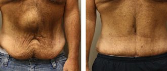
The most radical and effective way to treat a postoperative hernia is to perform a repeat operation (hernioplasty) aimed at strengthening the abdominal wall in the area of the scar. This method will prevent relapses and eliminate possible complications in the future.
Types of hernioplasty surgery
Suturing the hernial opening
The simplest and fastest operation for eliminating a ventral hernia is suturing the defect by overlapping the muscles located along the edges of the hole. Before suturing, it is necessary to reduce the hernial contents into the abdominal cavity. The operation is performed only if the size of the hernial orifice does not exceed 5 cm, and there are no inflammatory or other pathological changes in the wound area. The method has a drawback - the possibility of a recurrence of a hernia in the same place (relapse).
Postoperative abdominal hernia, tension-free hernioplasty operation
The method is used in most cases of ventral hernias. The tissue in the area of the hernial opening is duplicated with a mesh implant. The material does not cause allergic reactions. An implant flap of the required size, like a patch, is sutured to the tissues around the hernia (muscles, aponeurosis). Due to its structure, the implant firmly fuses with the surrounding tissues and significantly increases the strength of the abdominal wall where there was previously a hernia. The use of a mesh allows you to avoid excessive tension of the abdominal wall in the area of the scar, which promotes better healing and avoids relapses in the future.
Laparoscopic hernioplasty
This is an operation in which surgeons bring a mesh implant to the hernia defect through 2-3 mini-incisions, just a couple of centimeters long.
This modification of hernioplasty has all the advantages of laparoscopy: rapid recovery of the patient after surgery, minimal scars after the intervention and low trauma. Qualified surgeons at our clinic will always correctly and adequately select the most optimal methods for treating a postoperative hernia or give detailed recommendations for treating a postoperative hernia without surgery. Such non-surgical methods are available to doctors and, in some cases, they can temporarily improve the patient’s condition and prevent complications and progression of the disease. But only hernioplasty surgery will help you cure a ventral hernia forever.
Treatment without surgery
If the doctor decides to postpone the hernioplasty operation for any reason, the following measures are recommended for patients to prevent the hernia from enlarging and complications:
- Wearing a bandage is a temporary measure. A bandage (a wide band of elastic fabric will relieve some of the load from the abdominal muscles) is applicable only in the initial stages of hernia development.
- Avoid physical activity, as it increases intra-abdominal pressure and increases the hernia defect and the size of the protrusion.
- Monitor bowel functions, avoid constipation and flatulence (bloating), as they also increase pressure inside the abdominal cavity.
Postoperative hernia: the diet provides for a limitation on the amount of food taken at the same time (you need to eat in small portions, but more often). You should not eat foods that cause increased gas formation - cabbage, legumes, fatty, spicy and spicy foods, carbonated and alcoholic drinks. You should also avoid foods that contribute to constipation - flour and bakery products made from premium flour, confectionery, saturated meat broths, rice, strong tea and coffee, astringent fruits and berries.
Prevention of hernia formation after surgery
Such a protrusion occurs only when appendicitis is removed using the traditional method - by cutting the abdominal wall. When the procedure is performed using laparoscopy, the threat of this pathology is significantly reduced.
To prevent the formation of a hernia, you should prevent this phenomenon:
- Adhere to the lifestyle prescribed by your doctor.
- Control bowel function.
- Wear a special bandage.
- Try to avoid respiratory illnesses and severe coughing.