The internal organs are usually located in the hernial sac - part of the peritoneum (the inner membrane lining the abdominal cavity from the inside), which has fallen out along with the contents. The hernial sac can consist of several sections - chambers and contain any internal organs of the abdominal cavity inside. Symptoms of an umbilical hernia depend on its volume, the size of the hernial orifice, the presence of adhesions and obesity in the patient. Umbilical hernias can be reducible or unreducible; in the latter case, the walls of the hernial sac are fused to the surrounding tissues.
Small umbilical hernias with wide hernial orifices and the absence of adhesions usually do not bother patients too much - they quickly learn to repair it themselves. Large hernias with a narrow orifice (especially unreducible ones and in patients suffering from concomitant obesity) are an obstacle to the movement of feces through the intestines, which causes habitual constipation, pain, nausea and vomiting.
Often, umbilical hernias begin in women after childbirth. In this case, loops of the small and large intestines, the stomach and any other abdominal organs enter the hernial sac. With large hernias, the hernial sac is usually divided by septa into several sections (chambers).
A symptom of an umbilical hernia is swelling in the area of the anterior abdominal wall, which occurs when straining or when the patient is standing. In this case, in the lying position the swelling disappears. If there are no adhesions, then the patient can repair such a hernia himself, and it does not cause him much concern (including his ability to work), but sometimes pain occurs. This often asymptomatic course leads to the fact that patients consult a doctor when the hernia becomes unreducible or strangulated.
With a large hernia with a narrow hernial orifice, patients are often bothered by constipation and other discomfort from the gastrointestinal tract. In this regard, large hernias are an obstacle to physical labor. Such hernias are often complicated by strangulation and intestinal obstruction.
Causes of hernia
The cause of a hernia is weakness of the abdominal wall, which is not able to compensate for the increase in intra-abdominal pressure (for example, during heavy lifting, childbirth, defecation, strained cough, etc.). Defects in the abdominal wall resulting from surgery or injury can also lead to the formation of a hernia. The likelihood of a hernia increases with age-related thinning of muscle tissue and loss of elasticity. Congenital defects and hereditary predisposition matter.
Why is pathology dangerous?
In most cases, the prognosis is favorable. During pregnancy, complications occur relatively rarely.
The main danger is strangulation of the protrusion, when the organs cannot be moved into the hernial sac. As a result, the blood circulation of the intestine or greater omentum is disrupted, which can lead to tissue necrosis.
The risk of strangulated hernial protrusion is higher after childbirth. If pathology is present, it is imperative to see a surgeon. If necessary, elective surgery is indicated.
Types of abdominal hernias
Depending on the location, there are:
Inguinal hernia
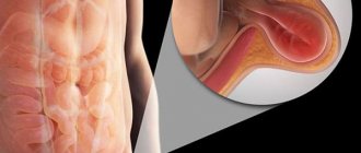
An inguinal hernia is the most common type of abdominal hernia. It is a pathological protrusion of the intestine or greater omentum into the cavity of the inguinal canal. In men, inguinal hernia occurs 5 times more often than in women, which is explained by the peculiarities of the anatomical structure of the groin area in both sexes. In men, the spermatic cord is located in the inguinal canal; in women, the round ligament of the uterus is located.
An inguinal hernia, if it is not strangulated, usually does not cause pain. The only sign of such a hernia is a protrusion in the lower abdomen. If you cough and put your hands on the hernia, you can feel the shock.
Femoral hernia
A femoral hernia is a protrusion of internal organs through the femoral canal.
Normally, the femoral canal does not exist, there is only a femoral ring filled with fatty tissue, loose enough to make this place vulnerable to protrusion of the hernial sac. Since women tend to have larger pelvises, femoral hernias are 4 times more common in them than in men. A femoral hernia goes through several stages in its development - initial, canal (when the protrusion has already led to the creation of the femoral canal, but the hernia has not yet penetrated the skin and has not become noticeable), complete. In the first two stages, a symptom of hernia formation is pain in the groin and upper thigh, which intensifies with coughing, straining and long walking. At the last stage, a characteristic swelling the size of a walnut or larger appears in the area of the femoral-inguinal flexion.
Umbilical hernias
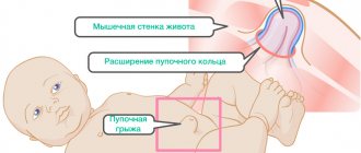
The navel is the place where the umbilical cord falls off, connecting the baby to the mother’s body. The muscles around the navel form an umbilical ring, which should contract fairly quickly. However, the umbilical ring remains a “weak” anatomical formation and protrusion of internal organs - the intestines or the greater omentum - can occur through it.
In newborns, weakness of the abdominal wall muscles quite often leads to the formation of an umbilical hernia (detected in 20% of infants). Sometimes such a hernia is noticeable only when the baby is upright or when he strains or screams. In most cases, an umbilical hernia in newborns goes away on its own, as the muscles of the abdominal wall strengthen. However, observation by a surgeon is mandatory. The hernia should not increase or become strangulated. A massage may be prescribed.
In some cases, umbilical hernia occurs in adults. Causes: weakness of the abdominal wall, increased intra-abdominal pressure. Provoking factors are pregnancy, obesity, chronic constipation, etc. This hernia looks like a ball in the navel area. Sometimes it only appears when straining or coughing. If the hernia is large, pain may occur that intensifies after eating or during physical activity.
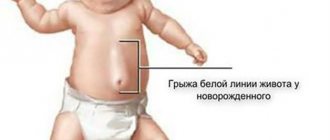
Hernia of the white line of the abdomen
The linea alba is a plate formed by intertwined tendons and separating the rectus abdominis muscles. It is called white because of the color of the tissue (it has few blood vessels). A white line runs down the middle of the abdomen - from the rib cage (xiphoid process of the sternum) through the navel to the pubis. Normally, its width is no more than 3 cm. But it can increase if the rectus muscles begin to diverge (for example, under the influence of high intra-abdominal pressure). This condition is called diastasis recti. In this case, hernias may form along the white line - above the navel (supra-umbilical hernia), in the area of the umbilical ring (peri-umbilical) or below the navel (sub-umbilical).
Alarming symptoms
There are some symptoms that are “alarming” in nature, i.e. may indicate the onset of some acute disease.
- Acute onset of pain (the so-called “dagger pain”) - occurs with a perforated ulcer or perforation of a hollow abdominal organ.
- Pain accompanied by an increase in body temperature occurs with acute appendicitis , cholecystitis, peritonitis, etc.
- Cramping pain - when the pain seems to “roll up” and “release” - is characteristic of intestinal obstruction.
- Black stools and vomiting “coffee grounds” are typical for gastrointestinal bleeding.
- Pain is accompanied by a severe general condition - it occurs in many acute surgical diseases.
- The pain is accompanied by repeated vomiting - it happens with acute pancreatitis, pancreatic necrosis, intestinal obstruction.
- The pain in the upper abdomen is of a girdling nature (i.e., radiates to the hypochondrium, back) - it happens with acute pancreatitis, pancreatic necrosis.
- Yellowness of the skin, accompanied by pain in the right hypochondrium, is characteristic of obstructive jaundice.
- The onset of pain in the upper abdomen, sometimes throughout the entire abdomen, followed, within several hours, by localization in the lower abdomen on the right - is characteristic of acute appendicitis.
- The pain is constant and severe and occurs in many acute diseases.
- Pain in the area of the hernial protrusion occurs with a strangulated hernia.
Methods for diagnosing hernia
Diagnosis of a hernia is carried out during examination of the patient. In this case, the doctor uses the methods of palpation (palpation), percussion (tapping) and auscultation (listening to the natural sounds of the body).
To obtain a more complete picture, instrumental studies are performed:
Radiography
Radiography of a hernia allows one to obtain additional information about the presence of adhesions, parietal strangulation of the hernia and partial intestinal obstruction.
More information about the diagnostic method
Ultrasound examinations
Ultrasound makes it possible to clarify the location of the hernia, the shape and size of the hernial orifice, assess the condition of the surrounding tissues (this allows you to choose the most effective technique for reducing the hernia), and determine the contents of the cavity of the hernial sac.
More information about the diagnostic method
Computed tomography (CT)
Computed tomography for hernia is used if ultrasound data is insufficient.
More information about the diagnostic method
Sign up for diagnostics To accurately diagnose the disease, make an appointment with specialists from the Family Doctor network.
What to do?
If suspicious symptoms appear and there is a sharp deterioration in the condition, you should immediately call an ambulance. Before the doctors arrive:
- it is necessary to ensure complete rest for the patient;
- It is forbidden to give a person food or water; if you are thirsty, you can only wet your lips with a damp cloth;
- You should not give painkillers or other medications so as not to blur the clinical picture;
- It is strictly forbidden to try to straighten the pinched tissues yourself;
- Do not use heating pads or warm compresses to relieve pain.
Hernia treatment methods
You can’t delay treating a hernia. If signs of a hernia are detected, you should immediately consult a surgeon.
The only way to treat a hernia is surgery. This type of operation is called hernioplasty.
.
Terms such as hernia repair
and
hernia removal
. But hernioplasty is a more correct name, since in most cases the hernia is not removed, but reduced.
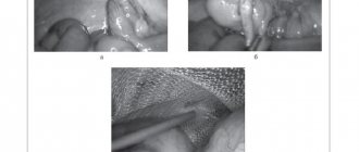
Hernia removal – laparoscopic hernioplasty
The hernial contents are immersed through the hernial orifice back into the abdominal cavity, after which plastic surgery of the hernial orifice is performed. At this stage, the problem of preventing the re-formation of a hernia is solved. The area of the hernial orifice is strengthened either by the body’s own tissues or by implants (polymer meshes). Within a month, the mesh grows into body tissues. Such a frame reliably holds the organs in place. When using your own tissues, relapses may occur (in 25% of cases).
At Family Doctor, hernia removal (hernioplasty) is performed laparoscopically - through small punctures in the abdominal wall, which allows for faster healing and avoidance of conspicuous scars. Laparoscopic hernioplasty is performed in a surgical hospital, under local or spinal anesthesia. The use of polymer implants almost completely eliminates the possibility of re-formation of a hernia.
More information about the treatment method
Make an appointment Do not self-medicate. Contact our specialists who will correctly diagnose and prescribe treatment.
Rate how useful the material was
thank you for rating
Surgery for umbilical hernia according to compulsory medical insurance at the Central Clinical Hospital of the Russian Academy of Sciences
The clinic of the Central Clinical Hospital of the Russian Academy of Sciences provides the full range of medical services for umbilical hernia: from examination and diagnosis to high-quality rehabilitation after surgery.
- An experienced doctor performs an examination, assesses the patient’s condition and, if the diagnosis is confirmed, prescribes a planned herniotomy operation.
- Surgical treatment is carried out in a modern operating room using high-quality instruments and drugs;
- The operation is performed by experienced, highly qualified surgeons.
Hernia treatment under compulsory medical insurance is a service that every citizen of the Russian Federation can receive at the Central Clinical Hospital of the Russian Academy of Sciences. In this case, the treatment will be carried out at the highest level with a caring and attentive attitude of all medical staff towards the patient.
The clinic has a rehabilitation treatment department, where you can undergo high-quality rehabilitation, which will allow you to return to your normal lifestyle as soon as possible.
The advantages of contacting the Central Clinical Hospital of the Russian Academy of Sciences for the treatment of umbilical hernia:
- consultations with experienced specialists;
- carrying out an operation under compulsory medical insurance;
- careful and attentive attitude towards the patient;
- use of modern surgical techniques, high-quality equipment, safe drugs;
- opportunity to receive rehabilitation treatment.
To make an appointment for examination and treatment at the Central Clinical Hospital of the Russian Academy of Sciences and clarify the cost of services, you can call or use a special form on the website.