- home
- general surgery
- Leiomyoma of the esophagus
Fig. 1 Histological structure of esophageal leiomyoma
Fig. 2 Location of leiomyoma in the wall of the esophagus (1 - tumor in the submucosal layer, 2 - esophagogastric junction)
Rice. 3 Excision of the tumor, without opening the esophageal mucosa
Rice. 4 Stitched wound on the esophagus after removal of leiomyma
Leiomyoma of the esophagus
is a benign tumor arising from its smooth muscle membrane or from the muscular elements of the mucous membrane. They usually have the appearance of a single node with smooth contours, less often they consist of several nodes, sometimes interconnected and entwining the esophagus circularly or over a considerable length. Located in the thickness of the muscular wall of the esophagus, the tumor pushes it apart, while the unchanged mucous membrane becomes thinner and stretches. As the size of the formation increases, it prolapses into the lumen of the esophagus, causing narrowing and dysphagia. Leiomyoma consists of bundles of smooth muscle alternating with areas of fibrous connective tissue (Fig. 1).
Leomyoma (50-70% of all benign esophageal tumors) most often develops in the middle and lower part of the esophagus. This is explained by the fact that in the distal half of the esophagus, the striated muscles of the esophageal wall are gradually replaced by smooth muscle fibers, which are the sources of this type of tumor (Fig. 2).
Clinical picture and symptoms of esophageal leiomyoma.
With esophageal leiomyoma, patients complain of dysphagia, which progresses slowly, is variable and is most pronounced when the tumor is large (up to 4-8 cm). Dysphagia in small tumors is explained by spasm of the esophagus due to irritation of the branches of the vagus nerve and the nerve plexus of the esophageal wall.
It is not uncommon for patients to complain of pain. It comes in varying intensity and is localized behind the sternum, in the pit of the stomach, and in the back. More often, pain occurs during or shortly after eating. It is not uncommon for patients to complain of regurgitation, vomiting, nausea, belching, loss of appetite, heartburn and drooling. Do not forget that these symptoms sometimes depend on concomitant diseases (peptic ulcer of the stomach and duodenum, hiatal hernia, cholelithiasis, etc.).
Sometimes patients with large leiomyomas may experience cough, shortness of breath, palpitations, arrhythmia, and cyanosis. These clinical manifestations occur when the esophageal tumor is located at the level of the tracheal bifurcation and at the site of the vagus nerve.
Symptoms of a benign esophageal tumor
Small benign tumors of the esophagus are quite common. They do not cause clinical manifestations and are often unexpectedly discovered at autopsy. The disease manifests itself with the onset of dysphagia. Benign tumors rarely cause esophageal obstruction. Dysphagia was observed in only 50% of patients. With large tumors, in addition to dysphagia, patients experience a sensation of a foreign body in the esophagus, vomiting and nausea, and sometimes pain when eating. It happens that large tumors do not cause any symptoms and are accidentally detected during an X-ray examination.
Unlike esophageal cancer, dysphagia in benign tumors does not tend to steadily and rapidly increase and can remain unchanged for several months or even years. In the anamnesis of some patients, periods of improvement in the passage of food due to a decrease in spasms are noted. The course of benign tumors is long; with non-epithelial tumors of the esophagus, patients live a long time, and the tumor does not show a significant tendency to grow. The general condition of patients with an esophageal tumor does not suffer. Sometimes there is some weight loss due to eating disorders and natural anxiety in such cases.
Diagnosis of esophageal leiomyoma.
Leiomyoma of the esophagus may be suspected by a doctor based on the clinical picture. The clinical picture can consist of a variety of symptoms: dysphagia, regurgitation, vomiting, nausea, belching, loss of appetite, heartburn and drooling, and these complaints can also be combined with cough, shortness of breath, palpitations and arrhythmia.
In the diagnosis of a benign tumor of the esophagus, x-ray examination of the esophagus is of paramount importance. X-ray signs of leiomyoma are expressed in a sharply defined filling defect, displacement of the lumen of the esophagus at the level of the tumor, its narrowing, and in some sections, expansion of the lumen. Folds of the mucous membrane are detected only on the wall opposite the tumor.
At the second stage, esophagoscopy is necessarily performed (preferably under sedation - artificial sleep, as is done in the Swiss University Clinic, Moscow). In the case of an intramural tumor, esophagoscopy reveals a protrusion of a pale, smoothed mucous membrane over the tumor. In these cases, as a rule, we do not perform a biopsy, since damage to the normal mucous membrane may somewhat complicate the subsequent operation. A biopsy is taken only if the mucous membrane is changed to exclude malignant lesions of the esophagus.
In the differential diagnosis of esophageal tumors, one should remember the possibility of malignant neoplasms of the esophagus, tumors and cysts of the mediastinum and lung, which can also cause a similar clinical picture and often similar findings from instrumental examination. To exclude leiomyosarcoma of the esophagus, it is necessary to perform a nuclear MRI or MSCT of the chest with contrast, and based on the degree of accumulation of the contrast agent, one can judge a possible malignant formation in the wall of the esophagus. It is worth noting that the transition of leiomyoma to leiomyosarcoma is extremely rare.
Treatment of benign tumors and cysts of the esophagus
The main treatment method for benign tumors is surgery. The purpose of the operation is to remove the tumor and prevent possible complications. Small tumors on a thin stalk can be removed through an esophagoscope using special instruments or destroyed (electrocoagulation). Intraluminal tumors on a wide base are excised with a section of the esophageal wall. Intramural tumors and cysts of the esophagus can almost always be enucleated without damaging the mucous membrane. The long-term results of the operations are good.
Treatment of esophageal leiomyoma. Indications for surgical intervention.
To identify leiomyoma of the esophagus, its localization, as well as choose the correct surgical treatment tactics, you must send me a complete description of gastroscopy, X-ray of the esophagus and stomach with barium, preferably an ultrasound of the abdominal organs, by indicating your age and main complaints. Then I will be able to give a more accurate answer to your situation.
My experience in treating patients with esophageal leiomyomas is more than 15 years. During this time, I was able to successfully operate and treat more than 90 patients laparoscopically.
For esophageal leiomyoma, treatment should only be surgical. Due to the slow growth of this tumor, surgical treatment is indicated only in cases of dysfunction of the esophagus and severe symptoms (listed above), provided there is no increased risk of surgery. If there is no clinical picture of esophageal leiomyoma, and the size of the tumor does not exceed 5 cm (and according to MSCT with contrast, there is no accumulation of it in the tumor tissue), then you can refrain from surgical treatment in this situation. Dynamic observation is permissible if regular endoscopic and x-ray examinations are possible in one medical institution, so that if the size of the formation increases or complaints appear, indications for surgery can be established in a timely manner. When planning treatment, it is necessary to take into account that the final conclusion about the benignity and malignancy of the tumor can be judged only after histological examination, which is possible only after a biopsy of the formation.
Surgical technique for esophageal leiomyoma.
Watch a video of operations performed by Professor K.V. Puchkov. You can visit the website “Video of operations of the best surgeons in the world.”
Due to the location of the tumor outside the mucosa, in most cases it is possible to remove the leiomyma from the wall of the esophagus. Traditionally, thoracotomy is used (left- or right-sided, depending on the level of localization of the leiomyoma). Which leads to major trauma to the chest wall, poor cosmetic effect and a long recovery period.
Puchkov K.V., Ivanov V.V. and others. Technology of dosed ligating electrothermal effects at the stages of laparoscopic operations: monograph. - M.: ID MEDPRACTIKA, 2005. - 176 p.
Puchkov K.V., Bakov V.S., Ivanov V.V. Simultaneous laparoscopic surgical interventions in surgery and gynecology: Monograph. - M.: ID MEDPRACTIKA, 2005. - 168 p.
Puchkov K.V., Rodichenko D.S. Manual suture in endoscopic surgery: monograph. - M.: MEDPRACTIKA, 2004. - 140 p.
Our clinic has accumulated experience in video-assisted thoracoscopic and laparoscopic operations when removing esophageal leiomyomas. With this method, leiomyoma is removed without thoracotomy, through 4 punctures on the chest or abdominal wall.
The essence of laparoscopic surgery for esophageal leiomyoma is as follows.
Very important!
Unlike other clinics, I use a minimally invasive laparoscopic approach, which allows, without opening the chest cavity, to isolate the required area of the esophagus, remove the tumor and reliably close the esophagus wound. Since in 99% of cases I manage to isolate leiomyoma without opening the mucous membrane (Fig. 3), I apply a single-row interrupted suture with a synthetic absorbable thread "Polysorb" (Switzerland). This technique allows you to reliably close the wound of the esophagus and not cause deformation of its wall, so as not to cause stenosis (Fig. 4).
The basis of the precision technique for bloodless isolation of leiomyoma from the esophageal wall is the use of the LigaSure dosed vessel ligation system (Switzerland).
With a thin 1 mm electrode, under high magnification, I carry out layer-by-layer dissection of muscle fibers and isolation of the tumor without opening the mucous membrane. This surgical technique allows the patient to begin eating food within 2-3 days.
Very important!
I always remove esophageal leiomyoma under the intraoperative control of fibroesophagoscopy; this allows during the operation (by means of illumination) to give the surgeon an excellent reference point in the surrounding tissues and, most importantly, to excise the tumor under double visual control (laparoscopy and fibroesophagoscopy). This double control guarantees a narrowing of the lumen of the esophagus at the end of the operation.
Sometimes there are indications for resection of a section of the esophagus. As a rule, this occurs in the following cases: - giant leiomyoma, which not only narrows the lumen of the esophagus, but also causes compression of surrounding organs, - circular spread of leiomyoma in the area of the cardio-esophageal junction, often with ulceration, - extensive damage to the mucosa during removal of leiomyoma, — according to an urgent study, a diagnosis of leiomyosarcoma was established.
In the case of a combination of esophageal leiomyoma, hiatal hernia, and chronic reflux esophagitis, I perform simultaneous simultaneous surgical intervention: tumor removal and correction of the hiatal hernia.
Diseases of the esophagus
The most common diseases of the esophagus are gastroesophageal reflux disease (GERD) and hiatal hernia. Much less common are achalasia cardia, varicose veins and esophageal diverticula.
In addition, tumors, both benign and malignant, can develop in the esophagus.
Gastroesophageal reflux disease (GERD)
, often also called reflux esophagitis, occurs due to the regular reflux of acidic stomach contents into the esophagus and damage to the lower esophagus under the influence of hydrochloric acid and the protein-digesting enzyme pepsin.
The causes of reflux are damage or functional insufficiency of special obturator mechanisms located at the border of the esophagus and stomach. Factors contributing to the development of the disease are stress; work associated with constant downward bending of the body; obesity; pregnancy; as well as taking certain medications, fatty and spicy foods, coffee, alcohol and smoking. GERD often develops in people with a hiatal hernia.
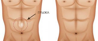
Hiatal hernia
- this is a displacement into the chest cavity through the esophageal opening of the diaphragm of the lower part of the esophagus, part of the stomach, and sometimes intestinal loops. The disease promotes the reflux of acidic stomach contents into the esophagus, so its main symptom is heartburn. A hernia occurs in every twentieth adult, and in every second person over the age of 50.
The cause of a hernia may be a weakening of the ligamentous apparatus. It is present in 5% of the entire adult population and in approximately 50% over the age of 50 years (age-related weakening of the ligamentous apparatus), and is more common in untrained, asthenic people. Another factor that provokes the development of this disease is a significant increase in intra-abdominal pressure due to severe flatulence, pregnancy, trauma or large tumors of the abdominal cavity, attacks of uncontrollable vomiting or persistent cough (for example, in patients with chronic obstructive bronchitis). Dyskenisia (impaired peristalsis) of the digestive tract, in particular the esophagus, which is often observed against the background of chronic inflammatory diseases (ulcerative)
disease of the stomach and duodenum, gastroduodenitis, pancreatitis, cholecystitis), can also lead to the development of a hernia.
Achalasia cardia
is a chronic neuromuscular disease in which there is no reflex opening of the opening at the border of the stomach and esophagus during swallowing. As a result, peristalsis and tone of the esophagus and the passage of food through it are disrupted.
The main symptoms of the disease are difficulty swallowing food (dysphagia), regurgitation and esophageal vomiting and chest pain. Dysphagia initially occurs sporadically due to stress or during hasty eating, then it becomes permanent and prevents the passage of not only solid food, but also liquid food (broth, juice, water) through the esophagus. The inability to swallow food leads to esophageal vomiting - the return of food into the pharynx and mouth when bending over and lying down, especially at night during sleep. This is dangerous, as there is a risk of the contents of the esophagus entering the respiratory tract, which can cause coughing, asthma attacks, and aspiration pneumonia.
Varicose veins of the esophagus
occurs due to disturbances in the outflow of blood from the veins of the esophagus. The veins of the esophagus dilate, twist and lengthen, forming varicose veins; the walls of such vessels become thinner and can rupture, causing bleeding.
The cause of this disorder is most often portal hypertension, that is, an increase in pressure in the portal vein. Its causes may be liver disease (cirrhosis, chronic hepatitis, tumors, tuberculosis, echinococcosis, etc.), thrombosis or compression of the portal vein (tumors, cysts, adhesions, bile duct stones.
Esophageal diverticulum
- This is a sac-like protrusion of its wall. Inflammation of diverticula (diverticulitis) can cause their suppuration, perforation and, consequently, bleeding, stenosis (narrowing of the lumen) of the esophagus, the formation of fistulas and their degeneration into a malignant tumor.
These formations can be congenital or develop as a result of chronic inflammation in the mediastinum, lesions of the lymph nodes, tuberculosis or histoplasmosis - a fungal disease of the lungs. Diverticula of the lower part of the esophagus are frequent complications of reflux esophagitis and achalasia cardia.
Malignant neoplasm of the esophagus is the sixth most common cancer among cancer diseases. It develops most often from the epithelial cells of its mucous membrane (carcinoma), squamous cell carcinoma is less common, adenocarcinoma is rare, and other types of malignant neoplasms are extremely rare.
Since cancer occurs, as a rule, against the background of chronic esophagitis, diseases in which there is a long-term inflammatory process in the esophagus are considered by modern medical science as predisposing to cancer, or precancerous conditions. Such conditions include Barrett's esophagus, hyperplastic esophagitis, etc. Dietary habits contribute to the development of the disease, in particular, the consumption of hot and rough foods, marinades, and alcohol; deficiency of vitamins, especially B2 and A, as well as iron, copper and zinc; bad habits (smoking, alcohol, chewing tobacco). A great risk is associated with the combination of tobacco and alcohol.
Benign tumors of the esophagus include: polyps, papillomas and adenomas (growing from epithelial cells), lipomas and fibroids (growing from fatty and muscle tissues, respectively).
Benign neoplasms are found in all parts of the esophagus. Usually these are single tumors on a wide stalk, having a smooth or tuberous structure. Considering the rather slow growth of such neoplasms, they may not manifest themselves in any way, and when the tumor reaches a large size, signs of obstruction of the esophagus and compression of the mediastinal organs appear. However, most often benign tumors of the esophagus are an incidental finding during esophagogastroduodenoscopy.
Esophagogastroduodenoscopy
allows not only to identify diseases of the esophagus, but also to monitor the treatment process, and sometimes directly treat some diseases of the esophagus. For accurate diagnosis, it is necessary that the procedure be performed by a highly qualified specialist using modern equipment. In our Clinic we use the most modern equipment and invite leading endoscopists to work. In addition, if you wish, you can undergo the procedure of esophagogastroduodenoscopy under general anesthesia without experiencing the slightest discomfort.
Postoperative follow-up
After the operation, 3-4 incisions 5-10 mm long remain on the skin of the abdomen. From the first day, patients begin to get out of bed, drink, and on the second day take liquid warm food. Discharge from the hospital is carried out on 3-4 days, depending on the severity of the disease. The patient can begin work in 2 - 3 weeks. A strict diet should be followed for one and a half to two months, a softer diet should be followed for 2-3 months. Further, as a rule, the patient leads a normal lifestyle - without medications or diet.
In the postoperative period, gastroesophageal reflux often develops, which may require repeated antireflux surgery. Unfortunately, it is extremely rare (due to the presence of many sutures on the esophagus and the danger of their failure) that it is possible to perform antireflux surgery (fundoplication) immediately during tumor removal. During surgery for esophageal leiomyoma, I always perform cardiopexy. In most cases, this intake is enough to prevent the onset of GERD symptoms.
At the request of patients, the clinic can undergo a full examination before surgery to determine the optimal treatment tactics and choose the method of surgical intervention.

