For a long time, acute pancreatitis remains one of the most problematic diseases in emergency abdominal surgery. Today, mortality in necrotizing acute pancreatitis tends to decrease, but is still consistently high throughout the world. Ten years ago, in order to identify the main patterns and the most typical diagnostic and treatment errors, we analyzed the structure of mortality in acute destructive pancreatitis [4]. Then the most significant factors influencing the outcome of this disease were the following:
— early diagnostic errors leading to a delay in the initiation of treatment for pancreatitis;
— lack of uniform tactics for the use of minimally invasive surgical methods, a clear tendency to exceed the indications for their use;
— lack of use of extracorporeal detoxification methods in the early stages;
- high incidence and late diagnosis of pneumonia;
— relatively high incidence of purulent abdominal complications;
— high incidence and lack of prevention of DIC syndrome and hemorrhagic complications in patients.
Over the past decade, the management of patients with acute necrotizing pancreatitis, including surgical aspects, has undergone great changes [7]. The result was not only a decrease in mortality, but also a change in the structure of its causes. This determined the relevance of conducting a new similar study with a comparative analysis of the results and the search for possible ways to further improve the treatment of patients with acute necrotizing pancreatitis.
Introduction
According to WHO, about 1.3 million people die from injuries on the streets and roads every year. In the general structure of mechanical injuries in peacetime, the share of abdominal wounds and injuries accounts for 5%, and in wartime - from 3% to 8% [1, 5, 6, 28, 31]. Isolated pancreatic injuries are rare and cause death in 3-9% of cases [4, 9]. If there are two additional injuries, mortality increases to 12%, with four - up to 40-50% [3, 12, 16, 23]. Shock, reflecting the severity of associated injuries, significantly affects the early mortality rate, which reaches 38% [5, 14, 19, 29].
The most common complications of pancreatic trauma in the late period are pancreatitis, retroperitoneal phlegmon, abscess of the omental bursa, erosive bleeding, sepsis, pancreatic fistula, pseudocyst. Mortality in the development of such complications reaches 22-80% [12, 13, 15, 17, 25, 27].
A distinctive feature of acute pancreatitis of traumatic origin is the high proportion of necrotic (up to 76%) and purulent-necrotic (up to 30%) forms of complications [4, 8, 12, 24]. The rapid progression of pancreatic necrosis and the high prevalence of the destructive process in pancreatic tissue are explained by simultaneous damage to acinar tissue and the microvasculature [2, 7, 11, 21, 26].
Often, traumatic pancreatitis is recognized when pancreatogenic complications have already developed, which is due to the peculiarities of the postoperative period in victims with damage to other abdominal organs, as well as associated trauma [4, 12, 16, 22]. From our point of view, the tactics of surgeons who begin intensive treatment of traumatic pancreatitis from the moment the fact of damage to the pancreas is established are logical.
The main tasks that intensive therapy should solve during the treatment of patients with post-traumatic pancreatitis are a decrease in the secretory function of the pancreas, antibacterial effects, ensuring adequate central and microhemodynamics, and correction of metabolic disorders [5, 7, 8, 11, 15, 20]. Severe trauma to the pancreas and developed post-traumatic pancreatitis lead to depressurization of the organ’s ductal system, which obliges the surgeon to adequately drain the damaged area and retroperitoneal tissue [1, 3, 8, 10, 30, 32].
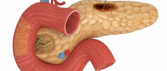
What is pancreatic necrosis?
The pancreas produces so-called pancreatic enzymes, which are normally involved in the digestion of food and the breakdown of proteins. If the pancreas becomes inflamed, the mechanism for protecting its own tissues from the destructive effects of secreted enzymes may be disrupted. The gland begins to deteriorate, which becomes the cause of pancreatic necrosis. We can say that with pancreatic necrosis, the gland gradually digests itself, its individual sections become necrotic, that is, they die. If the disease is not treated in time, areas of dead cells appear on the pancreas. The development of an abscess, which threatens the patient’s life, is possible.
Material and methods
The results of treatment of 139 patients with closed pancreatic trauma were analyzed based on materials from the municipal health care institution “City Clinical Hospital No. 7 of Krasnoyarsk”.
Among the victims, men predominated - 105 (75.5%) people, women were 34 (24.5%), ages ranged from 18 to 65 years, the maximum number of victims - 44.6% - was between the ages of 20-29 years. More than half of the patients were intoxicated at the time of injury. The time of admission to the hospital varied from 2 hours to 5 days from the moment of injury. The main cause of damage to the pancreas was motor vehicle trauma - 56.1% of cases, less common were a direct blow to the stomach - 32.4%, a fall from a height - 11.5%. Isolated pancreatic trauma was diagnosed in 50.5% of victims, associated trauma - in 25.9%, combined - in 23.6%.
There were 29.4% of patients with degree I damage to the pancreas, II - 40.2%, III - 28.1%, and IV degree - 2.3% (classification
D. Smego et al., 1985). Damage localized in the body of the pancreas was detected in 44.6% of victims, in the tail - in 18%, in the head - in 14.4%, in the isthmus - in 5%. Polyfocal damage to the pancreas occurred in 18% of victims.
All patients were divided into two groups.
Group 1 included 95 patients with pancreatic trauma who were treated from 1985 to 1999. The results of treatment were assessed based on a retrospective analysis of patient records. Group 2 included 44 patients who were treated for closed pancreatic injury from 2000 to 2012. In patients of group 2, original methods of diagnosis and treatment were used, and a prospective analysis of the results was carried out. The groups were comparable in age, gender, and duration of pancreatic injury. In victims of group 2, motor vehicle injuries were more common.
The extent of surgical treatment depended on the degree of damage to the pancreas. In case of I degree of organ damage, no intervention was performed on the parenchyma of the gland; the operation was completed with omentobursostomy, flow-through drainage of the omental bursa, and drainage of the abdominal cavity. For stage II damage, various options for hemostasis were used. In the victims of group 1, suturing of the bleeding vessel was used; in some cases, tamponade was used with an omentum on the feeding pedicle or with a gauze swab. In patients of group 2, in addition to suturing the vessel, electrocoagulation and tachocomb application were used. In case of injury to the head or body of the pancreas with damage to the main pancreatic duct, distal resection was performed. In case of IV degree pancreatic injury in combination with injury to the duodenum and biliary tract, pancreaticoduodenal resection was performed.
Specific therapy in the postoperative period involved the creation of functional rest of the pancreas, inhibition of pancreatic enzymes, and prevention of purulent-septic complications.
Patients of group 1 were treated with 5-fluorouracil at a dose of 10 mg/kg body weight to suppress secretory function. To inhibit proteolytic enzymes in the bloodstream, protease inhibitors were used - gordox, contrical intravenously, as a bolus 6-8 times a day. H2 receptor blockers were used as stress-protective therapy.
Victims of the 2nd group, on the 3rd-5th day of hospital stay, received roncoleukin in a dose of 500,000 units intravenously through an infusion pump (over 4-6 hours) three times with an interval of 48 hours. If purulent complications developed, the administration of roncoleukin was repeated for 10 -14th day and 24-28th day of observation. Secretolytic therapy was carried out using a synthetic analogue of somatostatin (Octreotide ZAO Pharm-Sintez), starting from the moment the organ injury was established. In the postoperative period for mild to moderate pancreatitis, the dose of the drug was 300 mcg per day subcutaneously for 5-7 days. In case of severe pancreatitis, the dosage reached 1200 mcg/day, the drug was administered intravenously using an infusion pump at an infusion rate of 50 mcg/h.
General clinical parameters were assessed in all patients. Biochemical studies included determination of the level of protein, urea, creatinine, bilirubin, amylase activity, aspartate and alanine aminotransferase, alkaline phosphatase, glucose-6-phosphate dehydrogenase using standard methods. Instrumental diagnostic methods included ultrasound scanning of the hepatopancreatoduodenal zone, computed tomography, magnetic resonance imaging, pulse oximetry, laser Doppler flowmetry (BLF 21 TransonicSystems Inc.). The frequency and severity of complications encountered and the causes of unfavorable outcome were analyzed.
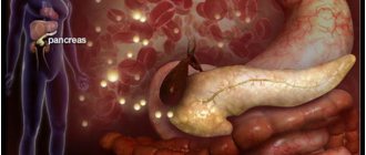
How to recognize pancreatic necrosis?
As a rule, the disease begins acutely: the patient feels sharp pain in the abdominal area, reminiscent of pain during an attack of acute appendicitis. In this case, a person may remember that on the eve of the attack he drank alcohol or ate fatty foods. More than half of patients diagnosed with pancreatic necrosis are admitted to the hospital in a state of acute alcohol intoxication.
Sometimes pain stops bothering the patient. This is considered an unfavorable sign and indicates that necrotic processes have spread to the nerve endings located near the pancreas.
After the onset of pain, the patient begins to vomit severely. One of the main symptoms of pancreatic necrosis (as well as other acute diseases of the pancreas) is that vomiting does not bring relief. Sometimes blood and bile are visible in the vomit. Due to excessive vomiting, symptoms of dehydration may appear.
Also, in patients with pancreatic necrosis, symptoms of intoxication of the body are noticeable: tachycardia, pallor of the skin, rapid breathing. As a result of encephalopathy, the patient may be disorganized or agitated. In approximately 30% of cases with acute pancreatic necrosis, a coma develops.
results
Among the victims of the 1st group there were 30 patients with grade I pancreatic trauma who were operated on through a laparotomy approach. Of the 30 victims, 11 died, the mortality rate was 36.7%: when only drainage of the omental bursa was performed, the mortality rate was higher; when pancreatic hematomas were opened and the organ was abdominized, the mortality rate was lower. 15 (50%) patients in group 1 developed post-necrotic and purulent complications, and 30% required repeat operations.
In 11 patients of the 2nd group with grade I pancreatic trauma, video laparoscopic operations were performed, in 7 patients the omental bursa was drained, in 2 patients the hematomas were opened and the pancreas was abdomenized. Laparoscopic operations were accompanied by fewer complications and lower mortality compared to group 1. The overall incidence of traumatic pancreatitis did not differ from group 1 and was determined mainly by the impact of primary traumatic factors, but the severity of pancreatitis and the number of purulent-necrotic complications in patients operated on with a minimally invasive method were significantly less. No reoperations were required in group 2. The mortality rate in patients of the 2nd group with stage I damage to the pancreas after videoendoscopic operations was 18.2%, which is significantly lower than the mortality rate in the 1st group ( p
<0,01).
In case of damage to the pancreas of the second degree, in victims of the 1st group, after applying hemostatic sutures to the parenchyma of the organ, the mortality rate was 28.6%, after tamponade of the pancreatic wound with an omentum or gauze turundas - 52.6%, after monopolar electrocoagulation - 33.3%. The incidence of post-traumatic pancreatitis was 78.2%; post-necrotic complications developed in 39.1% of victims, purulent peritonitis - in 4.3%, wound infection - in 17.4%.
Analysis of the results of victims of the 1st group showed that suturing the pancreatic tissue is accompanied by additional trauma to the organ, causing disruption of the blood supply and outflow of secretions and, thus, leading to an increase in the area of enzymatic damage. Tamponade of a gland wound increases the risk of infection and sequestration, allowing, on the one hand, the formation of a wound channel, and on the other hand, impeding the outflow of inflammatory exudate.
Videolaparoscopic operations were performed on 10 patients of group 2 with degree II damage to the pancreas. Coagulation hemostasis was used in 20% of patients, with a mortality rate of 50%; In 70% of cases, a tachocomb application was used to stop bleeding; the mortality rate was 14.3%. In all cases, the use of endoscopic application of tachocomb was accompanied by stable hemostasis. The overall mortality rate in patients of group 2 with stage II pancreatic injury reached 20%, which is significantly lower than the same figure in group 1 ( p
<0,05).
We analyzed the treatment results
42 victims with damage to the main pancreatic duct. In both groups, among the procedures performed, the majority of distal resections of the pancreas were performed - 57.9% in group 1 and 82.7% in group 2; in 4 patients of group 2, distal resection was performed with preservation of the spleen and traditional stump processing.
Mortality after distal resection of the pancreas in the 1st group was 36.4%, in the 2nd group after operations with spleen preservation - 25%, after treatment of the stump using tachocomb - 13.3%. Lower mortality rates among victims of group 2 after distal resection were due, in our opinion, to a significant decrease in the number of cases of severe post-traumatic pancreatitis. The use of laser Doppler flowmetry during surgery made it possible to objectively assess the viability of organ tissue and, thus, determine the optimal level of resection and remove the distal part of the gland, preserving tissue with minimally compromised microcirculation. The use of a tachocomb made it possible to reliably seal the pancreatic stump without disturbing the microblood flow in its distal part.
The average number of complications in patients of the 1st group with degrees III and IV damage to the pancreas was 2.3 conventional units, in patients of the 2nd group - 1.0 conventional units. Overall mortality in pancreatic trauma with damage to the pancreatic duct among patients of the 1st group - 52.6%, 2nd - 21.7% ( p
<0.05).
95 victims of group 1 developed 162 complications. Without taking into account the severity of pancreatic injury, early mortality in the group was 7.3%, late - 33.7% and overall - 41%. Traumatic pancreatitis occurred as the most common complication (72.6%). Retroperitoneal phlegmon was diagnosed in 11.6% of victims of group 1, purulent peritonitis - in 7.4%, wound infection - in 25.3%, eventration - in 1%, sepsis - in 15.8% ( see table
).
In patients of group 2, postnecrotic purulent complications occurred significantly less frequently than in patients of group 1 ( see table
). More than half as often in group 2, the course of the postoperative period was complicated by the development of retroperitoneal phlegmon (4.5%), purulent peritonitis (2.3%), wound infection (6.8%), sepsis (4.5%) , false cyst (4.5%). In our opinion, the results obtained are due to the use of a timely (early) pathogenetic approach to the treatment of post-traumatic pancreatitis, using secretolytic and immunocorrective therapy.
The use of minimally invasive surgical technologies in combination with specific drug therapy made it possible to reduce mortality in group 2. Early mortality in group 2 was 11.5% ( p
>0.05), late - 9% (
p
<0.01), general - 20.5% (
p
<0.01).
In conclusion, it should be noted that when choosing a method of surgery in a patient with a closed pancreatic injury, one should strive not for radicalism, but for the adequacy of the operation, and make wider use of minimally invasive surgical technologies and new methods of biological hemostasis. Every victim with a pancreatic injury, regardless of the severity of the organ damage, has a real risk of developing traumatic destructive pancreatitis and post-necrotic purulent complications, which dictates the need to prescribe pathogenetically targeted therapy.
conclusions
1. Operations from the laparotomy approach in patients with stage I damage to the pancreas are accompanied by a high mortality rate - 36.7%; drainage of the omental bursa using video laparoscopic technologies can reduce the rate to 11.1%. The results of surgical treatment of patients with stage II organ damage depend on the method of stopping bleeding from the organ parenchyma. The use of endoscopic application of tachocomb is accompanied by a decrease in the rate to 14.3%.
2. The use of laser Doppler flowmetry makes it possible to determine the optimal level of resection and remove the distal part of the gland, preserving tissue with minimally compromised microcirculation. The use of a new developed technology for the treatment of pancreatic injuries significantly reduces the number of purulent-necrotic complications and reduces mortality from 52.6 to 21.7%.
3. Timely use of secretolytic and immunoactive therapy can reduce the risk of developing severe post-trauma
matic pancreatitis, purulent-necrotic complications and improve the results of treatment of victims with closed pancreatic trauma.