The pancreas is an important organ of the digestive and endocrine system. It produces amylase, lipase and other enzymes for digesting proteins, fats and carbohydrates, as well as the hormones insulin, glucagon and somatostatin.
Under the influence of unfavorable internal and external factors, inflammatory processes occur in the pancreas, and benign and malignant neoplasms develop. Diagnosis of the pancreas allows you to detect pathologies, prescribe adequate treatment and monitor the results of therapy.
Together with our experts, we found out in what cases pancreas diagnostics is required in adults. They also clarified what laboratory and instrumental methods modern medicine uses to diagnose pancreatic diseases.
Why and when to check the pancreas
Disturbances in the functioning of the pancreas lead to digestive disorders, and often to endocrine pathologies, in particular diabetes mellitus. This disease develops when there is a deficiency of insulin, which regulates blood glucose levels. One of the most dangerous diseases of the pancreas is acute pancreatitis. It often causes peritonitis, pancreatic abscess and other serious complications that can be fatal and require urgent medical attention.
Timely diagnosis of pancreatic diseases allows timely treatment to be started and the development of complications to be prevented. Doctors recommend undergoing examination in the following cases:
- with pain in the left hypochondrium and in the upper abdomen or girdle pain, nausea, vomiting, the appearance of “fatty” mushy stools with an unpleasant odor;
- in the presence of diseases of the liver, gall bladder and biliary tract, as well as peptic ulcer of the stomach and duodenum;
- people who smoke and abuse alcohol;
- to monitor the results of therapy for pancreatitis and other pancreatic pathologies;
- if you have close relatives with pancreatic diseases;
- as part of a preventive examination - for men and women. over 35 years old.
The diagnostic examination plan is drawn up by the doctor after examination and history taking. Therefore, the set of methods will differ depending on the health status, age and other characteristics of the patient.
On a note
Weekly menu for those diagnosed with pancreatitis
How are diagnostic tests performed?
Examination of the pancreas, if necessary, can be supplemented with diagnostic tests to identify non-hormonal functions of the organ (exocrine). All methods are divided into:
- those requiring the use of an intestinal tube;
- non-invasive (probeless).
The advantage of tests (especially probeless ones) is convenience for the patient and low cost.
The disadvantage of the tests is that results appear only with a significant decrease in the secretory capacity of the pancreas, so they are considered insensitive
The following tests are used in practice:
- pancreozymin-secretin;
- Lund test;
- hydrochloric acid;
- elastase.
Pancreozymin-secretin test
For the patient on an empty stomach, a probe is inserted into the duodenum with two holes. Aspiration of gastric and duodenal secretions is carried out step by step. Then secretin and pancreozymin are administered intravenously. After the injection, new samples are taken to study the concentration of bicarbonates and trypsin activity. The rate of secretion is calculated.
Pancreatitis is characterized by a decrease in secretion, a decrease in the level of bicarbonates, and an increase in the concentration of enzymes. It is possible to identify false-positive data in patients with diabetes mellitus, biliary tract dysfunction, hepatitis and cirrhosis of the liver.
Lund test
It is distinguished by the use of a standard food mixture as a food irritant to the gland. In the morning, the patient is inserted into the duodenum with a weight attached to the end, through which a food mixture (vegetable oil, milk powder with dextrose) is inserted. Sample aspirates are collected within two hours. Then the level of amylase in them is determined. The option is simpler and cheaper, and does not involve injections.
Hydrochloric acid test
It differs from the previous ones in the type of irritant used - a solution of 0.5% hydrochloric acid in olive oil. Further actions are the same. The disadvantages are general.
Elastase test
Based on the study of elastase in the patient’s stool. Gives a positive result for chronic pancreatitis, liver diseases, gallstones, diabetes.
Methods for diagnosing the pancreas
During the initial examination, the doctor palpates and taps certain points on the abdomen. Normally, the pancreas cannot be felt, and palpation will be painless. For more accurate diagnosis of pancreatic diseases, instrumental and laboratory methods are used.
Instrumental methods for diagnosing the pancreas are used to assess the size and anatomical features of the organ, determine the condition of the ducts, the presence of tumors and stones.
Using laboratory tests, the hormonal and enzymatic activity of the gland is studied. Next, we will talk about the most common research methods.
Ultrasound of the pancreas
Ultrasound examination is the simplest and safest diagnostic method. With its help, you can determine the size, location and structure of the organ, see unevenly dilated ducts, detect neoplasms and the inflammatory process, identify calcification and the presence of stones. Ultrasound is used as part of standard primary diagnosis for suspected pancreatic diseases. To make a more accurate diagnosis, additional studies are prescribed, including CT and MRI of the pancreas.
MRI of the pancreas
Thanks to magnetic resonance imaging, it is possible to determine changes in the density of the gland, calcifications, pseudocysts, tumors, and inflammatory processes. Most often, MRI is prescribed for suspected tumors and injuries to the abdominal organs in order to assess the extent of damage to the gland.
Ultrasound is performed for the primary diagnosis of pancreatic diseases. Photo: Globallookpress
CT pancreas
Computed tomography is used if it is not possible to make an accurate diagnosis using other methods. In addition, this diagnostic method allows one to distinguish from each other such diseases and conditions as acute and chronic pancreatitis, cysts and pseudocysts, malignant and benign tumors. Multislice CT is the method of choice for the primary diagnosis of chronic pancreatitis (its sensitivity and specificity are 75–90%).
Esophagogastroduodenoscopy (EGDS)
EGDS is an endoscopic examination to study the mucous membrane of the stomach and duodenum. With pancreatic pathologies, changes are observed in the mucosa of these organs, and the organs themselves are deformed under the pressure of an enlarged pancreas. The study is carried out using an endoscope, which is inserted through the oral cavity. The image from the camera is transferred to a computer and decrypted.
Endoscopic retrograde cholangiopancreatography (ECPG)
This is an endoscopic method for examining the upper and middle parts of the digestive tract. It is used to determine the condition of the pancreatic ducts, the presence of stones and neoplasms. During the examination, an endoscope - a special tube with a camera - is inserted into the oral cavity. As soon as the endoscope reaches the duodenum, a catheter is inserted into it, through which a radiopaque substance enters. After this, a series of images are taken using X-rays, which give a visual representation of the condition of the structures being studied.
Ultrasound capabilities
Ultrasound examination has found wide application in practical healthcare. It is especially significant with the ability to check the pancreas without any tests when the organ is located deep.
The complexity of ultrasound is due to the individual characteristics of the location and size of the gland, the presence of gases in the intestines. Therefore, in 10% of subjects it is not possible to identify the organ. This is especially true for overweight patients. The method confirms the presence and location of the tumor in 80% of cases, and diagnoses cysts in almost 100% of cases if they are more than 15 mm in size.
Preparing an adult for a pancreatic diagnosis
Preparation depends on the type of diagnostic procedure and the person’s condition. The doctor who prescribes instrumental studies and laboratory tests should tell you about the features of the preparatory stage. But there are still a few general rules of preparation:
- a general and biochemical blood test is taken on an empty stomach, no intravenous drips are given on the day of the examination, and some diagnostic procedures, such as CT with contrast and fibrogastroduodenoscopy;
- before submitting the biomaterial for coprogram, do not take sorbents, laxatives and enzyme preparations, do not use rectal suppositories and enemas;
- before an ultrasound scan of the pancreas, refuse food, water and smoking for 6–8 hours; for 2–3 days before the examination, exclude fried and fatty foods and foods that cause gas formation from the diet;
- MRI is performed on an empty stomach; for 2–3 days before the procedure, do not consume foods that cause bloating; before the examination, remove jewelry and metal parts of clothing;
- before a CT scan with contrast, they are tested for creatinine and urea, do not drink alcohol and gas-forming products for several days, and refuse food and water 8 hours before the procedure;
- on the day of the ECP, they do not eat or drink; the night before, they do a cleansing enema and take sedatives.
Taking some medications may interfere with an accurate diagnosis. Therefore, the patient should tell the attending physician about the medications he takes on a regular basis.
It is important to know
Bacteria or poor diet: what actually causes gastritis?
Clinical blood test
The general blood test will also be slightly modified:
- the level of leukocytes increases (more than 8 * 109/l, which indicates an inflammatory process in the body;
- an increase in ESR (from 15 mm/h and above) indicates the same;
- the number of red blood cells and the level of hemoglobin decreases (observed in the case of hemorrhagic complications of the disease);
- decrease in the level of eosinophils (a subtype of granucytic leukocytes).
Indicators of a general blood test do not indicate a specific disease, but are only general. The results obtained are considered complementary to the biochemical analysis. However, the data obtained helps to assess the general health of the patient.
Popular questions and answers
Diseases of the pancreas are one of the most common pathologies of the digestive system in adults. In addition, there is a tendency for morbidity to increase - for example, the number of patients with acute and chronic pancreatitis has doubled in the world over the past 30 years. However, many diseases can be prevented, while others can be successfully treated at an early stage. Therefore, the issues of timely diagnosis of pancreatic diseases concern many. Our experts tell you how often you need to undergo examinations and which doctor to contact.
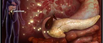
Where is the pancreas located?
Contrary to its name, this organ is not located under the stomach, but behind it on the back wall of the abdominal cavity, next to the kidneys. The pancreas is projected onto the anterior part of the abdominal wall in the left hypochondrium and epigastric region, therefore, during the pathological process, pain and discomfort are observed in this area. The pancreas consists of a tail, body and head and has an average size of 18 to 22 cm.
Which doctor checks the pancreas?
This is usually done by a therapist or gastroenterologist - a specialist in the diagnosis and treatment of the digestive system. Sometimes an examination by another specialist, such as a surgeon, endocrinologist or oncologist, is required. Instrumental examinations of the pancreas are performed by ultrasound diagnosticians, radiologists, and MRI diagnosticians.
How often should you have your pancreas checked?
Patients with pancreatic diseases should undergo examinations 1 or 2 times a year or more often depending on their condition. In addition to undergoing an ultrasound, they need to undergo a general and biochemical blood test with a study of serum amylase, protein and protein fractions, as well as a urine test for alpha-amylase. Healthy people over 35 years of age are recommended to have a screening examination of the pancreas at least once every 2–3 years.
Sources:
- Acute and chronic diseases of the pancreas. Educational and methodological manual for 4-5 year students of medical universities. Department of Faculty and Hospital Surgery. Krasnodar. 2020
- Introduction to ultrasound diagnosis of pancreatic diseases. https://clck.ru/aeyyn
- Recommendations of the Russian Gastroenterological Association for the diagnosis and treatment of chronic pancreatitis. year 2014.
- Outpatient diagnostics and treatment of the biliary system and pancreas. Educational and methodological manual. Belarusian State Medical University. 2005 year. https://rep.bsmu.by/bitstream/handle/BSMU/8715/Zabol_biliarn_sist.pdf?sequence=2&isAllowed=y
Squirrels
When the first symptoms of pancreatitis appear, the protein content changes due to inflammatory processes:
- A sharp increase in reactive protein. It is noted with any inflammation. After stopping the source of inflammation, the indicators decrease.
- Decrease in albumin concentration, as well as total protein. The presented problem arises due to changes in metabolic processes and digestive disorders. Food is not fully broken down due to a lack of enzyme, and this does not allow beneficial proteins to be absorbed into the blood. This factor is especially evident in chronic disease.