AEROPHAGIA
(
aerophagia
; Greek aer air + phagein eat, eat) - swallowing excess air and then regurgitating it.
Swallowing some air is a physiological mechanism by which intragastric pressure is regulated, and belching is usually not observed due to the passage of gases from the stomach to the intestines. However, when the stomach is quickly filled with gas, for example, when drinking carbonated water, beer, or when there is increased formation of CO2 in the stomach after drinking soda, belching is also observed normally.
Increased and systematic swallowing of air and frequent belching are observed either as a result of violation of the rules of eating (fast food, talking while eating), or as a pathological symptom.
Aerophagia can occur with difficulty in nasal breathing, with diseases of the digestive system (diseases of the teeth, oral cavity, excessive salivation with frequent swallowing of saliva, etc.) or is a pathological conditioned reflex (neurosis). In the latter case, swallowing air often occurs outside the eating process.
Aerophagia often develops with disorders of gastric motility and tone, pyloroduodenal stenosis, chronic gastritis, peptic ulcer disease (especially with a high location of the ulcerative lesion). Sources of pathological irritation leading to aerophagia are often cardiovascular failure, aneurysms of the descending aorta, cardiospasm, and narrowing of the esophagus.
Clinical picture
Patients complain of frequent empty and loud odorless belches; sometimes the belching is “multi-story”, and in patients suffering from hysteria, in some cases it is accompanied by a loud scream. Belching is observed both after eating and regardless of food intake; in more severe cases it is almost constant and disappears only during sleep. Patients are concerned about feelings of heaviness and fullness, localized mainly in the epigastric region, but often gastric gases, penetrating through the pylorus into the intestines, lead to bloating of the entire abdomen, in severe cases very significant, which resembles the picture of intestinal obstruction. In some cases, gastrocardial syndrome occurs (see). In this case, extrasystole and unpleasant pain in the heart area occur, leading to angina attacks. These symptoms are observed more often in people suffering from coronary heart disease. Less commonly, with aerophagia, difficulty breathing is observed, sometimes reaching attacks of suffocation - asthma dyspepticum (old name).
Preventing the development of weak sucking reflex
You can avoid weakening or loss of the sucking-swallowing reflex if you adhere to the following recommendations:
- prevention of fetal hypoxia or birth injuries;
- regular observation by a gynecologist throughout pregnancy, timely diagnosis of intrauterine diseases;
- carrying out sanitation of the birth canal before childbirth to avoid infection of the child;
- timely treatment of stomatitis, rhinitis, acute respiratory viral infections in infants that make sucking difficult;
- acquiring the skills of putting a baby to the breast (taking special courses for pregnant women).
If you notice that your child is not suckling well, a pediatric neurologist at the Edkarik clinic in Kaliningrad will help you find out the cause of the baby’s weak sucking reflex. Don’t hesitate - sign up for a consultation by calling the numbers listed on our website.
Diagnosis
The diagnosis of aerophagia is based on medical history and objective examination data. Often, a typical belching occurs during a medical examination, and it is typical that it stops when the patient’s attention is diverted; sometimes the doctor observes preparations for swallowing air: the patient pulls his head forward, presses his chin to his chest and makes empty swallowing movements.
On examination, the abdomen is distended, often more in the upper half. There are no signs of peritoneal irritation. An X-ray examination reveals a high position of the diaphragm, especially the left dome, due to the large air bubble of the stomach and the abundant accumulation of gases in the intestines (mainly in the area of the splenic angle). In connection with severe pneumatosis, a functional gastric cascade is often observed.
How to determine why a baby is not breastfeeding well?
The mother and the neonatologist can detect the weakness of unconditioned reflexes in the maternity hospital. If the child is healthy, the doctor pays attention to the breastfeeding process. Perhaps the reason is due to the fact that the child cannot grasp the nipple with his lips due to the awkward position in the mother’s arms. If the weakening of the sucking reflex is due to flat nipples, the mother can use corrector pads when feeding or perform special exercises that will help stretch the nipples.
In the case where the suspected cause of weakness of the sucking-swallowing reflex is neurological pathology, a number of studies are recommended to detect it:
- neurological examination,
- electroencephalography;
- echoencephalography;
- radiography of the skull bones;
- magnetic resonance and computed tomography of the brain;
- angiography of cerebral vessels.
If infectious and inflammatory diseases are suspected, the baby is prescribed general clinical blood and urine tests. If necessary, children consult with other specialized specialists (endocrinologist, infectious disease specialist, geneticist, neurosurgeon, dentist, ENT).
Treatment
With symptomatic aerophagia, the main attention is paid to the disease that caused its occurrence. With the neurotic nature of aerophagia, psychotherapy is of primary importance. The patient should be explained the essence of the disease. It is necessary to mobilize his will to suppress the pathological conditioned reflex. This is a difficult task, since usually swallowing air occurs involuntarily and unnoticed by the patient. But even here, self-education can lead to certain successes. It is much easier to suppress semi-voluntary belching: patients with aerophagia usually strive to induce it, hoping in this way to get rid of the painful feeling of bloating. The cessation of belching violates the integrity of the pathological conditioned reflex and contributes to its attenuation; at the same time, one of the mechanisms promoting the swallowing of air is eliminated. All types of psychotherapy are acceptable, including hypnotic suggestion. The patient is advised to spit rather than swallow saliva, eat slowly, and avoid talking while eating.
A diet is prescribed to reduce the feeling of heaviness and fullness associated with eating. Drinks containing carbon dioxide and foods that linger in the stomach for a long time are excluded. Food should be taken often, in small portions. In more severe cases, they resort to separate intake of solid and liquid foods. Gastric lavage is sometimes beneficial, probably due to the psychotherapeutic effect of this procedure, especially in cases where a large amount of air is noisily expelled during pumping. Systematic breathing exercises and physical education are advisable. Treatment of the underlying disease that contributed to the development of aerophagia is of great importance. With aerophagia, as well as with other neuroses, the use of all treatment methods aimed at calming and strengthening the nervous system is indicated.
Vomiting and regurgitation are one of the most common problems in the first months of a child’s life. This syndrome can be based on different conditions - both functional and organic. They differ in mechanism and require different treatment approaches.
Definition and mechanisms of development
Vomiting is the involuntary and rapid expulsion of stomach contents as a result of active contraction of the abdominal muscles and diaphragm. Vomiting is a complex reflex act that is carried out with the participation of the vomiting center. This center is localized in the medulla oblongata in the region of the reticular formation. Near it are the respiratory, vasomotor, cough and other autonomic centers, closely connected not only anatomically, but also functionally. As a result, vomiting may be accompanied by reactions caused by irritation of these centers: changes in breathing, circulatory disorders, salivation. Near the vomiting center, outside the blood-brain barrier, is the chemoreceptive trigger zone. Excitation of the vomiting center occurs with an increase in intracranial pressure, under the influence of impulses from the labyrinth, with irritation of reflexogenic zones (pharynx, coronary vessels, peritoneum, mesenteric vessels, bile ducts). Increased pressure in the stomach and various parts of the small and large intestines can also cause vomiting. The chemoreceptive trigger zone reacts directly to the effects of chemicals (drugs, toxins), impulses from there are transmitted to the vomiting center. Sometimes vomiting impulses are combined with painful ones; very severe pain of any origin can cause vomiting.
Afferent impulses reach the vomiting center through the vagus and sympathetic nerves, efferent impulses go along the V, IX, X pairs of cranial nerves to the palate, pharynx, larynx, esophagus, stomach, diaphragm, and through the spinal cord to the striated muscles of the chest and abdominal wall. The neurotransmitters of emetic reactions are dopamine and enkephalins.
Vomiting may be preceded by nausea, accompanied by vegetative-vascular reactions. In children in the first months of life, it can manifest as anxiety, refusal to eat, protruding the tip of the tongue, pushing out the pacifier, pallor, and tachycardia.
Regurgitation differs from vomiting in that with it the food eaten is released effortlessly, without pronounced contractions of the muscles of the abdominal wall. The general condition of the child is not disturbed, the release of gastric contents is less energetic, the child seems to “drain” milk from the mouth immediately or after a short period of time after feeding. Regurgitation is not accompanied by vegetative symptoms and does not affect the child’s behavior, appetite, or mood.
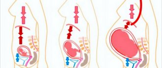
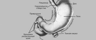
The tendency to regurgitate is one of the characteristic features of newborns and children in the first months of life. This is due to certain structural features of the upper digestive tract at this age. The lower esophageal sphincter (LES) is a physiological rather than anatomical formation and is characterized by a zone of increased pressure extending from the stomach 1–2 cm above the diaphragm. A number of components are involved in the formation of the LES obturator mechanism: muscular, diaphragmatic, vascular, Gubarev valve, angle of His. Regarding LES pressure in newborns, there are conflicting data in the literature: manometry showed that LES pressure after birth is 20–21 mmHg. Art., which is only slightly lower than in adults, however, the esophageal-gastric junction in newborns is located at the level of the legs of the diaphragm, the closure of the cardia is ensured by the Gubarev valve apparatus, the main role is played by the angle of His. In healthy infants, the angle of His is less than or equal to 90°. An increase of more than 90° leads to disruption of the closure of the cardia and causes insufficiency of the gastroesophageal junction. The angle of His is affected by the level of the gas bubble in the stomach, the shape and position of the stomach, and the location of the internal organs.
Esophageal motility is determined by the balance between inhibitory NO-ergic and stimulating cholinergic innervation. Insufficiency of the LES in newborns may be a consequence of the immaturity of the intramural ganglia (especially in premature infants) and impaired innervation as a result of traumatic-hypoxic damage to the brain and spinal cord. An imbalance of gastrointestinal hormones (gastrin, secretin, cholecystokinin, motilin, vasoactive intestinal peptide), as well as an increase in intra-abdominal and intragastric pressure in a number of diseases, is important. If the LES is not closed, the gastroesophageal barrier is disrupted, which leads to gastroesophageal reflux (GER).
Functional regurgitation in children in the first months of life may be based on [1]:
- impaired coordination of swallowing and esophageal peristalsis;
- insufficient peristalsis of the stomach and intestines;
- delayed gastric emptying;
- increased postprandial gastric distension;
- pylorospasm.
In most cases, these mechanisms are combined and are the result of immaturity of both the neurovegetative and intramural and hormonal systems for regulating motor function. As these systems mature, functional vomiting and regurgitation resolve spontaneously, usually during the first six months of life.
On the other hand, regurgitation can be a manifestation of organic diseases such as gastroesophageal reflux disease (GERD).
Under physiological conditions, the pH in the esophagus is neutral or slightly alkaline (6.5–7.5); when acidic stomach contents enter the esophagus, the pH decreases; a decrease in pH in the esophagus below 4 is considered critical, because in this case, pepsin is formed from pepsinogen-1, the most aggressive enzyme of gastric juice. However, in newborns, the pH in the stomach is usually 5.5–6.0, so even in the presence of GER, the pH in the esophagus may remain at subnormal values. Nevertheless, taking into account the predominant production of pepsinogen-2 at this age, an aggressive effect on the mucous membrane of the esophagus can be carried out even at slightly acidic pH values of the refluxate.
There are physiological and pathological GER. Physiological GER occurs as a result of observed and normally spontaneous relaxations (SR), increased pressure in the stomach after eating, has a short duration and is not accompanied by clinical symptoms. Spontaneous relaxations of the LES normally occur with a frequency of 5–6 episodes per hour, at which time the LES relaxes completely, the pressure in it is compared with that in the stomach, the duration of the relaxations is 5–35 seconds. After eating, the frequency and duration of spontaneous relaxations may increase, but at the same time, milk with a neutral pH is thrown from the stomach into the esophagus, which does not cause irritation of the esophagus and can be considered as acceptable physiological GER [3]. Pathological GER causes GERD and is accompanied by clinical symptoms.
It is caused by a decrease in basal pressure in the LES area, more frequent and prolonged episodes of spontaneous relaxation, increased pressure in the stomach due to impaired postprandial accommodation and slower evacuation. With inflammatory changes in the mucous membrane of the esophagus, the amplitude of its contractions and clearance decrease. This creates conditions for longer contact of gastric contents with the mucous membrane of the esophagus. The degree of damage to the latter is stronger, the longer the duration and frequency of GER, the higher the aggressive properties of the refluxate (hydrochloric acid, pepsin, bile) and the lower the protective mechanisms (salivation, mucous barrier, epithelial regeneration, blood supply).
In general, regurgitation and vomiting can be one of the important symptoms of many organic diseases (Table 1).
According to this principle, vomiting and regurgitation can be divided into two groups:
- Primary – when the pathology as the cause is in the gastrointestinal tract (GERD, gastrointestinal defects).
- Secondary – when the cause is outside the digestive tract. These causes can be divided into three groups: 1) infectious, 2) neurogenic, 3) dysmetabolic.
Clinic and diagnostics
From a clinical point of view, it is important to divide the gastrointestinal forms of regurgitation and vomiting into: 1) functional and 2) organic. The latter at an early age are reduced to malformations of the gastrointestinal tract.
Functional regurgitation should include conditions in which the disturbances in motor function underlying regurgitation are not associated with an organic obstacle to the passage of food.
Regurgitation more than once a day is observed in 67% of healthy children under 4 months of age. Many parents consider regurgitation to be a manifestation of pathology; 24% consult a doctor about this in the first 6 months of the child’s life. However, in most healthy children they resolve spontaneously during the first year of life, persisting in only 5% of children 10–12 months. Therefore, in cases of normal development of the child as a whole, in the absence of any other pathological symptoms, regurgitation can be regarded as functional.
According to the Rome III Criteria [2], a diagnosis of functional regurgitation can be made in an otherwise healthy child aged 3 weeks to 12 months if the following features are present:
Regurgitation occurs ≥ 2 times per day for ≥ 3 weeks.
Absence of belching, vomiting with blood, aspiration, apnea, developmental delay, difficulties in swallowing food and feeding, unusual postures.
In cases where these criteria are met, the diagnosis of functional regurgitation is established without additional examination.
GERD manifests itself in three groups of clinical symptoms and signs [4]: 1) regurgitation and eating disorders, 2) signs of inflammation in the esophagus (esophagitis), 3) respiratory disorders (Table 2).
Regurgitation with GERD usually occurs soon after feeding the child in a horizontal position, frequent and not abundant. However, their persistent nature does not fit into the physiological norm; systematic loss of nutrients usually leads to a decrease in weight gain and the formation of malnutrition of I–II degrees.
A characteristic manifestation of GERD is esophagitis. Sometimes it can cause paroxysmal pain and anxiety in the child, unusual “squirming” movements of the neck with the head thrown back (Sandifer syndrome), but is more often detected during endoscopic examination. Changes in the mucous membrane of the esophagus are characterized by hyperemia, fibrinous deposits, erosions and ulcers are less often found. Erosive-ulcerative esophagitis can cause chronic blood loss, sometimes blood in the vomit. As a result of blood loss, persistent iron deficiency anemia may develop. In most children, endoscopic examination does not reveal visual signs of inflammation; however, inflammation can be detected during morphological examination of esophageal biopsies. It should be taken into account that esophagitis may have a different etiology (infections, allergies); in this case, the results of 24-hour pH measurements remain normal, and the esophagus, as a rule, is affected along its entire length. The diagnosis is confirmed morphologically and immunohistochemically [4].
Respiratory manifestations of GERD may include apnea, cough, obstructive airway disease, and aspiration pneumonia.
By acting on the receptors of the middle and upper thirds of the esophagus, GER of acidic contents can lead to reflex laryngospasm, manifested by apnea. Laryngospasm that occurs at the end of the expiratory phase can cause severe hypoxia and sudden death syndrome. Typically, children who have apnea when waking up have GERD.
GER can cause not only apnea, but also reflex bronchospasm, while broncho-obstruction attacks are more often observed at night.
Aspiration of acid or gastric contents may be complicated by pneumonia, and children are often preceded by a persistent cough at night or awakening with laryngospasm. Pneumonia usually has a protracted or recurrent course. More rare extraesophageal manifestations of GERD may include heart rhythm disturbances.
Thus, with the same type of manifestations in the form of persistent regurgitation, GERD differs from functional regurgitation by the presence of additional clinical symptoms and signs. Impaired weight gain, the combination of regurgitation with respiratory symptoms, the appearance of anxiety symptoms such as blood in the vomit, occult blood in the stool, anemia or difficulty swallowing should lead the doctor to evaluate in favor of GERD. The diagnosis of GERD is confirmed endoscopically by detecting signs of esophagitis with a predominant lesion of the lower third of the esophagus. Daily pH-metry allows you to determine the frequency and duration of acid reflux; a decrease in intraesophageal pH below 4.0 for 7% of the time of day or more also confirms the diagnosis of GERD. In young children, GERD is often combined with a sliding hiatal hernia; in these cases, the disease is especially persistent and severe, and can be complicated by bleeding, anemia, and esophageal stenosis. The diagnosis of such a hernia is established endoscopically and radiologically.
Prematurity, developmental delay, congenital anomalies of the oropharyngeal zone, chest, lungs, central nervous system, heart, and other gastrointestinal defects are risk factors for GERD. Regurgitation, combined with vomiting, increasing and persistent, requires the exclusion of congenital anomalies: diaphragmatic hernia, malrotation and anthropopyloric obstruction. To establish a diagnosis, an x-ray examination is necessary.
An allergy to cow's milk protein can also cause frequent regurgitation in children. This is usually combined with symptoms of atopic dermatitis, but can also be an isolated manifestation.
Treatment
For a correct assessment of the clinical situation, a carefully collected history and assessment of the nature of the natural course of regurgitation syndrome are very important. In particular, spontaneous improvement with age is characteristic of functional regurgitation. Therefore, treatment begins with recommendations for care and feeding and therapy is intensified in accordance with the phase of the pathological process (Table 3).
The first task when a diagnosis of functional regurgitation is established is to reassure parents and explain the essence of the symptoms. Effective reassurance involves being sensitive and attentive to parents' expressed and unspoken concerns and fears. The doctor needs to give accurate answers to their questions, such as: what happened to my child, is it dangerous, will it go away, what can we do? He must promise to continue dynamic monitoring and assessment of the child’s condition.
Symptoms of RF can be reduced with some changes in care. More frequent feeding should be recommended, always in a sitting position with the baby in your arms, when his head is elevated by 45–60°. After feeding, you should hold the baby in an upright position. If these measures are insufficiently effective for breastfed children, breast milk thickeners can be recommended. The thickener is diluted in a small amount of expressed milk and given at the beginning of breastfeeding.
When artificially feeding, the child is prescribed mixtures with thickeners. Rice or potato starch (Enfamil AR, NAN AR, Nutrilak AR) or Mediterranean acacia bean gum (Frisov 1 and 2 with prebiotics, Nutrilon AR) are used as thickeners in milk formulas. Mixtures with starch are prescribed in full, since starch has nutritional value; these mixtures are preferable for children with suboptimal weight gain. Gum is a soluble polysaccharide that is not digested by gastrointestinal enzymes. It has a natural, thicker gel-like consistency that thickens further in the stomach and remains the same in the upper small intestine. Mixtures with gum are given in a separate horn with a large hole in the nipple at the end of feeding - after taking the standard mixture, starting from 30.0 ml. If necessary, the dose is increased and selected individually. The Frisov mixture 1 and 2 with prebiotics can be given either separately or in one bottle with the standard mixture.
Reaching the colon unchanged, the gum is hydrolyzed only by the intestinal microflora, thereby exerting a prebiotic effect. As a result of hydrolysis, short-chain fatty acids are formed, which have an additional osmotic effect, softening the stool and promoting freer bowel movements. Frisovo 1 and 2 with prebiotics also contains galacto-oligosaccharides, which have a predominantly bifidogenic effect. The combination of gum and galactooligosaccharides together provides a more effective prebiotic and osmotic effect.
Mixtures with thickeners also differ in protein quality. Some of them are predominantly casein with a casein to whey protein ratio of 80:20. This makes it possible to achieve a denser consistency of the food bolus in the stomach after the casein has curdled. However, from a physiological point of view, the ratio 40:60, as in human milk, is more optimal. This allows you to create a fine suspension in the stomach after curdling, which is evacuated faster.
A meta-analysis of 14 randomized trials of formulas with thickeners for functional regurgitation conducted in Europe confirmed their moderate effectiveness, regardless of the type of thickener [5].
If mixtures with thickeners are insufficiently effective, positional therapy may be recommended. It turned out that positioning the child on the left side, as well as on the back with the head end elevated by 30°, reduces regurgitation. Due to the increased risk of sudden death syndrome, the prone position is not currently used. Medications, incl. prokinetics usually do not reduce symptoms of functional regurgitation, so they are prescribed only if a diagnosis of GERD is established.
Drug therapy for GERD includes effects on upper gastrointestinal motility (prokinetics) and reduction of acid aggression (antisecretors). Prokinetic therapy is aimed at increasing the tone of the cardiac sphincter, propulsive movements of the esophagus and accelerating gastric emptying. For this purpose, dopamine receptor blockers are prescribed: domperidone (Motilium) 1–2 mg/kg/day or metoclopramide (Cerucal) 1 mg/kg/day in 3 doses 20–30 minutes before feeding.
In case of established mild esophagitis (catarrhal), to protect the mucous membrane of the esophagus, gel-like antacids (Gaviscon, Phosphalugel) are prescribed, ½ teaspoon 5-6 times a day after feedings. For erosive-ulcerative forms of esophagitis, the use of antisecretory drugs is indicated: H2-histamine blockers (ranitidine 5–8, famotidine 1–2 mg/kg/day) or proton pump inhibitors (esomeprazole 1 mg/kg/day).
Apnea, attacks of laryngospasm, recurrent pneumonia, as well as stricture and shortening of the esophagus may be indications for surgical treatment (fundoplication), which is used for persistent GER in the absence of effect from conservative therapy. Indications for surgical treatment include both hiatal hernia and neurological pathology against the background of persistent symptoms of GERD.