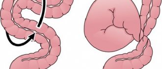
Dolichosigma is a special lining of the sigmoid part of the intestine of the human body. Lengthening of the human sigmoid colon with the formation of its additional loop is a congenital condition, and can be completely asymptomatic and not require any medical correction.
The causes of dolichosigma have not been established, but it is worth knowing that this disease is intrauterine, that is, even before the birth of the child, the formation of its organs occurs incorrectly. After birth, the development of dolichosigma is no longer possible.
If dolichosigma is detected by chance, during an examination for any condition not associated with complaints from the colon, you should not be upset and pay much attention to it. Such a “find” can be treated like a too long nose or protruding ears. This is just a feature of your body.
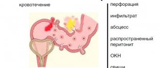
In cases where the patient experiences regular constipation, bloating and abdominal pain, and other causes of such complaints have been excluded, dolichosigma will require treatment.
Symptoms of dolichosigma, which require the help of a proctologist.
Often the disease does not manifest itself symptomatically, and people suffering from this pathology do not even suspect its existence and live a full life. But, in most cases, the disease makes itself felt:
- Constipation. Stagnation of feces can be minor (1-2 days) or serious (several months), causing discomfort. In such cases, patients are recommended to use enemas to cleanse the body mechanically.
- Intoxication process, that is, poisoning that occurs as a result of stagnation of harmful substances in the intestines during constipation.
- Decreased appetite, which occurs due to intoxication (poisoning) of the body.
- Pain in the lower abdomen, which has no specific localization and is aggravated by constipation. The patient feels pain throughout the entire perimeter of the abdomen and cannot show where exactly it hurts.
- Rumbling and bloating are a harmless symptom, which, it turns out, is also considered one of the manifestations of this pathology.
- Increased gas formation, that is, flatulence.
If one or more of these symptoms appears, you must immediately contact a proctologist who will prescribe an examination, consultation and the necessary treatment.
Proctologists at the Mediland clinic will conduct high-quality diagnostics and formulate effective treatment. To diagnose dolichosigma, an examination of the rectum and sigmoid colon will be carried out using digital examination and palpation of the abdomen, and X-rays, ultrasound, colonoscopy and sigmoidoscopy are also used in the diagnosis.
Make an appointment
CHOOSE A PROCTOLOGIST
Treatment methods for dolichosigma at Mediland MC
Conservative treatment of dolichosigma in adults
In cases where the patient experiences regular constipation, bloating and abdominal pain, and other causes of such complaints have been excluded, dolichosigma will require treatment.
If one or more of these symptoms appears, you should immediately contact a proctologist who will prescribe an examination, consultation and the necessary treatment. Treatment of dolichosigma can be conservative and surgical. The essence of conservative treatment of dolichosigma comes down to:
- choosing the right diet;
- normalization of digestive processes in the intestine and its motility;
- elimination of inflammation of the intestinal mucosa and dysbacteriosis, if present.
| To main |
(Radiation diagnostic news 2002 1-2: 32-34)
Ultrasound examination in the diagnosis of small intestinal obstruction.
Kushnerov A.I.
Research Institute of Oncology and Medical Radiology named after. N. N. Alexandrova.
Diagnosis and treatment of acute intestinal obstruction continues to be one of the pressing problems of modern surgery. The clinical picture of intestinal obstruction (severe pain, belching, nausea, vomiting, flatulence, collapse) develops relatively late into a clinical complex that is typical enough for correct recognition. This circumstance in some cases causes a delay in surgical intervention and its unsuccessful outcome. Early surgical intervention based on knowledge of the exact level of obstruction improves the prognosis and results of surgical treatment. No less important is the differential diagnosis of mechanical and dynamic obstruction, which often presents great difficulties.
Recently, ultrasound examination (US) has become increasingly important in the diagnosis of acute intestinal obstruction as a simple, accessible, non-invasive and objective method that is not associated with radiation exposure and has already taken a certain place in the diagnosis of this pathology [1-5]. A number of authors have developed an ultrasound technique using duplex scanning and color Doppler mapping. This technique allows you to determine the presence or absence of blood flow in the intestinal wall in patients with acute small intestinal obstruction and determine the presence of a necrotic area of the small intestine. As a result of determining the hemodynamic parameters of intramural blood flow, values characteristic of simple and strangulation forms of acute small intestinal obstruction are identified [6,7]. However, due to the lack of reliable ultrasound semiotics of intestinal obstruction, further targeted studies of the intestine using the ultrasound method seem to be an urgent problem.
The purpose of this work is to increase the efficiency of ultrasound diagnostics and differential diagnosis of small intestinal obstruction.
Materials and methods.
The results of ultrasound examination of 60 patients with mechanical small intestinal obstruction were analyzed, of which 33 had acute obstruction, 19 had subacute obstruction, and 8 had chronic obstruction. The age of the patients ranged from 10 to 78 years. There were 28 women, 32 men. The causes of acute and subacute small intestinal obstruction were abdominal adhesions in 51, volvulus - 2, strangulated hernia - 3, chronic - Crohn's disease in 4 patients.
We also analyzed the results of ultrasound of 84 patients with functional intestinal obstruction, the causes of which were acute pancreatitis - 7, acute cholecystitis - 20, acute appendicitis - 11, purulent peritonitis - 5, impaired mesenteric circulation - 4, renal colic - 12, blunt abdominal trauma - 4 , as well as as a complication in the early postoperative period after operations on the abdominal organs and retroperitoneal space - 21. The age of the patients ranged from 10 to 78 years.
Ultrasound research technique.
The study was performed with the patient lying on his back using a convex sensor with a frequency of 3.5 and 5 MHz. In some cases, ultrasound was performed in a sitting position, leaning against a wall, or, if the patient’s condition permitted, standing. A polypositional study of all parts of the abdominal cavity was carried out, including a polypositional study and dosed compression of both parenchymal organs and the small, large intestines and stomach. In the early postoperative period, in patients with severe and multiple postoperative scars, as well as in cases of limited contact access, a sector mechanical sensor was used. In addition, a mandatory stage of the study of patients with suspected acute intestinal obstruction, especially in conditions of severe flatulence, was a study along the mid-axillary line towards the central parts of the abdominal cavity.
During the dynamic study of mechanical small intestinal obstruction, attention was paid to the following criteria: 1. The presence or absence of a pathological symptom of an “affected hollow organ” with an analysis of the condition of the above and underlying sections of the intestine. 2. The internal contents of the intestine and its character. 3. Intestinal diameter. 4. Thickness of the intestinal wall. 5. Structure of the intestinal wall. 6. The nature of the folds of the mucous membrane (pronounced, smoothed, absent). 7. The nature of peristalsis. 8. Anatomical areas occupied by dilated intestinal loops and localization of identified changes. 9. Motility of intestinal loops. 10. Change in the diameter of intestinal loops. 11. The level where compression of collapsed intestinal loops by dilated and filled with liquid contents is determined. 12. Presence of viscero-parietal adhesions. 13. Presence of fluid in the interloop space and abdominal cavity. 14. Stomach dysfunction.
Results.
Obstructive small bowel obstruction
was caused by the presence of a mechanical obstacle to the movement of intestinal contents and was observed in 60 patients.
Of the listed criteria, during primary ultrasound, these patients more often observed heterogeneous liquid contents due to intracavitary fluid deposition (45), impaired peristalsis in the form of its intensification and, especially, active antiperistalsis and segmental dilatation of the intestine (49). As the condition worsened, the nature of the liquid contents became more homogeneous, acquired a mushy character, and then, as intestinal obstruction progressed, a decrease in the echogenicity of the contents down to anechoicity was observed. This period was usually combined with a decrease in the intensity of contractile movements of the intestinal wall due to the antiperistaltic pause (12).
In addition, attention was paid to the state of Kerkring's folds: when the obstruction was localized within the jejunum, its folds turned out to be preserved (20), when the obstruction was localized within the ileum, the mucous loops of the latter turned out to be devoid of folds (29). The closer to the site of obstruction, the more pronounced the thickening of the walls and folds was due to edema and fibrin overlay ( Fig. 1–4
).
[Increase]
| Rice. 1. An echogram of a patient with mechanical small intestinal obstruction shows an adhesive process at the level of the proximal ileum. When longitudinally scanning at the level of the jejunum, intraluminal deposition of fluid, expansion of the lumen of the small intestine, and thickening of the folds are noted. | |
[Increase]
| Rice. 2. The same patient. Transverse scanning. | |
The more distal the obstruction was located along the intestinal tube, the more anatomical areas (right and left epi-, meso- and hypogastric regions) in which intestinal loops with signs of intestinal obstruction were visualized during ultrasound. In the case of intestinal obstruction at the level of the ileocecal valve, the dilated and filled with liquid intestinal contents loops of the small intestine filled almost all parts of the abdominal cavity and corresponded in diameter to the large intestine (4 patients). With an average level of small intestinal obstruction, pathologically altered loops were localized in the mesogastric region. In cases of high proximal small bowel obstruction, small bowel loops occupied only the left half of the abdominal cavity (9). In this case, an excess amount of fluid was determined in the lumen of the duodenum and stomach. An increase in the size of the gallbladder indicated the presence of intraintestinal hypertension.
The definition of viscero-parietal adhesions in the abdominal cavity was based on the presence of intestinal loops fixed to the anterior abdominal wall, not displaced relative to it during active respiratory movements, as well as on sharp changes in the diameter of intestinal loops (13). In case of adhesive obstruction with the patient in the lateral position, fixation of the loops of the small intestine and their fusion with each other were detected. Moreover, a thorough examination of the intestinal loops made it possible to identify the difference in intestinal diameters, as well as the place of compression by the expanded loops of the collapsed “tangle” and, as a result, more accurately state the presence of a level of obstructive obstruction ( Fig. 3
).
In addition, immediately at the site of obstruction, antiperistalsis was reduced or absent, and the intestinal walls were significantly thicker than in the proximal sections. However, distended loops of small bowel could cover the site of obstruction (6). [Increase]
| Rice. 3. Echogram of the same patient. The dilated loops of the small intestine compress the collapsed loops of the ileum located distal to the site of obstruction. | |
[Increase]
| Rice. 4. Plain radiograph of the abdominal cavity of the same patient after oral contrast. | |
Dynamic ultrasound monitoring of the patients' condition made it possible to determine the prognosis and effectiveness of the treatment.
Functional intestinal obstruction
during ultrasound was characterized by significant polymorphism. There was an increase in pneumatization and swelling of intestinal loops, a significant decrease until the complete disappearance of intestinal motility, expansion and accumulation of a small amount of fluid mainly in the small intestine, transverse colon and in the right parts of the colon, depending on the cause of intestinal paresis. For example, in acute pancreatitis, a limited accumulation of echo-negative fluid was determined in the transverse colon (7), and in acute appendicitis - in the area of the dome of the cecum (11). In contrast to mechanical obstruction, antiperistaltic movements and fluid transfusion from one loop of intestine to another were not observed. Only pendulum-like movements of the intestine were noted within a limited segment containing fluid.
The most pronounced functional disorders of gastrointestinal motility were observed with diffuse purulent peritonitis (5), impaired mesenteric circulation (4), with renal colic (11) and in the early postoperative period after operations on the abdominal organs and retroperitoneal space (15). In these cases, standard visualization of solid organs was difficult due to interposition of gas in the intestine. Conclusion As our experience has shown, the use of ultrasound in the diagnosis of acute small intestinal obstruction is justified from a tactical point of view, since it does not take too long, does not aggravate the patient’s condition, is sufficiently informative and does not incur radiation exposure. Due to its safety and ease of use, this technique can be used many times during conservative treatment to determine further treatment tactics.
Literature.
- Buyanov V.M., Ishutinov V.D., Doroshev I.A. Ultrasound examination in the diagnosis of mechanical intestinal obstruction // Medical Radiology. - 1993. - N8. — pp. 11–13.
- Kirillov S.V. Ultrasound diagnostics and monitoring of acute intestinal obstruction // Russian Journal of Gastroenterology, Hepatology, Coloproctology. Materials of the 3rd Russian Gastroenterological Week. - 1997. - T.7. — N5. — 258 p.
- Gimondo P. Doppler sonography of hemodynamic changes of the inf. Meseteric artery in inflammatory bowel disease: preliminary data // Am. J. Roentgenol. - 1999. - Vol.173. — P.381–387.
- Ko YT, Lim JH, Lee DH et al. Small Bowel Obstruction: Sonographic Evaluation // Radiology. - 1993. - Vol.188. — P.649–653.
- Kong M., Wang K. A Prune-Induced Small Intestinal Obstruction: Sonographic Appearance // J. Clin. Ultrasound. - 1995. - Vol.23. — N10. — P. 558–560.
- Babkova I.V., Mishukova L.B., Larichev S.E. Ultrasound diagnosis of intramural blood flow disturbances in acute small intestinal obstruction using Dopplerography // Medical visualization. - 2000. - N3. — P.5–9.
- Kuntsevich G.I. Ultrasound diagnostics in abdominal and vascular surgery. M., 1999.