GASTRIC JUICE
- a product of the activity of the gastric glands and the integumentary epithelium of the gastric mucosa.
The main principles of the separation of Zh. were most fully studied by I. P. Pavlov et al. in experiments.
Clean J. s. is a colorless, slightly opalescent, odorless liquid with suspended lumps of mucus. It contains hydrochloric acid, enzymes, minerals, water, special physiologically active substances and mucus. J. s. has an acidic reaction. Its daily amount is approx. 2 l.
One of the most important components of Zh. is a hydrochloric acid. In 1897, I.P. Pavlov discovered the secretion of a solution of hydrochloric acid of constant concentration by the parietal cells of the gastric glands. Various assumptions have been made regarding the mechanism of formation of hydrochloric acid. Most authors accept a certain general scheme of this process, which is as follows. In the membrane of the intracellular tubules of the parietal cells there is a specific carrier of hydrogen ions, which are released from organic substrates, in particular glucose, mainly during metabolic transformations. The carrier, phosphorylated due to ATP hydrolysis and loaded with hydrogen ions, transports them to the outer surface of the membrane and, after additional conversion, releases them into fluids. In this case, ATP serves as a source of energy necessary for the transport of hydrogen ions against the concentration gradient (see Ion transport). Free electrons in the cell are transferred to oxygen using a special mechanism, and ADP is rephosphorylated in the usual metabolic way. A necessary condition for intense secretion of hydrochloric acid is intracellular respiration. In the absence of oxygen, secretion decreases sharply.
The bicarbonate buffer system also takes part in the formation of hydrochloric acid, preventing pH shifts in the secreting cell towards the alkaline side. Bicarbonates are also involved in maintaining ionic balance in the cell. The enzyme carbonic anhydrase (see), present in the parietal cells, ensures the regulation of this process. It has been established that the introduction of a carbonic anhydrase inhibitor completely suppresses the secretion of hydrochloric acid.
The specific mechanisms of these processes are poorly understood. There are two hypotheses that explain them: according to the first, the so-called. According to the redox hypothesis, the secretion of hydrogen ions in the parietal cells is directly related to the transfer of an electron through the Fe++-Fe+++ system to the final acceptor - molecular oxygen. The second hypothesis, the so-called ATPase, explains the transfer of hydrogen ions across the intracellular membrane by a specific ATPase stimulated by bicarbonate. This ATPase acts like a similar enzyme in the sodium-potassium pump, which functions in many cells. It binds hydrogen ions, simultaneously hydrolyzes ATP to ADP, transfers hydrogen to the outside of the membrane, and ADP is reduced to AMP in the mitochondrial respiratory chain. At the same time, it is believed that the intensive transfer of hydrogen ions is not a function of a simple ATPase pump, but includes additional factors that ensure the conjugation of the process of hydrochloric acid secretion and oxidative metabolism in mitochondria.
Composition and properties of gastric juice
Gastric juice is produced by the secretory glands of the gastric mucosa. Pure gastric juice is a colorless transparent liquid. One of the components of gastric juice is hydrochloric acid, so its pH is 1.5-1.8. The concentration of hydrochloric acid in gastric juice is 0.3-0.5%; the pH of the stomach contents after eating can be significantly higher than the pH of pure gastric juice due to its dilution and neutralization by alkaline food components. The composition of gastric juice includes inorganic (ions Na+, K+, Ca2+, Cl-, HCO3-) and organic substances (mucus, metabolic end products, enzymes). Enzymes are produced by the main cells of the gastric glands in an inactive form - in the form of pepsinogens
, which are activated when small peptides are cleaved from them under the influence of hydrochloric acid and are converted into pepsins.
The main proteolytic enzymes of gastric juice include pepsin A, gastrixin, parapepsin (pepsin B). Pepsin A
breaks down to oligopeptides at pH 1.5-2.0.
The optimal pH of the gastricsin
is 3.2-3.5.
It is believed that pepsin A and gastrixin act on various types of proteins, providing 95% of the proteolytic activity of gastric juice. Pepsin B
plays a less important role in gastric digestion and primarily breaks down gelatin. The ability of gastric juice enzymes to break down proteins at different pH values plays an important adaptive role, as it ensures the effective digestion of proteins under conditions of qualitative and quantitative diversity of food entering the stomach.
The composition of gastric juice also includes a small amount of lipase, which breaks down emulsified fats (triglycerides) into fatty acids and diglycerides at neutral and slightly acidic pH values (5.9-7.9). In infants, gastric lipase breaks down more than half of the emulsified fat contained in breast milk. In an adult, gastric lipase activity is low.
The role of hydrochloric acid in digestion
- activates gastric juice pepsinogens, converting them into pepsins;
- creates an acidic environment that is optimal for the action of gastric juice enzymes;
- causes swelling and denaturation of food proteins, which facilitates their digestion;
- has a bactericidal effect;
- regulates the production of gastric juice (when the pH in the antrum of the stomach becomes less than 3.0, the secretion of gastric juice begins to inhibit);
- has a regulatory effect on gastric motility and the process of evacuation of gastric contents into the duodenum (with a decrease in pH in the duodenum, a temporary inhibition of gastric motility is observed).
Ions
Chlorine ions are also actively transported. It is believed that on the basal surface of the parietal cells in the membrane there is a mechanism that acts as a neutral pump, pumping chloride ions from the blood into the cell while simultaneously transferring carbon dioxide ions in the opposite direction. Due to this, chlorine enters the cell against the concentration gradient and further into the body. Water, which is easily permeable to cell membranes, follows actively transported ions due to osmotic relationships. Parietal cells secrete a solution containing approx. 160 mEq/L HCl and 7 mEq/L neutral chloride as KCl. This solution is isotonic with blood. The concentration of hydrochloric acid at the time of formation is usually referred to as primary acidity. Part of the substance, after being released from the glandular cells, is neutralized by alkaline components and mucoproteins of the secretions of other cells, Ch. arr. mucous cells of the cervical parts of the gastric glands and cells of the integumentary epithelium.
The concentration of neutral chloride (chlorine ions in the composition of KCl and NaCl) in the secretions of acid-forming cells is very low; it is much higher in the secretions of mucous and integumentary epithelial cells. Therefore, there is an inverse relationship between neutral chloride and the latter: the higher the acidity of the juice, the lower the content of neutral chloride in it, and vice versa. The content of total chloride (hydrochloride and neutral chloride), on the contrary, is slightly higher in the secretion of parietal cells than in the secretion of cells that do not form acid (100 meq/l). In this regard, the intense secretion of hydrochloric acid is accompanied by a certain increase in the concentration of total chloride in the juice.
To quantify the content in liquids. hydrochloric acid is determined by the total acidity (the total content of all acidic components of the liquid), free (dissociated) and bound (interacting with protein) hydrochloric acid. General acidity of J. s. varies depending on the strength and nature of the stimulus, in the experiment it can reach 0.58% HCl; the content of free HCl is 0.50%, and the pH can be 0.9 (in pure dog fluid). The presence of a bound substance, determined by the difference between general and free, is due to Ch. arr. the presence of substances with a buffering effect of a protein nature, neutralizing part of the hydrochloric acid.
Solyanaya k-ta J. s. plays an important role in digestion. It creates conditions for the autocatalytic activation of proenzymes secreted by the glands and determines the necessary pH of the environment for the action of gastric proteinases on protein substrates. Under its influence, proteins are denatured to a certain extent, which facilitates their hydrolysis by enzymes. Together with proteolytic enzymes, hydrochloric acid imparts the bactericidal properties of gastrointestinal tract, inhibits the release of gastrin (see), and participates in the reflex regulation of the function of the pyloric sphincter. Hydrochloric acid entering the intestines. is the main factor in the release of secretin (see), which stimulates the secretory activity of the pancreas and liver, and is involved in the release of some other hormones.
Functions of gastric mucus
The mucus that is part of the gastric juice, together with HCO3- ions, forms a hydrophobic viscous gel that protects the mucosa from the damaging effects of hydrochloric acid and pepsins. The mucus produced by the glands of the fundus of the stomach includes a special gastromucoprotein, or intrinsic Castle factor,
which is necessary for the complete absorption of vitamin B12. It binds to vitamin B12, which enters the stomach as part of food, protects it from destruction and promotes the absorption of this vitamin in the small intestine. Vitamin B12 is necessary for the normal functioning of hematopoiesis in the red bone marrow, namely for the proper maturation of red blood cell precursor cells.
A lack of vitamin B12 in the internal environment of the body, associated with a violation of its absorption due to a lack of intrinsic Castle factor, is observed when part of the stomach is removed, atrophic gastritis and leads to the development of a serious disease - B12-deficiency anemia.
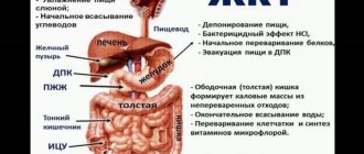
The stomach performs the following functions:
- Depositing . Food remains in the stomach for several hours.
- Secretory. The cells of its mucosa produce gastric juice.
- Motor . It ensures mixing and movement of food masses into the intestines.
- Suction. It absorbs a small amount of water, glucose, amino acids, and alcohols.
- excretory.
Some metabolic products (urea, creatinine and heavy metal salts) are removed into the digestive canal with gastric juice.
- Endocrine or hormonal . The gastric mucosa contains cells that produce gastrointestinal hormones - gastrin, histamine, motilin.
- Protective. The stomach is a barrier to pathogenic microflora, as well as harmful nutrients (vomiting).
Composition and properties of gastric juice: 1.5-2.5 liters of juice are produced per day.
Outside of digestion, only 10-15 ml of juice is released per hour.
Do you have the right breakfast?
A diet for atrophic gastritis with low acidity also does not require strict restrictions. However, you need to know that vegetables and fruits stimulate gastric secretion, and protein foods, which must be digested in the stomach, reduce it.
I will dwell on the peculiarities of nutrition in our case, when appetite may worsen, selectivity in food may appear, chewing and swallowing may be impaired, and forgetfulness may occur—sometimes a person does not remember what and when he ate.
Such patients require high-quality, balanced nutrition to provide the body with all the necessary components: proteins, fats, carbohydrates, minerals and vitamins.
You should eat often, slowly, in small portions, 5-6 times a day, do not overeat, chew well. Food should be warm. Last meal 2-3 hours before bedtime.
As you know, according to the recommendations of nutritionists, 30% of the daily diet should be for breakfast, 50% for lunch and 20% for dinner. But since patients with chronic atrophic gastritis need fractional, frequent meals, the portions should accordingly be small and balanced in composition.
For example, for breakfast, eat some porridge with a spoon of butter, 50 g of cottage cheese, a fresh or baked apple, drink tea or coffee. Such a breakfast can be called correct, because it contains all the components necessary for life - proteins, fats, carbohydrates. And after 3 hours, eat a sandwich with cheese, an egg, some dried fruit, and drink a glass of kefir.
For more information about proper nutrition, see my YouTube “Eat to Live.” Here are videos about proteins, about fats, about carbohydrates.
Quantity, composition and properties of gastric juice
This juice has a neutral reaction and consists of water, mucin and electrolytes. When eating, the amount of juice produced increases to 500-1200 ml. The juice produced in this case is a colorless transparent liquid of a strongly acidic reaction, since it contains 0.5% hydrochloric acid. The pH of digestive juice is 0.9-2.5. It contains 98.5% water and 1.5% solids.
Of these, 1.1% are inorganic substances, and 0.4% are organic. The inorganic part of the dry residue contains cations of potassium, sodium, magnesium and anions of chlorine, phosphoric and sulfuric acids. Organic substances are represented by urea, creatinine, uric acid, enzymes and mucus.
Gastric juice enzymes include peptidases, lipase, and lysozyme.
Pepsins are classified as peptidases. This is a complex of several enzymes that break down proteins.
Hydrochloric acid is formed in parietal cells. Hydrochloric acid dissolved in gastric juice is called free. When combined with proteins, it determines the associated acidity of the juice. All the acidic products in the juice contribute to its overall acidity.
Hydrochloric acid value of juice:
- Activates pepsinogen.
- Creates an optimal reaction environment for the action of pepsins.
- Causes denaturation and loosening of proteins, providing access for pepsins to protein molecules.
- Promotes curdling of milk.
- Has an antibacterial effect.
- Stimulates gastric motility and secretion of gastric glands.
- Promotes the production of gastrointestinal hormones in the duodenum.
Mucus is produced by accessory cells. Some vitamins (groups B and C) accumulate in the mucus
Food coming from the oral cavity is located in the stomach in layers and is not mixed for 1-2 hours.
Therefore, carbohydrate digestion continues in the inner layers under the action of salivary enzymes.
Posted in Uncategorized by admin.
The main cells of the gastric glands synthesize pepsinogen, an inactive precursor of pepsin, which is the main hydrolytic enzyme of gastric juice. The proenzyme synthesized on ribosomes accumulates in the form of zymogen granules and is released into the lumen of the gastric gland by exocytosis. In the stomach cavity, the inhibitory protein complex is cleaved from pepsinogen and the proenzyme is converted into pepsin.
Activation of pepsinogen is triggered by HCl, and subsequently proceeds autocatalytically: pepsin itself activates its proenzyme.
The term pepsin currently refers to a mixture of several proteolytic enzymes. In humans, 6-8 different enzymes have been found that differ immunohistochemically. At an optimal pH value, pepsin hydrolyzes proteins by breaking peptide bonds in the protein molecule formed by groups of phenylamine, tyrosine, tryptophan and other amino acids.
As a result, the protein molecule breaks down into peptones and peptides. Pepsin ensures the hydrolysis of basic protein substances, especially collagen, the main component of connective tissue fibers.
The main pepsins in gastric juice include the following:
- pepsin
A - group of enzymes that hydrolyze proteins at an optimum pH of 1.5-2.0;
- gastrixin (pepsin C),
hydrolyzing proteins at an optimum pH of 3.2-3.5;
— pepsin B (parapepsin)
breaks down gelatin and connective tissue proteins (at pH 5.6 and higher, the proteolytic effect of the enzyme is weakened);
— rennin (pepsin D, chymosin)
breaks down milk casein in the presence of Ca2+ ions.
Gastric juice contains a number of non-proteolytic enzymes.
Among them are gastric lipase,
breaking down fats that are in food in an emulsified state (milk fats) into glycerol and fatty acids at a pH of 5.9-7.9.
In infants, gastric lipase breaks down up to 59% of milk fat. There is little lipase in the gastric juice of adults. Therefore, the bulk of fats are digested in the small intestine.
Cells of the surface epithelium of the gastric mucosa produce lysozyme (muromidase).
Lysozyme determines the bactericidal properties of gastric juice.
Urease
breaks down urea in the stomach at pH 8.0.
The ammonia released during this process neutralizes hydrochloric acid and prevents excess acidity of the chyme entering the duodenum from the stomach.
Enzymes
The main enzymes of Zh. are pepsin (see) and gastrixin. The main cells of the gastric glands produce an inactive precursor of pepsin - pepsinogen, which is a protein with a molecular weight of approx. 42,000, which differs from pepsin in a number of properties, including stability in neutral and slightly alkaline solutions. Pepsinogen and other proenzymes accumulate in glandular cells in the form of secretory granules, which are partially dissolved during secretion. In this case, the proenzymes pass into the lumen of the glandular tubes. A certain amount of proenzymes is secreted not only during digestion, but also outside of it, as well as during periods of hunger, which explains the constant presence of pepsinogen in the gastric contents. Pepsinogen is also found in blood, bile, and urine. Activation of pepsinogen occurs in the acidic environment of gastric juice autocatalytically; in this case, peptide fragments are separated from the pepsinogen molecule, constituting 22% of its nitrogen value. Fragment with a pier weighing 3000, located in the N-terminal position in the pepsinogen molecule, after release it plays the role of a pepsin inhibitor. Maximum pepsin activity occurs in the pH range from 1.5 to 2.5.
Another proteinase J. s. is gastrixin, which is a protein with a mol. weighing 31,500. This enzyme is close in its substrate specificity to pepsin. Gastricin is produced as an inactive precursor, which is activated in the environment of acidic gastric contents. In J. s. it accounts for about a quarter of all proteolytic activity.
The main difference between gastrixin and pepsin is its ability to break down proteins in a less acidic environment: its optimum action is near pH 3.2. This plays a significant physiol role: as a result of the neutralizing effect of food on the hydrochloric acid in the stomach, the pH of the gastric contents in the initial period of digestion may not reach the optimal value for the action of pepsin. In this case, the digestion of proteins by gastricsin, due to the resulting protein breakdown products, can contribute to the release of gastrin in the antrum of the stomach and, therefore, participate in the self-regulation of gastric secretion. Although gastrixin, like pepsin, acts on most food proteins, it differs markedly in its effect on synthetic substrates. In addition, it has a slightly different amino acid composition, a different electrophoretic mobility and is characterized by greater thermal stability and greater stability in a neutral solution.
In J. s. just like in the gastric mucosa, other enzymes close to pepsin are also contained in small quantities. Pepsin B (parapepsin I) was isolated in its pure form, characterized by a pronounced ability to break down gelatin and probably corresponding to the gelatinase enzyme, as well as pepsin C (parapepsin II), apparently corresponding to gastricsin. In addition, another component, somewhat different from pepsin, was found in pepsin preparations and in the contents of the stomach - pepsin D, fiziol, the role of which has not been clarified. In J. s. young ruminants contain a specific enzyme, rennin (see Chymosin), which curdles milk. This enzyme has not been found in humans.
In J. s. lipase is present (see Lipases), capable of breaking down triglycerides in a sharply acidic environment. Although the activity of lipase J. s. small, it apparently has a certain significance in digestion, causing the formation in the stomach of a certain amount of triglyceride breakdown products, which then contribute to the processes of fat emulsification in the intestine.
J. s. contains a significant amount of calcium - up to 5 mg%. It is released as part of mucoid secretions (in particular, in insoluble gastric mucus). Calcium is a secretion product of the integumentary epithelial and mucous cells of the cervical sections of the gastric glands. Its definition in Zh. s. is used as an indicator of the secretory activity of these two types of glandular cells. The presence of calcium in the stomach. just as in saliva, apparently, it has a known significance for enzymatic processes occurring in the intestines: it has been shown that even very small amounts of calcium affect the course of activation of trypsinogen by enterokinase (see), increasing the yield of active trypsin (see) and reducing the proportion of inert protein.
In J. s. Potassium is present in amounts of approx. 20 mg%. Its concentration is higher than the sodium concentration and exceeds the potassium content in the blood serum. Potassium is secreted both by parietal cells and by cells that do not form potassium. Its concentration in liquids. changes little and does not depend on the acidity of the juice.
A permanent component of housing. is ammonia. Ammonia concentration L. s. higher than in blood serum and amounts to 2-8 ml%, increasing as a result of intense activity of the gastric glands. It is possible that it is a product of biochemical processes occurring in the gastric glands. In J. s. Magnesium (sulfate and, in very small quantities, phosphate) was also found.
J. s. contains specific substances that have important physiological significance. First of all, this is the so-called. Castle internal factor (see Castle factors), which is a specific mucoprotein. Studies conducted with 57Co-labeled vitamin B12 have shown that intrinsic factor is concentrated in the parietal cells of the gastric glands, which apparently secrete it. In the stomach, this mucoprotein forms a complex with vitamin B12, due to which the vitamin is protected from destruction in the intestine and becomes able to be actively absorbed in the ileum. In J. s. just as in the gastric mucosa, fiziol was found, an active substance of glycoprotein nature - deli. It is a specific agent that, when introduced into the blood in a relatively small amount, sharply inhibits gastric secretion. The presence in the housing estate has also been established. lysozyme (see).
In addition to the listed compounds, in Zh. A number of biologically active substances have been discovered that have an anticoagulant effect, lipotropic activity and stimulate erythropoiesis. Since the stomach has an excretory function, the composition of the stomach. may include end products of protein metabolism, some vitamins, parenterally administered drugs, etc.
Stomach mucus and its meaning
An important organic component of gastric juice are mucoids produced by mucocytes of the surface epithelium, neck of the fundic and pyloric glands (up to 15 g/l).
Gastromucoprotein (Castle's intrinsic hematopoietic factor, necessary for the absorption of vitamin B12) also belongs to mucoids.
Mucus is represented mainly by two types of substances - glycoproteins and proteoglycans. Mucin is secreted through the apical membrane of the mucocyte, forms a layer of mucus 0.5 - 1.5 mm thick, it envelops the gastric mucosa and prevents the damaging effects of hydrochloric acid and pepsins on the cells of the mucous membrane and irritants received with food.
These same cells simultaneously produce bicarbonate along with mucin. The mucosobicarbonate barrier formed by the interaction of mucin and bicarbonate protects the mucous membrane from autolysis under the influence of hydrochloric acid and pepsins.
Composition and properties of gastric juice. The meaning of its components
1.5 - 2.5 liters of juice are produced per day. Outside of digestion, only 10 - 15 ml of juice is released per hour. This juice has a neutral reaction and consists of water, mucin and electrolytes. When eating, the amount of juice produced increases to 500 - 1200 ml. The juice produced in this case is a colorless transparent liquid of a strongly acidic reaction, since it contains 0.5% hydrochloric acid. The pH of digestive juice is 0.9 - 2.5.
It contains 98.5% water and 1.5% solids. Of these, 1.1% are inorganic substances, and 0.4% are organic. The inorganic part of the dry residue contains cations of potassium, sodium, magnesium and anions of chlorine, phosphoric and sulfuric acids. Organic substances are represented by urea, creatinine, uric acid, enzymes and mucus.
Gastric juice enzymes include peptidases, lipase, and lysozyme.
Pepsins are classified as peptidases. This is a complex of several enzymes that break down proteins. Pepsins hydrolyze peptide bonds in protein molecules with the formation of products of their incomplete cleavage - peptones and polypeptides. Pepsins are synthesized by the chief cells of the mucosa in an inactive form, in the form of pepsinogens. The hydrochloric acid in the juice splits off the protein that inhibits their activity. They become active enzymes. Pepsin A is active at pH = 1.2 - 2.0. Pepsin C, gastrixin at pH = 3.0 - 3.5.
These 2 enzymes break down short chain proteins. Pepsin B, parapepsin is active at pH = 3.0 - 3.5. It breaks down connective tissue proteins. Pepsin D hydrolyzes milk protein casein. Pepsins A, B and D are mainly synthesized in the antrum. Gastricsin is formed in all parts of the stomach. Digestion of proteins occurs most actively in the mucosal layer of mucus, since enzymes and hydrochloric acid are concentrated there.
Gastric lipase breaks down emulsified milk fats. In an adult, its significance is not great.
Herring instead of pickled cucumber
A diet for atrophic gastritis necessarily involves reducing salt intake. Up to a third of a teaspoon per day is acceptable. I advise you to add salt to your food at the table, and cook without salt - foods already contain 2-3 grams of it. Instead of salt, add spices or aromatic oils to your dishes. Limit pickles and marinades. But it is not forbidden to eat a piece of herring - it contains vitamins D and B, selenium, omega-3 fatty acids, which is not found in pickled cucumber.
Try to cook foods less. Steam or boil. Avoid fried foods. Make salads and vinaigrettes from fresh vegetables and fruits.
As you know, water dulls your appetite. Therefore, if you are underweight, drink only when thirst appears, and it is better that the water is warm.
The atmosphere at the dinner table is very important. During a meal, there is no need to watch TV, discuss various problems, be nervous, much less sort things out - this affects your well-being, appetite and absorption of food.
How much gastric juice is secreted per day?
In children, it hydrolyzes up to 50% of milk fat. Lysozyme destroys microorganisms that enter the stomach.
Hydrochloric acid is formed in parietal cells due to the following processes:
1.Transition of bicarbonate anions into the blood in exchange for hydrogen cations.
The process of formation of bicarbonate anions in parietal cells occurs with the participation of carbonic anhydrase. As a result of this exchange, alkalosis occurs at the height of secretion.
2. Due to the active transport of protons into these cells.
3.With the help of active transport of chlorine anions in them.
Hydrochloric acid dissolved in gastric juice is called free. When combined with proteins, it determines the associated acidity of the juice. All the acidic products in the juice contribute to its overall acidity.
Hydrochloric acid value of juice:
1. Activates pepsinogens.
2.Creates an optimal reaction environment for the action of pepsins.
3. Causes denaturation and loosening of proteins, providing access for pepsins to protein molecules.
4.Promotes curdling of milk. Those. formation of insoluble casein from dissolved caseinogen.
5.Has an antibacterial effect.
6. Stimulates gastric motility and secretion of gastric glands.
7. Promotes the production of gastrointestinal hormones in the duodenum.
Mucus is produced by accessory cells.
Mucin forms a membrane tightly adjacent to the mucosa. Thus, it protects its cells from mechanical damage and the digestive action of the juice. Some vitamins (groups B and C) accumulate in mucus, and also contain intrinsic Castle factor. This gastromucoprotein is necessary for the absorption of vitamin B12, which ensures normal erythropoiesis.
Food coming from the oral cavity is located in the stomach in layers and is not mixed for 1 - 2 hours.
Therefore, carbohydrate digestion continues in the inner layers under the action of salivary enzymes.
Quantitative determination of acidity
Quantitative determination of acidity is based on titration of gastric juice with alkali using indicators that change color depending on the pH of the medium. An indicator for general acidity - phenolphthalein remains colorless in an acidic environment, but turns pink in an alkaline environment. The indicator dimethylamidoazobenzene turns bright red in the presence of free hydrochloric acid; in its absence it gives a yellow or orange color. Alizarin sulfonic acid sodium gives a slightly yellow color in an acidic environment, and violet when moving to an alkaline environment; in the presence of this indicator, all acidic valences are titrated, with the exception of the bound hydrochloric acid. The following methods are used in laboratory practice.
Michaelis method.
To 5 ml of filtered gastric juice add 1-2 drops of phenolphthalein and 1-2 drops of dimethylamidoazobenzene. Note the level of alkali in the burette and begin titrating with constant stirring. Then note the alkali level when the initial red color changes to yellowish-pink, the alkali level when a lemon yellow color appears, and the alkali level when the color changes to a permanent pink color. The difference between the second and first levels corresponds to the amount of free hydrochloric acid. The amount of alkali used for titration to a level corresponding to the arithmetic average between the third and fourth levels is equal to the total hydrochloric acid (the sum of free and bound). The amount of bound hydrochloric acid is determined by subtracting the figure of free hydrochloric acid from the figure equal to the total hydrochloric acid. The difference between the total acidity and the sum of free and bound hydrochloric acid is equal to the acid residue (organic acids and acid-reacting phosphates).
Tepfer method.
5 ml of gastric contents are poured into 2 cups. In the first portion, total acidity and free hydrochloric acid are determined using the Michaelis method.
Add 1 drop of sodium alizarin sulfonic acid to the second portion. Titrate until the yellow color changes to faint violet and calculate the amount of alkali used for titration. In the presence of this indicator, all acid-reacting substances are neutralized, with the exception of bound hydrochloric acid, the amount of the cut is determined by subtracting the amount of alkali used to titrate the second portion from the total acidity figure.
By multiplying by 20 all values in both methods, calculations are made in relation to 100 ml of gastric contents. The resulting digital values are expressed in titration units. If, when sodium alizarine sulfonic acid is added to the gastric contents, a violet color immediately appears, a conclusion is drawn about the absence of free and bound hydrochloric acid.
In the presence of a small amount of gastric juice, acidity is determined by titration with three indicators in one portion of microchemicals. method, in the presence of significant impurities in it (blood, bile, food) - by pH-metry. If there is no free hydrochloric acid in the gastric juice, the so-called salt deficiency.
To assess acid secretion, not only the acidity of the juice is determined, but also the absolute value of hydrochloric acid production over a certain period of time - the hydrochloric acid flow rate, which is expressed in milligrams (mg) or milliequivalents (meq) (1 meq corresponds to 36.5 mg HCl) . The flow rate can be calculated for total acidity, free and bound hydrochloric acid, as well as for different phases of secretion (basal, stimulated, etc.). To calculate the amount of acid secretion, special tables, formulas and nomograms have been proposed.
When studying the enzyme-forming function of the stomach in stomach. pepsin is determined by the methods of Pyatnitsky (see Pepsin) and Tugolukov (see Uropepsin). To determine the activity of pepsin and pepsinogen, the Anson-Chernikov method is used (see Anson-Chernikov method).
A certain idea about the excretory function of the stomach is given by the study of chlorides determined by the Volhard method.
To study the protein composition of Zh. the electrophoresis method is used (see). Of practical interest is the determination of gastromucoproteins using the Glass and Boyd method.
High molecular weight substances. determined using electrophoresis on paper. Of the several variants of the method, the simplified version is especially common: J. s. undergoes dialysis first against water, and then against carbovox solution. At the same time, J. s. simultaneously concentrates 20 times. This juice is centrifuged in the presence of a borate buffer, and the supernatant is applied to paper strips. After electrophoresis, the strips are dried at a temperature of 100-110°, stained with a dye (most often amido black), washed with methanol, dried in air and densitometrically. On the anodic side of the electropherogram, 4-5 fractions are detected, of which the most mobile corresponds to pepsin, and a number of others correspond to mucoproteins of the stomach. On the cathode side there are 4-6 fractions corresponding to Ch. arr. products of peptic breakdown of albumin, which passes into juice from the blood.
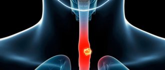
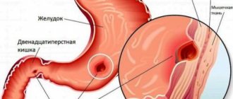
An increase in the acidity of the stomach contents (see Hyperchlorhydria) is associated with increased secretion of stomach acid, insufficient neutralization of hydrochloric acid by the alkaline components of the gastric contents, and a delay in the evacuation of gastric contents into the duodenum. This is most often observed with peptic ulcer disease, especially with duodenal ulcer (see Zollinger-Ellison syndrome).
With hypochlorhydria (see), the level of free hydrochloric acid after using a test breakfast in the gastric contents is below 20 wedge, units (1 ml of 0.1 N NaOH solution per 100 ml of contents); with achlorhydria (see), free hydrochloric acid is completely absent. Under normal conditions, the total acidity of the contents after a test breakfast is on average 65, and the free hydrochloric acid is 40, units in men and slightly lower in women. Reducing the acidity of gastric contents depends on Ch. arr. from a decrease in the concentration of hydrochloric acid in the stomach, which in this case, to a greater extent than normal, is diluted by other components of the stomach contents. Enhanced neutralization of free hydrochloric acid by components of the stomach contents is also possible. In other cases, the gastric glands (parietal cells) may completely lose their ability to secrete hydrochloric acid. In accordance with this, apparent and true achlorhydria are distinguished. With the first, free hydrochloric acid is not detected in the contents of the stomach after a test breakfast, but appears after an injection of histamine, i.e., under the influence of a stronger stimulant that directly acts on the gastric glands. With true achlorhydria, free hydrochloric acid is absent even after histamine injection. The pepsin content in these conditions decreases, as a rule, to a small extent. If it also drops sharply or there is no pepsin in the contents at all, they speak of gastric achylia (see).
Hypo- and achlorhydria are a symptom of the disease, although in some cases they are also observed in practically healthy people. Hypochlorhydria is typical for acute inflammatory diseases of the liver and gallbladder, nutritional disorders, severe vitamin deficiency, both forms of achlorhydria are for chronic diseases, gastritis, stomach cancer, and low acidity in these cases is often accompanied by increased formation of gastric mucus.
True achlorhydria and gastric achylia are observed in pernicious anemia, when not only the formation of acid and enzymes is disrupted, but also the secretion of intrinsic factor Castle, which is responsible for the absorption of vitamin B12.
In certain diseases (hypertrophic gastritis, some forms of stomach cancer), increased transudation of albumin from the blood into the gastric cavity occurs: in the stomach. there are products of albumin cleavage by proteinases or, under conditions of reduced proteolytic activity, unchanged albumin.
See also Stomach, physiology.
Daily amount, composition and properties of gastric juice
Cellular mechanisms of hydrochloric acid secretion. Features of gastric digestion in children.
Gastric juice is a secretion secreted by the glands of the gastric mucosa.
Colorless, slightly opalescent liquid. Density (specific gravity) of gastric juice - 1.006 - 1.009, pH = 1.5-2.0. The daily amount reaches 2 liters.
The gastric juice of a healthy person contains a small amount of mucus and undigested fiber.
When analyzing gastric juice, indicators such as total acidity, the amount of free hydrochloric acid, etc. are necessarily determined.
The gastric secretion consists of two components: the parietal secretion, secreted by the parietal cells and having an acidic reaction, and the non-parietal secretion, secreted by all other cells of the stomach and having an alkaline reaction. The lining secretion contains hydrochloric acid in high concentration.
The latter does not damage the gastric mucosa due to the presence of protective factors (non-lining secretion, mucus and buffering properties of food). The non-plate secretion contains pepsin, gastrixin, mucin, chlorides, bicarbonates, sodium and potassium phosphates. The main source of formation of non-plate secretion is the mucous membrane of the pylorus; pepsinogen (the precursor of pepsin, a protein-digesting enzyme) is produced by the chief cells in the body of the stomach.
The second protein-digesting enzyme is gastrixin. Its proteolytic activity is almost two times higher than that of pepsin. Human stomach glands can produce lipase and possibly other enzymes. In addition, gastro-mucoprotein, or internal Castle factor (see Castle factors), a group of biologically active substances in the blood, is secreted into the stomach.
The cells that produce these substances are still unknown. The regulatory mechanism of gastric secretion is complex and not fully disclosed. The participation in this process of the nervous and endocrine systems, as well as local regulatory mechanisms in the stomach and intestines, has been established.
The synthesis of HCl is associated with the aerobic oxidation of glucose and the formation of ATP, the energy that is used by the active transport system of H+ ions.
Built into the apical membrane is H+/K+ ATPase, which pumps H+ ions out of the cell in exchange for potassium. One theory suggests that the main supplier of hydrogen ions is carbonic acid, which is formed as a result of the hydration of carbon dioxide, a reaction catalyzed by carbonic anhydrase. The carbonic acid anion leaves the cell through the basement membrane in exchange for chloride, which is then excreted through the apical membrane chloride channels.
Function, composition and properties of gastric juice, how it is formed
Another theory considers water as a source of hydrogen (Fig. 7).
It is believed that the parietal cells of the gastric glands are excited in three ways:
the vagus nerve has a direct effect on them through muscarinic cholinergic receptors (M-cholinergic receptors) and indirectly by activating G-cells of the pyloric part of the stomach.
gastrin has a direct effect on them through specific G receptors.
gastrin activates ECL (mast) cells that secrete histamine.
Histamine activates parietal cells through H2 receptors.
Blockade of cholinergic receptors with atropine reduces the secretion of hydrochloric acid. H2-receptor and M-cholinergic receptor blockers are used in the treatment of hyperacid conditions of the stomach.
The hormone secretin inhibits the secretion of hydrochloric acid. Its secretion depends on the pH of the stomach contents: the higher the acidity of the chyme entering the duodenum, the more secretin is released.
Fatty foods stimulate the secretion of cholecystokinin (CC). CA reduces secretion of juices in the stomach and inhibits the activity of parietal cells. Other hormones and peptides also reduce the secretion of hydrochloric acid: glucagon, GIP, VIP, somatostatin, neurotensin.
Digestion in the stomach in children
In a newborn, the cardiac part of the stomach is well developed, the pyloric part is worse. The fundus of the stomach and the pyloric part develop sufficiently only by 10-12 years.
The entrance to the stomach is wide, the cardiac sphincter is poorly developed, but the muscular layer of the pylorus is pronounced, so regurgitation and vomiting are often observed in infants.
The capacity of a newborn's stomach is 40-50 ml, by the end of the first month 120-140 ml, by the end of the first year 300-400 ml.
The gastric mucosa contains the same glands as in adults, but the number of secretory cells is 10-12 times less than in adults, the glands are shorter and wider.
In early infants, the volume of gastric juice is not large, because
the brain phase of gastric secretion is weakly expressed, the receptor apparatus of the stomach is poorly developed, mechanical and chemical effects do not have a pronounced stimulating effect on the secretion of the glands.
The pH of the gastric contents of a newborn baby ranges from slightly alkaline to slightly acidic.
During the first day, the environment in the stomach becomes acidic (pH 4-6). The acidity of gastric juice is created not by HCl (there is a small amount of free HCl in the juice), but by lactic acid.
Activation of proteolytic enzymes is carried out mainly by lactic acid.
In the slightly acidic environment of the stomach of young infants, proteases are inactive, due to this, various immunoglobulins are not hydrolyzed and are absorbed in the intestines in their native state, providing the proper level of immunity.
Pepsinogens are activated by lactic acid. In the stomach of a newborn, 20-30% of incoming proteins are digested.
Under the influence of saliva and gastric juice in the presence of calcium ions, caseinogen protein dissolved in milk, lingering in the stomach, turns into insoluble loose flakes, which are then exposed to proteolytic enzymes.
Gastric lipase breaks down only emulsified milk fats; Breast milk lipase is activated by lipokinase from the baby's gastric juice.
In the slightly acidic environment of the stomach, the amylolytic activity of the baby’s saliva and mother’s milk can persist.
When breastfeeding, gastric juice is less acidic, with less enzymatic activity, than when feeding with cow's milk and nutritional formulas.
When switching to a mixed diet, the pH gradually decreases and reaches adult values only by 7-12 years.
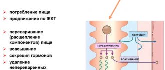
Food from the oral cavity enters the stomach, where it undergoes further chemical and mechanical processing. In addition, the stomach is a food depot. Mechanical processing of food is ensured by the motor activity of the stomach, chemical processing is carried out by enzymes of gastric juice.
Crushed and chemically processed food masses mixed with gastric juice form liquid or semi-liquid chyme.
The stomach performs the following functions: secretory, motor, absorption (these functions will be described below), excretory (secretion of urea, uric acid, creatinine, heavy metal salts, iodine, medicinal substances), endocrine (formation of the hormones gastrin and histamine), homeostatic (regulation pH), participation in hematopoiesis (production of internal factor Castle).
Secretory function of the stomach
The secretory function of the stomach is provided by glands located in its mucous membrane. There are three types of glands: cardiac, fundic (the stomach's own glands) and pyloric (pyloric glands).
The glands consist of main cells, parietal cells, accessory cells and mucocytes. Chief cells produce pepsinogens, parietal cells produce hydrochloric acid, accessory cells and mucocytes produce mucoid secretion. Fundic glands contain all three types of cells. Therefore, the juice of the fundus of the stomach contains enzymes and a lot of hydrochloric acid, and it is this juice that plays a leading role in gastric digestion.
Gastric juice is a complex digestive juice produced by various cells of the gastric mucosa.
The main components of gastric juice
Hydrochloric acid
The parietal cells of the fundic glands of the stomach secrete hydrochloric acid, the most important component of gastric juice.
Its main functions: maintaining a certain level of acidity in the stomach, ensuring the conversion of pepsinogen into pepsin, preventing the penetration of pathogenic bacteria and microbes into the body, promoting the swelling of protein components of food, its hydrolysis, stimulates the production of pancreatic secretions [ source not specified 1389 days
].
Hydrochloric acid produced by parietal cells has a constant concentration: 160 mmol/l (0.3–0.5%).
Bicarbonates
Bicarbonates HCO3− are necessary to neutralize hydrochloric acid at the surface of the mucous membrane of the stomach and duodenum in order to protect the mucosa from the effects of acid.
Produced by superficial accessory (mucoid) cells.
The concentration of bicarbonates in gastric juice is 45 mmol/l.
Pepsinogen and pepsin
Pepsin is the main enzyme that breaks down proteins. There are several isoforms of pepsin, each of which acts on a different class of proteins. Pepsins are obtained from pepsinogens when the latter enter an environment with a certain acidity.
The main cells of the fundic glands are responsible for the production of pepsinogens in the stomach.
Slime
Mucus is the most important factor in protecting the gastric mucosa. The mucus forms an immiscible layer of gel, about 0.6 mm thick, concentrating bicarbonates, which neutralize the acid and thereby protect the mucous membrane from the damaging effects of hydrochloric acid and pepsin. Produced by superficial accessory cells.
Internal factor
Intrinsic factor (Castle factor) is an enzyme that converts the inactive form of vitamin B12, supplied with food, into an active, absorbable form.
Secreted by the parietal cells of the fundic glands of the stomach.
Slime
A constant component of the liquid. is mucus. There are two types of mucus - insoluble and soluble.
Insoluble mucus (insoluble mucin) - a product of the activity of cells of the integumentary epithelium - contains mucoprotein, which, in addition to protein, includes mucoitinsulfuric acid, including acetylated hexosamine, sulfuric and glucuronic acid, a certain amount of sialic acid . After separation by the cells, the mucus binds hydrochloric acid and adsorbs pepsin. mucus also contains leukocytes and desquamated epithelial cells. It covers the entire inner surface of the stomach and is an important factor in protecting the gastric mucosa from the damaging effects of proteinases and hydrochloric acid, as well as chemicals. and mechanical effects of food. To a certain extent, insoluble mucus is digested very slowly by pepsin in the presence of HCl. A certain amount of it and its decay products may be in the liquid. in a dissolved state.
Soluble mucus (soluble mucin) is produced by Ch. arr. mucous cells of the neck of the gastric glands. It accumulates in them in the form of secretory granules, which, during the process of secretion, gradually dissolve in the liquid alkaline secretion secreted by these cells. Soluble mucin is also a mucoprotein containing predominantly mucoitinsulfuric acid in its carbohydrate part. Observations on the secretion of soluble mucin under the influence of various stimuli have shown that it is secreted in parallel with pepsin. In this regard, B.P. Babkin in 1960 suggested that the main cells of the gastric glands may also participate in the formation of soluble mucin, which they secrete either in the form of a complex with pepsin, or in a free state in parallel with the enzyme. The secretion of both types of mucus is regulated by impulses from the vagus nerves.
Some authors suggest that mucus containing pepsin, enveloping dense food particles, promotes the hydrolysis of proteins by the enzyme. Mucus has buffering properties. Thus, 1 g of mucin binds approx. 18 ml 0.1 n. saline solution or 12 ml of a similar alkali solution. J. s. separated under the influence of specific stimuli acting during the intake and digestion of food. In dogs, during hunger, secretion of only some components of the stomach is observed. (without hydrochloric acid), in particular pepsinogen and gastric mucus, the separation of which increases with periodic activity of the digestive tract. It has been established that in a person with a number of diseases, especially with peptic ulcer disease, there can be an abundant continuous secretion of active gastric juice even outside meals, including at night.
J. s. characteristic composition, containing hydrochloric acid, pepsin, mucoprotein and other components, is secreted only by the fundic gastric glands, which are located in the area of the fundus and body of the stomach. The glands of the pyloric and cardiac sections secrete a secretion rich in mucin, including a certain amount of pepsin, but not containing hydrochloric acid. This J. s. has less digestive capacity. There is evidence that pepsinogen, partly entering the blood from the fundic glands, can pass from the blood into the secretion of the pyloric glands.
Composition of J. s. varies depending on the phases of gastric secretion, on the nature of the stimuli prevailing in each given phase. Experiments carried out on dogs have shown that fat secreted in significant quantities upon stimulation of the vagus nerves is characterized by high total acidity, a high content of free hydrochloric acid and total chloride, and is quite rich in soluble and insoluble mucus, as well as calcium, has a very high concentration of pepsin and other proteinases. In contrast, the juice produced during stimulation with gastrin (see) and especially histamine (see), contains very few mucous substances, 2-3 times less calcium, is very poor in enzymes, although it is characterized by a high content of hydrochloric acid and total chloride. The juice in the first and second phases of gastric secretion also has the same features, only less pronounced.
The glands located on the lesser and greater curvature of the stomach secrete glands that are somewhat different in composition. In the activity of the glands of the lesser curvature, the reflex phase is more pronounced, and the juice secreted by them during this phase is slightly more acidic and has a higher pepsin content than the juice produced under the same conditions by the glands of the greater curvature.
In many patol conditions, the composition of the liquid and its acidity change sharply. To detect these changes, the contents of the stomach are examined, which in humans is obtained using a probe (see Probing of the stomach) using test breakfasts (see Test breakfast), injecting histamine (see Histamine test), as well as using probeless methods.
For the qualitative determination of free hydrochloric acid, indicators are used that change color in its presence. Thus, red Congo paper moistened with gastric juice turns blue in the presence of free hydrochloric acid. When adding 1 drop of 0.5% alcohol solution of dimethylamidoazobenzene to 1 ml of gastric juice in the presence of free hydrochloric acid, a red color appears. Yellow and pinkish-yellow coloring indicates the absence of free hydrochloric acid.