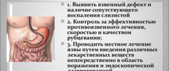
When a person suspects gastrointestinal diseases, proper diagnosis is required.
It helps to understand why a person experiences pain or other unpleasant symptoms, to select the right treatment, and to analyze the success of treatment or the recovery process after surgery.
There are two main diagnostic methods that allow you to check the current condition of the stomach. These include MRI and FGDS.
Which of the following methods should be used in order to obtain as much information as possible that is required for diagnosis? Let's look at each method and answer the question of what is best to use in this case.
FGDS
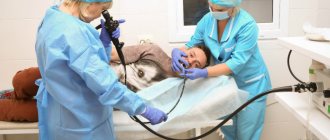
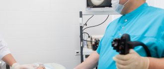
FGDS is one of the most informative methods that should be used specifically to determine the current state of the intestines. During the examination, a special probe with a camera installed on it is used.
When performing an FGDS, it is possible to examine the esophagus, check the condition of the stomach, and also take various types of biological material for testing.
There are many symptoms that indicate that a patient needs to undergo an FGDS. These include heartburn, abdominal pain, nausea, vomiting, belching, severe weight loss for no particular reason.
Typically, FGDS is prescribed by gastroenterologists based on the results of data obtained by other diagnostic methods. These include data such as ultrasound data of the abdominal organs, stool, urine, and blood tests.
The method helps to accurately determine the presence of many types of diseases of the gastrointestinal tract, as well as to find signs of the presence of Helicobacter in the human body.
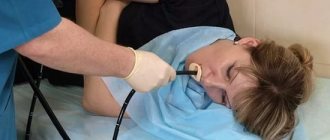
It is worth remembering that many patients try not to use FGDS due to the fact that the procedure itself causes severe discomfort. A long tube is inserted into the mouth - this provokes panic and a strong gag reflex.
There is a possibility of damage to the esophagus, as well as many other potential problems. For this reason, if symptoms permit, many choose an MRI.
Comparison of gastric ultrasound and FGDS: which is better? Pros and cons of both procedures
, only endoscopic and radiological methods were used in medicine to diagnose stomach diseases . Ultrasound diagnostics in gastroenterology was used for other purposes - examination of parenchymal organs (liver, kidneys, pancreas) and biliary tract. But, as experience accumulates and science develops, it has been proven that ultrasound can also be used to study the condition of the gastrointestinal tract, in particular the stomach. In this regard, the question arises: what is better: FGDS or ultrasound? How to choose the right method for examining the stomach? We will try to answer these questions in our article, and also consider:
- what is a stomach ultrasound?
- what is FGDS;
- how they are carried out and how to prepare for them;
- what are the indications and contraindications for these diagnostic procedures;
- what are the advantages and disadvantages of both methods;
- Let's briefly look at alternative research methods.
CONTENT
FGDS (fibrogastroduodenoscopy) is an endoscopic examination of the upper gastrointestinal tract. To better understand the meaning of the abbreviation “FGDS”, you should translate it into Russian: fibra – fiber, gastro – stomach, skopeo – watch.
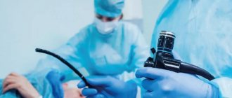
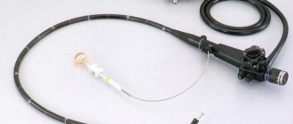
The essence of gastroscopy is of a fibrogastroscope into the digestive tract through the oral cavity . The latter has the form of a tube with a diameter of about 1 cm and is a flexible endoscope equipped with a fiber-optic system. Using this device, you can sequentially examine the esophagus, stomach, and duodenum from the inside.
tissues using ultrasonic waves, which have the ability to reflect from tissue separation boundaries with different acoustic impedance . As a result, an image of an organ (in this case, the stomach) is formed on the monitor screen, the parameters of which are analyzed by a specialist.
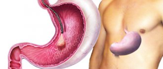
Ultrasound of the stomach (ultrasound examination of the stomach) is an instrumental diagnostic method that allows, using special equipment and an ultrasound sensor, to examine the stomach and, if necessary, nearby structures. This method involves the study of biological
Ultrasound of the stomach and FGDS are completely different diagnostic techniques, however, they have common goals:
- assess the condition of the stomach ;
- its functioning;
- identify the pathological process (if any).
Both methods have good resolution. The main difference between them is the main approach to research.
- FGDS allows the doctor to examine the organ from the inside and makes it possible to perform various diagnostic and therapeutic procedures (for example, take tissue for analysis or stop bleeding).
- Ultrasound visualizes the stomach through the anterior abdominal wall without penetrating the body cavities . With its help, the specialist receives a spatial image of the organ, can examine it in different projections and study the entire thickness of its wall.
When prescribing a particular test, the doctor always takes into account: what information it can provide, whether this information is sufficient to make a diagnosis, and how safe it is for the patient. As a result, the most optimal diagnostic procedure is selected for each patient.
Table 1. Advantages and disadvantages of FGDS and ultrasound of the stomach.
| Advantages | Flaws | |
| FGDS |
|
|
| Ultrasound of the stomach |
|
|
Examination of the stomach using instrumental methods is indicated if there is a suspicion of the presence of a pathological process in this organ, which can manifest itself with the following symptoms:
- pain in the upper abdomen, especially associated with eating;
- heaviness and discomfort after eating;
- nausea;
- single or repeated vomiting;
- belching;
- heartburn;
- bloating.
For patients with the above complaints, both ultrasound and FGDS are indicated. As a result, the following pathological conditions can be identified:
- erosive and ulcerative lesions (peptic ulcer) ;
- gastropathy;
- polyps;
- stomach tumors;
- duodeno-gastric reflux;
- stenosis of the gastric outlet, etc.
How to choose which study is best? First of all , it is more advisable to perform an ultrasound , because it is a safe and non-invasive diagnostic method. And then, if necessary, prescribe an FGDS. also performed if the patient refuses FGDS or if contraindications are identified.
Please note Despite the information content of ultrasound, according to the recommendations of the Ministry of Health, this diagnostic procedure is not included in the list of mandatory ones for stomach pathology.
If it is necessary to collect tissue for morphological examination (biopsy of a tumor or long-term non-healing ulcer, confirmation of the diagnosis of gastritis), an FGDS must be performed . This study is also extremely necessary if gastric bleeding or a foreign body in the stomach is suspected.
Gastroscopy is not used in all patients, even if indicated. For some of them, it cannot be used, as it can be harmful to health. Absolute contraindications to FGDS are:
- impaired swallowing due to diseases of the esophagus (burns, cicatricial strictures);
- acute complicated forms of ischemia of the heart muscle and brain (myocardial infarction, stroke);
- severe cardiac and respiratory failure;
- acute infectious processes;
- violation of the integrity of the digestive tract (perforation of the esophagus, stomach);
- a significant increase in the size of the thyroid gland (grade 3 goiter).
Ultrasound of the stomach can be prescribed to all patients , even pregnant women.
The amount of preparation for FGDS may vary among different patients.
Some of them require a diet, enzymes, and sedatives, while others do not require special preparation. Your doctor will tell you about these features. But in any case, the procedure is carried out only on an empty stomach, so the last meal is allowed 12-14 hours before the intended examination.
Ultrasound is also performed only on an empty stomach; preparation for it lasts 2-3 days and includes:
- a diet excluding products that increase gas formation (milk, legumes, cabbage, black bread, grapes, etc.);
- if you are prone to constipation, bowel cleansing (enema);
- for flatulence - sorbents, enzymes, espumizan.
You can read more about preparing for an ultrasound here, and about preparing for an FGDS here.
Please note: Preparation for an ultrasound scan in a child requires special attention. After all, the younger the baby, the more difficult it is for him to withstand the required period of forced fasting.
Ultrasound of the stomach is carried out in 2 stages:
- in natural conditions;
- after administration of contrast.
First, the patient is placed in a horizontal position (lying on his back); as the study progresses, the body position is changed. The sensor is installed in the projection area of the stomach, after lubricating the skin with a special gel, then it is moved in different directions, assessing the image on the monitor screen. In special cases (if indicated), the procedure is repeated after ingestion of a liquid containing contrast. In special cases, the doctor may supplement the ultrasound with stress.
Ultrasound of the stomach
To perform FGDS, the patient is placed lying on his left side. The endoscope is inserted into the oral cavity through a mouthpiece after preliminary anesthesia of the throat with lidocaine. After which the doctor carefully moves the device deeper into the digestive tract, conducts an examination and performs the necessary manipulations. There is also a transnasal method of administering EGD, you can read about it here.
If gastric ultrasound and endoscopy data are not enough to make a diagnosis or for some reason they cannot be performed on the patient, then gastric fluoroscopy with or without contrast can be used as an alternative method.
If ultrasound and FGDS rather complement each other, then x-ray may be an alternative to both of them. In this article, we take a closer look at the advantages and disadvantages of FGDS and x-rays in the diagnosis of gastrointestinal diseases.
FGDS and ultrasound are two wonderful methods that have long served medicine faithfully. How wonderful would it be if they could be combined? And you will be surprised, but such a procedure exists. And it is called endouzi, you can read about it in our article.
Ultrasound of the stomach and FGDS are studies that do not replace, but complement each other. Although the indications for them are not very different, the information obtained from these diagnostic procedures is very important. And if one method fails to identify the cause of poor health, then another comes to the rescue.
Before finishing the article, I would like to address the audience:
- Have you ever had a case where the conclusion of an FGDS was not enough for a specialist to make a correct diagnosis?
- Have you ever had to examine your stomach using an ultrasound? Was this research able to answer all the doctor’s questions and solve the problem?
Share with the community in the comments!
Advertising
Fecal occult blood test
Blood in the stool can indicate many serious diseases - not only polyps or ulcers, but also cancer (for example, colorectal cancer). Suspicion of these diseases may prompt your doctor to recommend a stool occult blood test. Stool samples are analyzed using a microscope and chemical and bacteriological tests.
Most often, a stool occult blood test is prescribed for suspected diseases of the digestive system. In people over 50 years of age, it is used as a screening test for colorectal cancer. It is also recommended for suspected diseases such as polyps, ulcers, adenomas or hemorrhoids.
Special preparation is required before taking this test. Three days before the examination, you should not eat meat or green vegetables. It is better to replace them with more fiber. You should also avoid taking oral medications such as vitamin C, iron, and aspirin. You should also not use laxatives. They will interfere with analyzing the condition of stool.
The accuracy of the test results depends on the type of test, whether it is the guaiacol gFOBT method, the iFOBT immunochemical method or the porphyrin test. In most cases, stool samples are collected three days in a row. The test is completely non-invasive. The normal fecal occult blood test result is 0.5 to 1.5 ml/day.
Not always gastroscopy. Examination for abdominal pain
I have a stomachache? Which doctor should I go to? What is the best research to determine the cause of this pain? Is there even a “best”?
Especially for readers of our site, we organized a consultation on diagnostic methods for abdominal pain with specialists from the Expert Clinic Kursk.
Gastroenterologist: Soshnikova Natalya Vitalievna
- The stomach can hurt for a variety of reasons. Natalya Vitalievna, tell us how to understand that abdominal pain is probably associated with problems in the gastrointestinal tract?
If the cause lies in problems of the gastrointestinal tract, then, in addition to pain, there may be other symptoms. Among them:
- nausea, vomiting;
- heartburn;
- belching;
- rumbling in the intestines;
- bloating.
These additional signs do not always occur in diseases of the digestive system. For example, during an acute process there may only be pain.
— What specialty should you consult with a doctor if you discover any abnormalities in the gastrointestinal tract?
If there is a gastroenterologist in a medical institution, then go see him. If not, see a surgeon or therapist. According to indications, other specialists can also be involved, since there are many diseases of the digestive system.
— What is included in the complex of patient examinations for suspected gastrointestinal diseases?
Primary screening includes:
— Ultrasound of the abdominal organs (more information about preparing for an ultrasound of the abdominal cavity can be read here);
— fibrogastroduodenoscopy;
- General and biochemical blood test.
Based on their results, the doctor decides whether in-depth studies are needed or not.
Are you afraid to undergo gastroscopy? Read our material on the topic: Where can you find the courage to make up your mind? Gastroscopy – without fear!
— Is there a uniform standard for examining patients with abdominal pain caused by diseases of the gastrointestinal tract or is the diagnostic complex selected individually in each specific case?
The standard includes the studies I mentioned above. If necessary, other examinations may be prescribed in each specific case. For example, stool analysis for occult blood, consultations with related specialists, etc.
CT specialist: Acting Head of the Department of Radiation Diagnostics Strokov Roman Aleksandrovich
— Roman Aleksandrovich, in what situations with abdominal pain can you not do without a computed tomography scan? When is this research method indicated?
In practice, the first will be an ultrasound. If the results obtained with its help are not enough or the cause of the complaints remains unclear, then more complex methods are used - such as computed tomography and magnetic resonance imaging.
Read the material on the topic: What does an MRI of the abdominal cavity show and what is included?
CT most often helps in diagnosing pathologies such as acute calculous cholecystitis, acute pancreatitis, abdominal aortic aneurysm, urolithiasis (it can also cause abdominal pain), chronic intestinal ischemia. In addition, this diagnosis can be prescribed to detect:
- inflammatory bowel diseases (including acute appendicitis);
- complications and outcomes of inflammatory processes of the pancreas;
- some neoplasms of the abdominal organs.
— Is CT an independent diagnosis or, in order for the picture to be complete, does the patient need to undergo other methods of examining the gastrointestinal tract?
It all depends on the specific clinical situation. It happens that a computed tomography scan is sufficient to make a diagnosis. But it is also possible that it - like, in principle, any type of diagnostics - is used to complement another research method.
Increased gas formation in the intestines can reduce the information content of ultrasound examination of the abdominal organs
Ultrasound doctor: Zotolokin Sergey Vladimirovich
— Sergey Vladimirovich, how can ultrasound examination help in identifying the causes of abdominal pain?
Ultrasound is one of the leading methods with which you can answer a number of diagnostic questions. The fundamental difference, for example, from gastroscopy is that we examine “dense” organs (liver, pancreas, spleen and a number of others), and not tubular ones and not containing mainly or only air.
Ultrasound examination can detect signs of inflammatory processes, neoplasms, stones in the gall bladder, kidneys and a number of others.
— Is there an ultrasound examination of the stomach? How informative is it?
It is described in the literature, but in practice it is of little use due to its low information content. If, for example, there is a large tumor in the stomach, we will be able to notice it. For small formations and inflammatory processes, the diagnostic value of ultrasound in relation to the stomach is minimal.
— In what situations will ultrasound not give a clear picture of the state of the gastrointestinal tract?
This is possible, for example, if:
— Ultrasound is used to evaluate organs for which it is not intended due to its limitations as a diagnostic method;
— there is gas in the path of the ultrasonic wave (for example, in the intestines).
Read the material on the topic: You have been prescribed an ultrasound. What do you need to know before the study?
Endoscopist: Kalinina Yulia Nikolaevna
— Yulia Nikolaevna, why do you need gastroscopy? When is it prescribed and what does it reveal?
It is used for early diagnosis of diseases of the upper gastrointestinal tract - esophagus, stomach, duodenum (for example, gastritis, erosive-ulcerative and tumor lesions).
It is prescribed for various symptoms:
- stomach pain;
- belching;
- nausea;
- loss of appetite.
A person can undergo this examination on his own initiative, without a doctor’s referral.
— In what situations is gastroscopy uninformative and cannot help identify the cause of abdominal pain?
This is possible when the cause or source of pain is not the esophagus, stomach, or duodenum, since this method is intended specifically for examining these organs. For other organs, it will not provide the necessary diagnostic information.
— Can capsule endoscopy replace conventional gastroscopy?
No. Firstly, the capsule cannot be controlled in any way while it moves through the digestive tube. In particular, in the stomach, she - purely statistically - may not “see” any pathological formations. Secondly (and this is a fundamental limitation of the method), with capsule endoscopy it is not possible to take a piece of tissue for histological examination.
When applied to the abdominal cavity, MRI has limitations in assessing hollow organs
MRI specialist: Acting Head of the Department of Radiation Diagnostics Strokov Roman Aleksandrovich
— Roman Aleksandrovich, how does magnetic resonance imaging help gastroenterologists diagnose patients with abdominal pain?
MRI is capable of detecting many pathologies of inflammatory, tumor nature, stones in the biliary tract, etc.
But, despite the fact that this method is good at identifying changes in parenchymal, solid organs, it has limitations when assessing the hollow organs of the abdominal cavity.
In many cases, MRI helps resolve questions that remain, for example, after an ultrasound.
— The condition of which organs of the gastrointestinal tract can be assessed by an MRI diagnostic physician during magnetic resonance imaging?
First of all, these are the parenchymal organs of the abdominal cavity (liver, pancreas, spleen), surrounding tissue, lymph nodes, and large vessels. The condition of hollow organs (intestines, stomach) is indirectly assessed: the prerogative in diagnosing their diseases belongs to endoscopic methods.
Acting Head of the Department of Radiation Diagnostics: Strokov Roman Aleksandrovich
— Of the methods we examined for diagnosing the causes of abdominal pain, which is the most informative?
If we talk about the number of situations where the method is the most informative, then this is MRI. However, one must always remember that depending on the organ, the disease and its stage, and the individual characteristics of the clinical situation, different methods may take precedence.
The research method(s) must be chosen by the doctor.
Gastroenterologist: Soshnikova Natalya Vitalievna
— Natalya Vitalievna, as a gastroenterologist, do you only need instrumental methods of examining the stomach to diagnose the patient?
No. Laboratory tests are also important. For example, with their help it is possible to establish the presence of the microorganism Helicobacter pylori (H. pylori), which plays a significant role in the development of a certain form of gastritis, erosive and ulcerative lesions and stomach cancer.
You also need to remember that instrumental diagnostic methods make it possible to evaluate the structure and structural changes of an organ, while laboratory methods make it possible to study its functions.
— Can a patient independently understand the variety of diagnostics of stomach pathologies and choose the method that is suitable for him or should a doctor do this?
The research method(s) should be chosen by the doctor, since the same symptom may indicate different diseases and organs. For example, if the upper half of the abdomen hurts, then the problem may not only be in the stomach. Whether the doctor will prescribe gastroscopy, ultrasound, CT, MRI, etc., or a combination of them, will depend on his diagnostic assumptions.
Other materials on topics:
X-ray of the stomach: an eternal classic or a step back?
What does an MRI of the intestine show?
What to do if your child has a stomach ache?
Our doctors
What is the difference between FGS and FGDS?
At first glance, both methods do not differ, since the examination technique is the same. However, FGD is carried out to study the stomach cavity, and FGDS aims to study the duodenum - duodenum. Therefore, FGS stands for fibrogastroendoscopy, and FGDS stands for fibrogastroduodenoscopy.
So, FGS differs in that the observation camera at the end of the probe is inserted exclusively into the stomach cavity. With its help you can:
- determine the condition of the internal walls of the gasrum (stomach);
- do a biopsy - taking a piece of tissue for examination;
- take a scraping from the walls of the gasrum.
This study is carried out if there is a suspicion of an ulcer, the presence of neoplasms or a disease of bacterial origin.
With FGDS, the probe is inserted a little further into the small intestine to examine the intestinal epithelium. In terms of technique and methodology, the procedure is not fundamentally different, so it is called gastroscopy. The difference between FGS and FGDS is in the time of manipulation: the intestinal examination is carried out after scanning the stomach, that is, the procedure takes place in two stages.
Methodology
Gastroduodenoscopy is performed on an outpatient basis or in a hospital, including in the intensive care unit, when the study is prescribed for the purpose of hemostasis and for other emergency indications. The patient lies on the couch on his left side. After local anesthesia, a plastic ring is placed between the patient’s teeth. The specialist inserts the endoscopic tube under visual control along the anatomical axis of the oropharynx. When the endoscope is inserted into the esophagus, air (and sometimes water) is insufflated, as a result of which it becomes possible to pass the first bottleneck of the esophagus in its proximal part. After reaching the gastroesophageal sphincter, the subject is asked to inhale while insufflating air.
Once in the stomach cavity, the endoscopic tube, under the control of a physician, passes along the lesser curvature, after which the distal section of the endoscope bends, and the specialist is able to examine the area of the greater curvature. If indicated, the study includes a visual examination of the duodenal bulb and post-bulb section. To conduct endoscopy with examination of the entire length of the intestine, it is necessary to use a special duodenoscope with lateral optics. Also in this case, special preparation of the patient in the form of premedication is required, since the passage of an endoscopic tube from the stomach into the duodenum can be painful even in the absence of stenosis and other pathologies of the pylorus.
The duration of gastroduodenoscopy depends on the purpose of the study and is partly determined by the results obtained during the examination. The minimum time allotted for the procedure is about 2-5 minutes. If a biopsy sample is required for further histological examination, the duration of endoscopy increases.
Endoscopic examinations during sleep without pain are a common practice for modern medical institutions. The NIIEKM clinic has created all the conditions for high-quality, safe diagnostics and a comfortable stay for patients.
| Esophagogastroduodenoscopy (abbreviated as GASTROSCOPY or EGDS) | 2500 ₽ |
| Urease test for Helicobacter Pylori | 500 ₽ |
| Esophagogastroduodenoscopy under anesthesia with the provision of a bed, without taking into account the Helicobacter Pylori urease test | 6500 ₽ |
For detailed information, call the toll-free number 8 (800) 222-60-86.
Contraindications
Contraindications to gastroscopy are divided into absolute and relative. The study is not possible in severe general conditions, in particular those caused by myocardial infarction and acute cerebrovascular accidents. The procedure is not performed in cases of severe cardiovascular and pulmonary insufficiency. The technique is not used for hemophilia due to the high risk of bleeding. Relative contraindications to endoscopy include acute inflammatory diseases of the tonsils, pharynx, mediastinum and other nearby organs due to the risk of spreading infection (except when the study is performed for health reasons). Esophagogastroduodenoscopy is limited in patients suffering from epilepsy and mental disorders. In this case, the decision is made individually; the use of general anesthesia is mandatory.
Gastroscopy results
The test result is ready 20-30 minutes after diagnosis. The result of the morphological study is available in 5-7 days.
At Best Clinic, gastroscopy is performed by experienced endoscopists using expert-class equipment, which has good resolution. Thin and flexible endoscopes and the use of intravenous sedation make the diagnostic process comfortable and painless.
Take care of the health of your digestive system - sign up for an endoscopic examination at a convenient time!
Indications
Gastroscopy (EGD) is performed for both diagnostic and therapeutic purposes. The study is carried out to identify and confirm a number of diseases of the esophagus, stomach, duodenum and some general somatic pathologies. In particular, we are talking about inflammatory processes (esophagitis, gastritis, bulbitis, etc.) of toxic, infectious and other origins. Esophagogastroduodenoscopy is one of the main methods for diagnosing gastric and duodenal ulcers. With this diagnosis, the procedure is also prescribed for therapeutic purposes; gastric endoscopy is used to administer various drugs that promote ulcer healing.
Another indication for endoscopy is tumor formations of the esophagus, stomach and duodenum. These can be benign processes (polyps) and malignant neoplasia. It is on the conclusion of the biopsy performed during the study that further tactics of patient management are based. Esophagogastroduodenoscopy allows you to visualize specific changes in the mucous membrane of the duodenum and collect material for histological examination in Whipple's disease, Crohn's disease and some other pathologies. Endoscopy makes it possible to reliably confirm the presence of esophageal or gastrointestinal bleeding. During the procedure, mechanical and chemical methods are used to stop bleeding, and hemostatic agents are administered.
Hematemesis and melena are observed in many nosologies, therefore esophagogastroduodenoscopy is performed for the purpose of differential diagnosis of diseases of the esophagus, stomach and other parts of the gastrointestinal tract that can provoke bleeding. In addition, endoscopy is used in some cases to identify the cause of intestinal obstruction (duodenal obstruction of various natures). The study is widely used for surgical removal of polyps, precancerous formations, bougienage in the presence of stenoses, removal of foreign bodies, etc. Esophagogastroduodenoscopy is also prescribed in the preparation of patients for surgical treatment of a number of diseases.