According to the anatomical structure and functional purpose, the stomach is divided into 3 parts:
- upper - connects to the esophagus, is called “cardial”, contains a dome or bottom, a raised formation;
- middle - body;
- the lower - pyloric, located on the border with the duodenum, in turn, is divided into the antrum and the pyloric canal, which ends with the muscular sphincter.
The antrum accounts for up to 30% of the volume of the stomach. It is impossible to visually determine exactly where the antrum is located, since the border is very arbitrary. Based on the histological picture of the epithelial layer, there is a better chance of establishing that the tissue belongs to a specific part of the organ.
The antrum of the stomach is involved in the general functions of the organ, but also has its own characteristics. Their violation causes various diseases. Therefore, it is worth dwelling on the specifics of the antrum’s work.
Physiological “duties” of the antrum
All functions of the antrum of the stomach are associated with the digestion process. Here's what happens:
- crushing food particles to 2 mm or less with simultaneous mixing, the result should be a homogeneous mass without separation of pieces;
- pushing the formed lump towards the pylorus and duodenum;
- preparation for further digestion in the intestine means a decrease in acidity, which was provided by the body of the stomach, because there must be an alkaline reaction in the small intestine, the maximum concentration of alkaline mucus is produced in the pyloric area;
- to eliminate hydrochloric acid in the cells of the mucous membrane there is a hormonal substance - gastrin, it is also called the “informant hormone”, since the action is associated with the transmission of an impulse to higher centers about the appearance of food;
- the production of serotonin allows for reliable evacuation of the food bolus by stimulating the muscular apparatus of the stomach;
- synthesis of somatostatin, which, if necessary, can suppress the secretion of enzymes.
What causes antrum diseases?
All variants of pathology of the antrum are united by a single most common cause - the presence of a special pathogen Helicobacter pylori or Helicobacter. The fact is that the antrum is the favorite location of this microorganism.
Human infection occurs through the mouth. And, once in the stomach, in the pyloric part the pathogen finds the most convenient conditions for life. It tolerates the acidity of gastric juice well. It independently neutralizes it with the help of enzymes that release ammonia. Actively reproducing.
Thanks to the presence of antennae, the bacterium is able to move in the gel-like environment of mucus.
Excessive alkalization is considered a mechanism that triggers pathological changes in the antrum area, which subsequently leads to stomach diseases.
In addition to Helicobacter, the following risk factors are involved in the pathology of the antrum:
- alcohol abuse;
- smoking;
- long-term use of irritating drugs (from the Aspirin group, non-steroidal anti-inflammatory drugs, against headaches, for the treatment of tuberculosis);
- commitment to violating the rules of nutrition (passion for spicy seasonings, fried and smoked foods, very hot or cold foods, fast food, disproportionately long breaks in eating);
- stressful conditions in the family, at work, contributing to the emergence of neuroses;
- general vascular changes with severe atherosclerosis, arteritis, diathesis, disrupting the nutrition of the stomach wall;
- infection with parasites (helminthiasis, amoebiasis, giardiasis);
- allergic reactions to food;
- exposure to viruses (HIV, cytomegalovirus);
- decreased immunity caused by various reasons;
- hereditary genetic predisposition.
There was a connection between the frequency of gastric damage and diseases of the endocrine organs, bronchi, lungs and heart, iron deficiency, diseases of the urinary system, as well as the presence of chronic foci of infection (tonsillitis, sinusitis, caries, adnexitis in women and others).
Chronic pathology is accompanied by suppression of the body's defenses. In combination with Helicobacter, these factors cause gastric damage of varying extent and depth. Let's consider the most common diseases with features of symptoms and treatment.
Diagnosis is based on identified morphological changes in tissues and endoscopic examination
Antral gastritis
The morphology of the inflammatory response includes step-by-step processes:
- infiltration of the antrum mucosa with lymphocytes, neutrophils, macrophages, plasma cells;
- formation of follicles from lymphoid tissue (lymphoid hyperplasia);
- destruction of the epithelium in the form of individual foci (focal gastritis) or massive areas of damage.
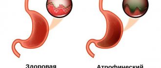
Antral gastritis is mainly a chronic disease. Unlike gastritis, the body of the stomach is rarely acute. It begins against a background of high acidity. The production of hydrochloric acid by parietal cells is stimulated by Helicobacter.
Gradually, the functions of the epithelium are depleted, and atrophy processes begin. This means replacing epithelial cells with non-functioning fibrous cells. Another option is the transformation of the gastric epithelium into the intestinal epithelium, which is atypical in location. The process is dangerous due to degeneration into a cancerous tumor.
Depending on the violation of secretion, there are:
- atrophic gastritis - accompanied by a gradual loss of the gastric mucosa's ability to synthesize acid, hormonal substances, mucus, death of the epithelium, thinning of the stomach wall, is considered a precancerous disease;
- hyperplastic - characterized by the formation of large folds, cysts, small polyps, and activation of the process of cell proliferation.
The type of antral gastritis depends on the depth of the lesion. Superficial is considered the most favorable form of the course; changes affect only the superficial layer of the mucosa and are not accompanied by the formation of scars or pronounced disorders of secretory function.
Fibrogastroscopy reveals hyperemic and edematous mucosa, and pinpoint hemorrhages are possible.
Superficial inflammation reaches the muscles, but does not touch them
Erosive gastritis - the inflammatory reaction goes deep into the stomach wall. As a result, surface erosions and cracks are formed first. Chronic erosion without treatment leads to the formation of ulcers. If the outcome is favorable, a scar appears at the site of inflammation.
Symptoms of antral gastritis with superficial damage may not bother a person or may occur after overeating or drinking alcohol. Other forms are more persistent. Most often patients are concerned about:
- pain of varying intensity immediately after eating or on an empty stomach;
- heartburn and belching;
- taste in mouth;
- smell when breathing;
- bloating;
- bowel dysfunction (diarrhea or constipation).
With massive damage, manifestations of general intoxication are possible: nausea and vomiting, weakness, loss of appetite, weight loss.
The appearance of blood in stool and vomit indicates an erosive form of gastritis. The addition of anemia is accompanied by increased weakness, headaches, and pallor. Persistent symptoms that respond poorly to treatment should cause alarm due to the transformation of gastritis into a peptic ulcer, tumor, inflammation of the pancreas, and bulbitis of the head of the duodenum.
Stomach
Stomach
(lat.
ventriculus
) - a section of the digestive tract following the esophagus and preceding the duodenum. Upon examination, the position, size, and shape of the stomach depend on the position of the patient, the filling of the stomach, as well as on the condition of the surrounding organs - the liver, spleen, and intestines. The stomach, 5/6 of its size, lies to the left of the midline and only the pyloric part lies to the right.
The upper part of the stomach, being a continuation of the esophagus, is tightly fixed to the diaphragm by connective tissue cords. The entrance to the stomach (cardia) is located 3 cm from the place of attachment of the VII left costal cartilage to the sternum or at the level of the X-XI thoracic vertebra in the back. The highest point of the gastric vault lies on the fifth rib on the left along the parasternal line. The greater curvature, as the most mobile part of the stomach, is located more in front, adjacent, together with part of the anterior surface of the stomach, to the anterior abdominal wall. On the left, the upper part of the greater curvature touches the spleen, and the lower part touches the transverse colon.
The level of the lower edge of the greater curvature is very variable and depends on the type of constitution, gender, position of the patient (horizontal, vertical), the size of the abdomen, the tone and filling of the stomach. In women it is 1-2 cm lower than in men. In a horizontal position of the patient with an average filling of the stomach, it is located 2-3 cm above the navel in men, at the level of the navel in women, and when the stomach is full, the level drops lower. In the vertical position of the subject, the lower edge of the stomach in men is 3-4 cm, in women - 2-3 cm above the iliac line. The swollen and overcrowded transverse colon pushes the greater curvature from the anterior abdominal wall backward and upward. The outlet of the stomach is located at the level of the first lumbar vertebra, 1-2 cm to the right of the midline (Ionov A.Yu. et al.).
The normal residence time of the contents (digested food) in the stomach is about 1 hour.
Anatomy of the stomach
Anatomically, the stomach is divided into four parts:
- cardiac
(lat.
pars cardiaca
), adjacent to the esophagus; - pyloric
or pyloric (lat.
pars pylorica
), adjacent to the duodenum; - body of the stomach
(lat.
corpus ventriculi
), located between the cardiac and pyloric parts; - fundus of the stomach
(lat.
fundus ventriculi
), located above and to the left of the cardiac part.
In the pyloric section there is a pyloric cave
(lat.
antrum pyloricum
), synonyms
antrum
or
anturm
and
pyloric
(lat.
canalis pyloricus
).
The figure on the right shows: 1. Body of the stomach. 2. Fundus of the stomach. 3. Anterior wall of the stomach. 4. Greater curvature. 5. Small curvature. 6. Lower esophageal sphincter (cardia). 9. Pyloric sphincter. 10. Antrum. 11. Pyloric canal. 12. Corner cut. 13. A groove formed during digestion between the longitudinal folds of the mucosa along the lesser curvature. 14. Folds of the mucous membrane.
The following anatomical structures are also distinguished in the stomach:
- anterior wall of the stomach
(lat.
paries anterior
); - posterior wall of the stomach
(lat.
paries posterior
); - lesser curvature of the stomach
(lat.
curvatura ventriculi minor
); - greater curvature of the stomach
(lat.
curvatura ventriculi major
).
The stomach is separated from the esophagus by the lower esophageal sphincter and from the duodenum by the pyloric sphincter.
The shape of the stomach depends on the position of the body, the fullness of food, and the functional state of the person. With average filling, the length of the stomach is 14–30 cm, width 10–16 cm, length of the lesser curvature 10.5 cm, greater curvature 32–64 cm, wall thickness in the cardiac region 2–3 mm (up to 6 mm), in the antrum 3 –4 mm (up to 8 mm). The stomach capacity is from 1.5 to 2.5 liters (the male stomach is larger than the female). The normal weight of the stomach of a “conditional person” (with a body weight of 70 kg) is 150 g.
The stomach wall consists of four main layers (listed from the inner surface of the wall to the outer):
- mucous membrane covered with single-layer columnar epithelium
- submucosa
- muscle layer, consisting of three sublayers of smooth muscle:
- inner sublayer of oblique muscles
- middle sublayer of circular muscles
- outer sublayer of longitudinal muscles
Between the submucosal base and the muscular layer there is a nervous Meissner (synonym submucosal; lat. plexus submucosus
) plexus, which regulates the secretory function of epithelial cells, between the circular and longitudinal muscles - Auerbach (synonym intermuscular; lat.
plexus myentericus
) plexus.
Lectures for medical university students (video)
Lecture for students of the medical university “Ulcers of the stomach and duodenum” by Professor Yu.T. Tsukanova, in which he touches on the anatomy of the stomach and talks about its mucous membrane
Still from a video lecture by Dr. O.S. Tarasova “Physiology of Digestion” for students at the Faculty of Bioengineering and Bioinformatics of Moscow State University. M.V. Lomonosov
Frame “Stomach in children” from a video lecture by Ph.D. Gostishchevoy E.V. Anatomical and physiological features of the digestive organs in children. Methods and methods for studying the digestive system in children
Still from a video lecture by Lebedev A.V. Acid-dependent diseases of the gastrointestinal tract
Still from a video lecture by Abdiev G.Kh. Surgical diseases of the stomach and duodenum
Stomach mucosa
The mucous membrane of the stomach is formed by a single layer of columnar epithelium, a layer of its own and a muscular plate that forms folds (relief of the mucous membrane), gastric fields and gastric pits, where the excretory ducts of the gastric glands are localized.
In the proper layer of the mucous membrane there are tubular gastric glands, consisting of parietal cells that produce hydrochloric acid; main cells producing the proenzyme pepsin pepsinogen, and accessory (mucosal) cells secreting mucus. In addition, mucus is synthesized by mucous cells located in the layer of the surface (integumentary) epithelium of the stomach. The surface of the gastric mucosa is covered with a continuous thin layer of mucous gel consisting of glycoproteins, and underneath is a layer of bicarbonates adjacent to the superficial epithelium of the mucosa. Together they form the mucobicarbonate barrier of the stomach, which protects epithelial cells from the aggression of the acid-peptic factor (Y.S. Zimmerman). The mucus contains antimicrobial activity immunoglobulin A (IgA), lysozyme, lactoferrin and other components.
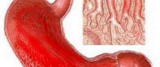
The surface of the mucous membrane of the body of the stomach has a pitted structure (see figure on the left, Matveeva L.V. et al.), which creates conditions for minimal contact of the epithelium with the aggressive intracavitary environment of the stomach, which is also facilitated by a thick layer of mucous gel. Therefore, the acidity on the surface of the epithelium is close to neutral. The mucous membrane of the body of the stomach is characterized by a relatively short path for the movement of hydrochloric acid from the parietal cells into the lumen of the stomach, since they are located mainly in the upper half of the glands, and the main cells are in the basal part. An important contribution to the mechanism of protecting the gastric mucosa from the aggression of gastric juice is made by the extremely rapid nature of gland secretion, caused by the work of the muscle fibers of the gastric mucosa. On the contrary, the mucous membrane of the antral region of the stomach (see the figure on the right) is characterized by a “villous” structure of the surface of the mucous membrane, which is formed by short villi or convoluted ridges 125–350 µm high (Lysikov Yu.A. et al.).
Still from video by Partsvania-Vinogradova E.V. The role of pH-metry in acid-related diseases
Stomach in children
In children, the shape of the stomach is not constant and depends on the constitution of the child’s body, age and diet.
In newborns, the stomach has a round shape; by the beginning of the first year it becomes oblong. By the age of 7–11, a child’s stomach does not differ in shape from an adult’s. In infants, the stomach is positioned horizontally, but as soon as the child begins to walk, it takes on a more vertical position. By the birth of a child, the fundus and cardiac part of the stomach are not sufficiently developed, and the pyloric part is much better, which explains frequent regurgitation. Regurgitation is also promoted by swallowing air during sucking (aerophagia), with improper feeding technique, short frenulum of the tongue, greedy sucking, and too rapid release of milk from the mother's breast.
| Age | Stomach volume, ml |
| newborns | 30–35 |
| 1 year | 250–350 |
| 2 years | 300–400 |
| 3 years | 400–500 |
| 8 years | 1000 |
Gastric juice
The main components of gastric juice are: hydrochloric acid secreted by parietal cells, proteolytic enzymes produced by chief cells and non-proteolytic enzymes, mucus and bicarbonates (secreted by accessory cells), intrinsic Castle factor (production of parietal cells).
The gastric juice of a healthy person is practically colorless, odorless and contains a small amount of mucus.
Basal secretion, not stimulated by food or otherwise, in men is: gastric juice 80-100 ml/h, hydrochloric acid - 2.5-5.0 mmol/h, pepsin - 20-35 mg/h. Women have 25–30% less. About 2 liters of gastric juice are produced in the stomach of an adult per day.
The gastric juice of an infant contains the same components as the gastric juice of an adult: rennet, hydrochloric acid, pepsin, lipase, but their content is reduced, especially in newborns, and increases gradually. Pepsin breaks down proteins into albumins and peptones. Lipase breaks down neutral fats into fatty acids and glycerol. Rennet (the most active enzyme in infants) curdles milk (Bokonbaeva S.D. et al.).
Stomach acidity
The main contribution to the total acidity of gastric juice is made by hydrochloric acid produced by the parietal cells of the fundic glands of the stomach, located mainly in the area of the fundus and body of the stomach.
The concentration of hydrochloric acid secreted by parietal cells is the same and equal to 160 mmol/l, but the acidity of the secreted gastric juice varies due to changes in the number of functioning parietal cells and neutralization of hydrochloric acid by alkaline components of gastric juice. Normal acidity in the lumen of the body of the stomach on an empty stomach is 1.5–2.0 pH. The acidity on the surface of the epithelial layer facing the lumen of the stomach is 1.5–2.0 pH. The acidity in the depths of the epithelial layer of the stomach is about 7.0 pH. Normal acidity in the antrum of the stomach is 1.3–7.4 pH.
On the graph: 24-hour pH-gram of the body of the stomach is normal (Storonova O.A., Trukhmanov A.S.)
Currently, the only reliable method for measuring gastric acidity is considered to be intragastric pH-metry, performed using special devices - acidogastrometers, equipped with pH probes with several pH sensors, which allows you to measure acidity simultaneously in different zones of the gastrointestinal tract.
Stomach acidity in relatively healthy people (who do not have any subjective gastroenterological sensations) changes cyclically during the day. Daily fluctuations in acidity are greater in the antrum than in the body of the stomach. The main reason for such changes in acidity is the longer duration of nocturnal duodenogastric reflux (DGR) compared to daytime, which throws duodenal contents into the stomach and, thereby, reduces the acidity in the lumen of the stomach (increases pH). The table below shows the average acidity values in the antrum and body of the stomach in apparently healthy patients (Kolesnikova I.Yu., 2009):
| Index | Day | Day | Night |
| Average acidity of the stomach body, units. pH | 3,2 | 3,1 | 3,3 |
| Average acidity of the antrum of the stomach, units. pH | 4,0 | 3,6 | 4,4 |
| Number of DGRs lasting more than 5 minutes | 29 | 12 | 18 |
| Number of DGRs reaching the body of the stomach | 11 | 5 | 6 |
The general acidity of gastric juice in children of the first year of life is 2.5–3 times lower than in adults. Free hydrochloric acid is determined during breastfeeding after 1–1.5 hours, and during artificial feeding – after 2.5–3 hours after feeding. The acidity of gastric juice is subject to significant fluctuations depending on the nature and diet, and the state of the gastrointestinal tract.
Gastric motility
In terms of motor activity, the stomach can be divided into two zones: proximal (upper) and distal (lower). There are no rhythmic contractions or peristalsis in the proximal zone. The tone of this zone depends on the fullness of the stomach. When food arrives, the tone of the muscular lining of the stomach decreases and the stomach reflexively relaxes.
| Motor activity of various parts of the stomach and duodenum (Gorban V.V. et al.) |
Gastric accommodation is a postprandial vagal reflex, leading to a decrease in the tone of the stomach (primarily in the proximal section) in response to food intake (see figure on the left). Gastric accommodation provides a reservoir for ingested food without a significant increase in intragastric pressure. Esophageal distension may also be accompanied by proximal gastric relaxation. So-called adaptive relaxation involves relaxation of the proximal stomach in response to stretching of the antrum. Adaptive relaxation ensures the creation of a pressure gradient inside the organ, which promotes mixing of food and its adequate grinding. Adaptive relaxation can be generated by both local (with the initiation of mechanoreceptors in the antrum) and vago-vagal reflexes.
| Application of electrodes to the patient's abdomen during electrogastrography |
In the proximal zone in the area of the greater curvature of the stomach, interstitial cells of Cajal are located, which form the rhythm of gastric contractions (on average 3 cycles per minute).
The resulting peristaltic waves are directed towards the duodenum. Their functional role is to push the contents of the stomach towards the pylorus. The motor function of the stomach is studied using electrogastrography. It is accepted that, depending on the frequency of the fundamental harmonic, the following occurs:
- normogastria (normal motility) - at a frequency of 2 to 4 cycles per minute
- bradygastria (reduced motility) - with a frequency of less than 2 cycles per minute
- tachygastria (increased motor skills) - at a frequency of 4 to 10 per minute.
The graph shows electrogastrograms of the stomach before (red curve) and after eating (green). The abscissa axis is seconds, the ordinate axis is millivolts (Popov A.I., Rudalev A.V.).
Using electroenterogastrography (which uses the same Gastroscan-GEM device, but the research methodology differs from electrogastrography), the following gastric motility disorders are detected:
- irritable stomach
- characterized by normal or reduced electrical activity of the stomach on an empty stomach with a subsequent increase in its electrical activity after eating by more than 1.5-2 times - lazy stomach
- characterized by normal electrical activity of the stomach on an empty stomach and a decrease in it after eating - asthenic stomach
- characterized by a high level of electrical activity of the stomach on an empty stomach and a decrease in it after eating (Rachkova N.S., Khavkin A.I.).
An important role in the implementation of the motor function of the stomach in children belongs to the activity of the pylorus, thanks to the reflex periodic opening and closing of which food masses pass in small portions from the stomach to the duodenum. In the first months of life, the motor function of the stomach is poorly expressed, peristalsis is sluggish, and the gas bubble is enlarged. In infants, there may be an increase in the tone of the stomach muscles in the pyloric region, the maximum manifestation of which is pyloric spasm. Cardiospasm sometimes occurs in older people.
Functional deficiency decreases with age, which is explained, firstly, by the gradual development of conditioned reflexes to food stimuli; secondly, the complication of the child’s diet; thirdly, the development of the cerebral cortex. By two years, the structural and physiological characteristics of the stomach correspond to those of an adult (Bokonbaeva S.D. et al.).
Stomach enzymes
Cells of the gastric mucosa secrete a large number of enzymes. The most important proteolytic enzymes of gastric juice: pepsin, gastrixin (pepsin C), and chymosin (rennin). The pepsin precursor (proenzyme) pepsinogen, as well as the proenzymes gastricsin and chymosin, are produced by the main cells of the gastric mucosa, and are subsequently activated by hydrochloric acid. Non-proteolytic enzymes of gastric juice are lysozyme, carbonic anhydrase, amylase, lipase and others.
Endocrine cells of the stomach
The gastric mucosa contains a large number of endocrine cells that produce a number of hormones.
35% of the endocrine cells of the stomach of a healthy person are enterochromaffin-like cells that secrete histamine, 26% are G-cells that secrete gastrin. In third place in number are D-cells that secrete somatostatin. The figure on the right shows a diagram of the fundic gland (Dubinskaya T.K.):
1 - mucus-bicarbonate layer 2 - surface epithelium 3 - mucous cells of the neck of the glands 4 - parietal cells 5 - endocrine cells 6 - chief (zymogenic) cells 7 - fundic gland 8 - gastric fossa
Microflora of the stomach
Until recently, it was believed that due to the bactericidal effect of gastric juice, microflora that penetrated the stomach died within 30 minutes. However, modern methods of microbiological research have proven that this is not the case. The amount of various mucosal microflora in the stomach of healthy people is 103–104/ml (3 lg CFU/g), including Helicobacter pylori
(5.3 lg CFU/g) detected in 44.4% of cases, 55.5% - streptococci (4 lg CFU/g), 61.1% - staphylococci (3.7 lg CFU/g), 50% - lactobacilli (3.2 lg CFU/g), 22.2% - fungi of the genus
Candida
(3.5 lg CFU/g).
In addition, bacteroides, corynebacteria, micrococci, etc. were sown in an amount of 2.7–3.7 lg CFU/g. It should be noted that Helicobacter pylori
was detected only in association with other bacteria.
The environment in the stomach turned out to be sterile in healthy people only in 10% of cases. Based on their origin, the microflora of the stomach is conventionally divided into oral-respiratory and fecal. In 2005, strains of lactobacilli that adapted (like Helicobacter pylori
) to existing in the sharply acidic environment of the stomach were discovered in the stomach of healthy people:
Lactobacillus gastricus, Lactobacillus antri, Lactobacillus kalixensis, Lactobacillus ultunensis
.
In various diseases (chronic gastritis, peptic ulcer, stomach cancer), the number and diversity of bacterial species colonizing the stomach increases significantly. In chronic gastritis, the largest amount of mucosal microflora is found in the antrum, in peptic ulcer disease - in the periulcerous zone (in the inflammatory ridge). Moreover, the dominant position is often occupied not by Helicobacter pylori
, but by streptococci, staphylococci, enterobacteria, micrococci, lactobacilli, and fungi of the genus
Candida
(Y.S. Zimmerman).
According to Engstrand L. (2012), in the stomach of a healthy person, in the absence of dominance of Helicobacter pylory
, the main volume of the gastric microbiota is represented by ten genera:
Prevotella, Streptococcus, Veillonella, Rothia, Haemophilus, Actinomyces, Fusobacterium, Neisseria, Porphyromonas
and
Gemella
, belonging to five types (lat.
phylum
) of bacteria:
Firmicutes, Proteobacteria, Bacteroidetes, Actinobacteria
and
Fusobacteria
.
Spectrum and frequency of occurrence of microorganisms in the mucous membranes of the esophagus, stomach and duodenum of healthy people (Julai G.S. et al.)
In addition to
Helicobacter pylori,
other representatives of the genus
Helicobacter
.
Patients in whom non-H. pylori Helicobacter species, suffered from gastritis, peptic ulcers, stomach cancer and MALT lymphoma. Although these bacteria are often incorrectly called " Helicobacter heilmannii
", a number of similar but distinct important bacterial species are actually implicated in the development of gastric pathologies, including
Helicobacter bizzozeronii, Helicobacter felis, Helicobacter heilmannii, Helicobacter salomonis
and
Helicobacter suis species.
Diagnosis of infections caused by non-H. pylori Helicobacter pylori agents is not always simple, partly due to their focal colonization in the human stomach (Starostin B.D. Treatment of Helicobacter pylori infection - Maastricht V / Florence consensus report).
Some diseases and conditions of the stomach
Some stomach diseases and syndromes (see):
- gastritis
Perforation of the stomach - a complication of peptic ulcer - acute gastritis
- chronic gastritis
- atrophic gastritis
- anacid gastritis
- superficial gastritis
- antral gastritis
- reflux gastritis
- Ménétrier's disease
- infectious gastritis*
- Helicobacter pylori
-induced gastritis - bacterial gastritis not associated with Helicobacter pylori
- Helicobacter heilmannii
- associated gastritis - enterococcal gastritis
- anisacidosis of the stomach
- strongyloidiasis of the stomach
- cryptosporidosis of the stomach
| Filippova M.P. Peptic ulcer disease. Lecture for the 2nd year of the FKP MGMSU named after. A.I. Evdokimova (video) |
- postprandial distress syndrome
*Classification of infectious gastritis follows the Kyoto global consensus.
Neoplasms:
- stomach cancer
- stomach polyps and polyp-like formations
- gastric adenomas
- Peutz-Jeghers syndrome
Gastric motility disorders:
- "irritable" stomach
- "lazy" stomach
Some symptoms that may be associated with stomach problems:
- stomach ache
- nausea
- vomit
- gastralgia
Acidity chart for duodenogastric reflux (Storonova O.A., Trukhmanov A.S.)
Patient Materials
- Vasilenko V.V. Stomach // www.gastroscan.ru. 2022.
- Agapkin S.N. The most important thing about the stomach and intestines. Peptic ulcer // Eksmo Publishing House LLC. Moscow. 2022. 53 p.
- Vasilenko V.V. Stomach and resorts / www.gastroscan.ru. 2018
The GastroScan.ru website contains: a subsection “Doctors' Advice” in the “Patients” section of the site, a subsection “Popular Gastroenterology” in the “Literature” section, as well as a subsection “Popular Gastroenterology” in the “Video” section, containing materials for patients on various aspects of gastroenterology.
Publications for healthcare professionals regarding gastric anatomy, physiology and diseases
- Matveeva L.V., Usanova A.A., Mosina L.M. Physiology of the stomach: monograph / Saransk, 2012. – 100 p.
- Butov M.A., Kuznetsov P.S. Examination of patients with diseases of the digestive system. Part 1. Examination of patients with stomach diseases. A textbook on propaedeutics of internal diseases for 3rd year students of the Faculty of Medicine. - Ryazan. — 2007.
- Rapoport S.I., Lakshin A.A., Rakitin B.V., Trifonov M.M. pH-metry of the esophagus and stomach in diseases of the upper digestive tract / Ed. Academician of the Russian Academy of Medical Sciences F.I. Komarova. — M.: ID MEDPRACTIKA-M. — 2005. – p. 208.
- Stupin V.A., Siluyanov S.V. Violation of the secretory function of the stomach in peptic ulcer // Russian Journal of Gastroenterology, Hepatology, Coloproctology. - 1997. - No. 4. - p. 23–28.
- Korotko G.F. Organization of gastric digestion // Bulletin of surgical gastroenterology. - 2006. - No. 1. - p. 17–25.
- Zimmerman Ya.S. Modern methods for studying the functions of the stomach and their diagnostic capabilities // RZHGGK. - 2011. - T.21. - No. 5. — P.4-16.
- Lysikov Yu.A., Goryacheva O.A., Tsvetkova L.N., Krasavin A.V., Gureev A.N., Tsvetkov P.M. Clinical and morphological features of duodenal ulcer in children // Pediatrics. 2011. Volume 90. No. 2. pp. 38–42.
- Levit R.M. Clinical, endoscopic and morphological characteristics of chronic gastroduodenitis in childhood. Abstract of dissertation. Doctor of Medical Sciences, 01/14/08 – pediatrics, YSMU, Moscow, 2016.
- Kolesnikova I.Yu., Volkov V.S. Diagnosis and treatment of acid-related diseases of the digestive tract. Guide for doctors / M.: Publishing House “Medical Information Agency”, 2014. - 432 p.
- Guseinov A.Z., Bronshtein P.G., Sazhin V.P. Stomach surgery monograph // St. Petersburg - Tula: Tula State University Publishing House, 2014. - 264 p.
- Maev I.V., Andreev D.N., Zaborovsky A.V. Fundamentals of acid production in the stomach. Medical advice. 2018;(3):7-14.
On the website GastroScan.ru in the “Literature” section there is a subsection “Diseases of the stomach and duodenum”, containing publications for healthcare professionals. Back to section
Ulcers
Ulcerative lesions of the antrum are possible after the stage of inflammation, when focal atrophy of the mucosa passes through the stage of erosion to deep damage to the submucosal and muscular layers.
Ulcers located in the antrum account for up to 10% of all gastric ulcers
In addition to inflammation, the mechanisms of the disease include:
- low contractile function of the antrum;
- stagnation and fermentation of the food bolus;
- increased enzyme production.
The presence of risk factors provokes the transition of inflammation into an ulcer. Typical symptoms:
- pain in the epigastric region, becoming more intense at night;
- constant heartburn;
- nausea and vomiting;
- belching after eating;
- blood impurities in stool and vomit.
Our services
The administration of CELT JSC regularly updates the price list posted on the clinic’s website. However, in order to avoid possible misunderstandings, we ask you to clarify the cost of services by phone: +7
| Service name | Price in rubles |
| Appointment with a surgical doctor (primary, for complex programs) | 3 000 |
| Fluoroscopy and radiography of the stomach | 4 800 |
| Gastroscopy (videoesophagogastroduodenoscopy) | 6 000 |
All services
Make an appointment through the application or by calling +7 +7 We work every day:
- Monday—Friday: 8.00—20.00
- Saturday: 8.00–18.00
- Sunday is a day off
The nearest metro and MCC stations to the clinic:
- Highway of Enthusiasts or Perovo
- Partisan
- Enthusiast Highway
Driving directions
Benign neoplasms of the antrum
Non-cancerous formations of the antrum include polyps and lymphofollicular hyperplasia. Polyps arise from the proliferation of glandular epithelial cells. The antrum accounts for 60% of all gastric polyps.
They are characterized by single growth or the formation of an entire colony. They differ in shape and size (up to 30 mm). Identified against the background of other stomach diseases. They pose a threat of cancerous degeneration. They practically do not give any symptoms. Pain is caused by eating disorders. They can become twisted or pinched, causing blood to appear in the stool.
Based on their origin, antral polyps are divided into three types:
- inflammatory - begin with lymphoid follicles (from 70 to 90%);
- adenomas - grow from the glandular epithelium;
- specific - neoplasms in Pattes-Jeghers-Touraine syndrome, which is a hereditary pathology including hyperpigmentation of the skin and polyposis of the intestines, stomach, differ in glandular structure, pigment content (melanin), and rarely - muscle fibers.
The first two types of polyps develop in old and senile age; specific ones are usually detected before the age of 30.
Pattes-Jeghers-Touraine syndrome is also characterized by the appearance of spots on the face (xanthomas), in which the melanin pigment is located at the level of the basal layer of the epidermis and in the mucous membrane. Pigmentation appears in childhood and may decrease or disappear with age.
Polyps can “sit on a stalk” or be attached with a wide base to the wall
Lymphofollicular hyperplasia is accompanied by the growth or formation of follicular tissue in the submucosal layer of the stomach. The disease has no age-related advantages. Among the reasons, in addition to those described above, a special place is given to:
- herpes infection;
- autoimmune diseases;
- endocrine disorders;
- contact with carcinogens.
It is important that, according to observations, this type of hyperplasia most often precedes the formation of polyps.
Treatment
The optimal treatment regimen for the patient is selected by a council of doctors based on:
- stages of the disease;
- general condition of the patient;
- the presence of concomitant pathologies;
- degree of lymph node damage;
- localization of metastases.
The most effective treatment is surgery. Operations are carried out in two ways:
- subtotal resection - cutting off the tumor along with the part of the organ in which it is located (capturing healthy tissue from 5 to 7 cm), the nearest lymph nodes;
- gastrectomy - removal of the stomach completely, with tumors, tissue, regional lymph nodes, metastases in other organs (when they exist). Doctors then reconstruct the digestive tract by connecting the esophagus to the small intestine.
Most patients are indicated for radical surgery. In rare cases, low-traumatic endoscopic intervention is possible. To do this, cancer must be “caught” at stages I, sometimes II. The tumor should be highly or moderately differentiated (when the cells retain most of their natural characteristics), without ulceration, and not exceed 2 cm in cross section.
Surgery for antral cancer is combined with radiation and chemotherapy.
These methods apply:
- before surgery - to reduce the tumor in size, stop the division of malignant cells;
- after the intervention - to destroy the remaining changed tissues, minimizing the risk of relapse of the disease.
The doctor develops a treatment regimen individually for each patient. Sometimes the use of targeted drugs or photodynamic therapy is justified.
Chemoradiation treatment is prescribed to patients who are contraindicated for surgery due to the presence of distant metastases or general health. At the same time, doctors provide symptomatic therapy aimed at improving the patient’s quality of life.
Cancer patients also undergo surgery for palliative purposes. For example, they cut off part of the tumor, metastases in case of stenosis of the lower part of the stomach, or create a bypass path (anastomosis) to the intestine if the natural one is blocked by the tumor.
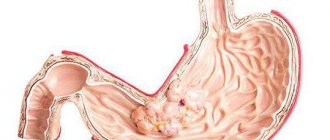
Cancer tumor
Cancer (cancer in Latin or abbreviated cr) of the antrum accounts for up to 70% of malignant tumors of the stomach. There are:
- adenocarcinoma - formed from glandular cells, the most common tumor (90%);
- solid cancer is a rare neoplasm, the structure is not related to glandular elements;
- scirrhus cancer is an even rarer form, formed from connective tissue.
Specifics of antral cancer localization:
- infiltrative growth without the formation of clear boundaries;
- aggressive course with rapid metastasis;
- frequent relapses after gastric resection.
This disease gives a disappointing prognosis.
The stage of cancer development is determined by the depth of the lesion and the presence of metastasis
The most common cause of cancer is chronic atrophic gastritis. It causes three types of morphological changes:
- glandular atrophy - disappearance of mucosal cells;
- dysplasia - the appearance in the stomach of epithelium characteristic of the intestine (intestinal metaplasia) with correspondingly impaired properties;
- neoplasia - transformation into malignant cells.
Symptoms of antrum cancer, compared to those described above, are different:
- constant feeling of fullness or distension in the stomach area;
- nausea and vomiting, which patients induce themselves to alleviate the condition;
- aversion to food;
- significant exhaustion;
- patient irritability;
- temperature rise.
Tumor infiltration of stomach tissue is accompanied by destruction of blood vessels. Therefore, one of the signs is gastric bleeding (vomiting blood, black loose stools).
Symptoms
This form of gastritis has both general and local symptoms. Common ones include:
• lethargy and drowsiness; • severe intoxication of the body; • irritability; • increased body temperature; • heaviness in the stomach.
As for local symptoms, this disease affects not only the stomach, causing severe pain, belching and heartburn, but also the intestines. During an exacerbation, patients experience:
• constipation; • flatulence; • diarrhea; • bloating; • rumbling.
Moreover, patients often confuse their condition with poisoning and hope for a quick recovery, but no matter how it turns out, gastritis lingers for a long time. Therefore, in this case, it is better not to self-medicate, but to immediately consult a doctor.
pixabay.com/
The role of diagnostics
In identifying diseases of the gastric antrum, diagnosis is crucial, since symptoms do not reflect the extent and severity of the lesion.
Fibrogastroduodenoscopy is an endoscopic method that allows you to visually examine all parts of the esophagus, stomach and the beginning of the duodenum; in addition, it is used when choosing a section of the mucosa for taking a cytological examination, microscopy to identify Helicobacter.
To detect the root cause of the lesion and prescribe treatment, it is necessary to confirm the presence of Helicobacter in the stomach. To do this, use the methods of enzyme immunoassay of blood, polymerase chain reaction for typical antibodies and immunoglobulins, urease breath test and stool analysis.
The presence of a bleeding ulcer or a disintegrating tumor can be indicated by a timely fecal reaction to occult blood.
Undeservedly, some doctors have forgotten the x-ray diagnostic method. It allows one to judge the ulcer niche or the presence of polyps or cancer by folding, deficiency or excess of tissue, and altered contours.
An x-ray can diagnose a benign tumor.
Using a daily urine test, you can determine the amount of uropepsin secreted and calculate the acidity of gastric juice. There are no characteristic signs in a general blood test. Anemia, eosinophilia, and leukocytosis may indicate a severe course of the disease. Analyzes are carried out both during the diagnostic period and to monitor the effectiveness of treatment.
Our doctors
Lutsevich Oleg Emmanuilovich
Chief Surgeon of CELT, Honored Doctor of the Russian Federation, Chief Specialist of the Moscow Department of Health in Endosurgery and Endoscopy, Corresponding Member of the Russian Academy of Sciences, Head of the Department of Faculty Surgery No. 1 of the State Budgetary Educational Institution BPO MGMSU, Doctor of Medical Sciences, Doctor of the Highest Category, Professor
43 years of experience
Make an appointment
Fedorova Elena Vladimirovna
Surgeon, Candidate of Medical Sciences, doctor of the highest category
22 years of experience
Make an appointment
Prokhorov Yuri Anatolievich
Surgeon, head of the surgical service of CELT, candidate of medical sciences, doctor of the highest category
33 years of experience
Make an appointment
Gordeev Sergey Alexandrovich
Surgeon, Candidate of Medical Sciences, doctor of the highest category
42 years of experience
Make an appointment
Features of the fibrogastroscopic picture: erosion and gastropathy
Consideration of erosive lesions of the antrum became possible only with the development of the fibrogastroscopic type of diagnosis. This pathology complicates gastritis and serves as the beginning of ulcers and tumor degeneration.
Erosion is a violation of the integrity of the mucous membrane without penetration into the deep parts of the stomach wall. The following varieties are distinguished:
- acute erosions - limited to compactions, cured in 10 days;
- chronic - look like spots with a diameter of up to 10 mm, require long-term treatment;
- hemorrhagic erosions - give a picture of the consequences of needle injections, the size of the lesion remains within 10 mm in diameter, the color takes on shades from scarlet to cherry-red (depending on the affected vessel, the vein gives dark spots), the mucous membrane surrounding the lesions is swollen, in it contains blood.
Erosion, unlike an ulcer, is located superficially
In the antrum, you can find a single erosion with all the described signs, as well as a complete type of erosion - characterized by spreading along the ridges of the folds of the stomach, the formation of a black coating. Antrum gastropathy is described by a doctor if there are no signs of inflammation, the entire mucous membrane is hyperemic, but the specific form of the disease is unclear.
Possible complications
During major operations to remove malignant tumors, the risk of complications is always present. They are associated both with the intervention itself and with the effect of anesthesia.
The main ones:
- heavy bleeding;
- cancer recurrence;
- purulent sepsis;
- intestinal obstruction;
- pneumonia;
- anemia;
- hypoxia;
- heart attack;
- peritonitis;
- pancreatic necrosis;
- thromboembolism;
- failure of the organ stump or the reconstructed path from the esophagus to the intestine.
The probability of death after surgery for antral cancer is 11-25%.
The degree of risk of postoperative complications is influenced by the correctness of the diagnosis, the qualifications of doctors, and the patient’s careful compliance with all recommendations.