What is the difference in the abbreviation
Many patients do not understand the difference between these methods. The methods for carrying them out are similar. However, there are some differences that can be found out while parsing the abbreviation.
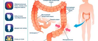
- FGDS is a fibrogastroduodenoscopy, which includes visualization of the duodenum and stomach.
- EGDS is an esophagogastroduodenoscopy that allows you to examine the upper parts of the gastrointestinal tract. That is, visualization occurs not only of the duodenum, stomach, but also of the esophagus.
From the decoding of the abbreviation it is clear that the difference lies in indicating which area needs to be examined. In addition, there is also the concept of “videogastroscopy”. During this diagnosis, a video recording is made during the procedure.
How is EGDS performed?
In order to undergo the EGDS or FGDS procedures, you need to prepare. There is no need to worry too much about the specifics of their implementation. In modern medicine there are methods that will eliminate pain during this procedure. Doctors make extensive use of anesthesia in this case.
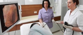
The doctor sprays the oral cavity and esophagus with a special antiseptic in order to suppress possible gag reflexes and numb the specific area. After the doctor has given painkillers, you need to proceed to carefully inserting a special flexible tube called an endoscope. It goes directly into the esophagus through the mouth.
There is one difficult point here - the procedure for swallowing an endoscope. As soon as the tube enters the gastrointestinal tract, the professional himself regulates the depth of its penetration. During the procedure, the person lies on his side. This makes it much easier to complete the research task.
Indications for research
To understand what such research methods are and what their differences are, it is necessary to understand the indications for their use. Esophagogastroduodenoscopy is indicated if there are suspected pathological foci of the esophagus, small intestine, or stomach. That is, if the patient has symptoms that indicate the presence of diseases of these organs:
- belching;
- heartburn;
- discomfort when swallowing food;
- sensation of cutting in the esophagus.
Using a probe, you can accurately determine the presence of scars, obstruction at the junction of the mouth into the stomach, fistulas, diverticula, and neoplasms. Further, as the probe passes through the digestive system, gastroscopy allows the detection of erosions, bleeding, ulcerative lesions, tumor processes, diverticula of the small intestine, and stomach. Fibrogastroduodenoscopy is carried out to determine pathological foci of the duodenum and stomach.
Most often this is:
- varicose veins;
- ulcerative lesions;
- gastritis;
- diverticula;
- duodenitis;
- esophagitis;
- reflux;
- neoplasms.
The most common symptomatology for which the study is carried out is the appearance of nausea after eating and pain in the stomach. As the disease progresses, the list of clinical pictures expands, adding:
- unexpected weight loss when eating a diet characterized by sufficient calorie content;
- loss of appetite;
- flatulence.
It turns out that the differences between these research methods lie in the advantage of endoscopy, which appears due to the diagnosis of the esophagus. Fibrogastroscopy does not provide this opportunity.
Esophagogastroduodenoscopy (EGD, gastroscopy) is an examination of the esophagus, stomach and duodenum for which it is easy to prepare.
Endoscopy is a fairly young branch of medicine. At the same time, its rapid development over the last decade has made it possible to diagnose diseases of the gastrointestinal tract (GIT) in the early stages, when even the most serious diagnosis can be treated, including using minimally invasive endoscopic methods.
The purpose of the procedure is to examine the mucous membrane of the walls of the gastrointestinal tract and, if indicated, the study is supplemented by performing a biopsy and other methods.
Each doctor in our department, when performing gastroscopy, is primarily guided by the main principle of medicine - do no harm. Therefore, a personal approach to each patient is used.
First there is a conversation:
- Collecting an anamnesis (history) of the disease (if any). In this case, it is very important to provide the doctor with the medical documentation you have (take extracts and protocols of previous endoscopic examinations with you).
- Collection of information about allergic reactions to medications (in particular lidocaine).
Then, if there is no allergy, the patient’s throat is irrigated with a special solution of local anesthetic to suppress the gag reflex, because the procedure itself is painless. After which the patient is placed on his left side and the gastroscope is inserted, alternately, into the esophagus, stomach and the initial parts of the small intestine. At the same time, the doctor assesses the condition of the mucous membrane, which makes it possible to identify diseases such as:
Gastroesophageal reflux disease (GERD) is a chronic, relapsing, multisymptomatic disease caused by spontaneous, regularly repeated reflux of gastric and/or duodenal contents into the esophagus, leading to damage to the lower esophagus.
There are two forms of GERD: Endoscopic-negative reflux disease, or non-erosive reflux disease (NERD). It accounts for about 70% of cases of the disease. Reflux esophagitis (RE) – about 30% of cases.
GERD manifests itself primarily as heartburn and sour belching, which often occur after eating, when bending the body forward or at night. The second most common manifestation of this disease is chest pain, which radiates to the interscapular region, neck, lower jaw, and left half of the chest.
Extraesophageal manifestations of the disease include pulmonary symptoms (cough, shortness of breath, most often occurring in the supine position), otolaryngological symptoms (hoarseness, dry throat) and gastric symptoms (rapid satiety, bloating, nausea, vomiting).
Barrett's esophagus is a condition of the esophagus in which the squamous stratified epithelium of the lower lining of the esophagus is replaced by columnar epithelium. Doctors call this metaplasia. Such replacement is usually caused by chronic damage to the mucous membrane of the esophagus by acid thrown up from the stomach, i.e. is a complication of esophagitis or gastroesophageal reflux disease. Barrett's esophagus is found in approximately 1% of the population. Barrett's esophagus is considered a precancerous condition and often leads to the development of esophageal cancer.
Gastritis is a term used to refer to inflammatory and dystrophic changes in the gastric mucosa (GMU) of different origins and course. There are two main forms of gastritis – acute and chronic.
Clinical manifestations of gastritis can differ significantly from each other. This is primarily due to differences in the level of hydrochloric acid, pepsin, and gastric motor activity. The nature of changes in the gastric mucosa is of decisive importance - the presence of erosions, atrophy, the presence of an infectious agent, etc.
Gastritis usually manifests itself as pain and dyspeptic digestive disorders.
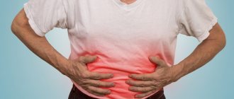
Pain syndrome. Pain with gastritis is usually localized in the epigastric region and appears 1.5 - 2 hours after eating. Often there are “hunger pains” (in the morning, on an empty stomach). Sometimes pain appears immediately after eating. The pain is often spastic in nature and can be sharp, intense, and cramping. In other cases, mild dull pressing pain in the epigastrium is observed.
Dyspeptic digestive disorders are observed in most patients. The appearance of heartburn and sour belching is characteristic, which indicates the appearance of gastroesophageal reflux (reflux of acidic stomach contents into the esophagus). Sometimes there is a feeling of discomfort in the epigastrium, a feeling of fullness. In case of exacerbation, nausea and vomiting may occur, bringing relief.
Gastric ulcer is a chronic relapsing disease that occurs with periods of exacerbation and remission. Its main symptom is the formation of a defect (ulcer) in the wall of the stomach. Other organs of the digestive system may also be involved in the pathological process, with the development of complications that threaten the patient’s life.
Duodenal ulcer (DU) is a chronic relapsing disease that occurs with periods of exacerbation and remission. The main symptom of a duodenal ulcer is the formation of a defect (ulcer) in its wall. Often, ulcers affect not only the duodenum, but also the stomach (stomach ulcer), and other organs of the digestive system with the development of dangerous complications.
The main symptom of exacerbation of a gastric ulcer and duodenal ulcer is pain in the upper abdomen, which, depending on the location of the ulcer, can radiate to the left half of the chest, scapula, thoracic and lumbar spine, left and right hypochondrium, and various parts of the abdomen. The time at which pain occurs also depends on the location of the ulcer.
Symptoms of a stomach ulcer. With an ulcer of the cardial part of the stomach, i.e. its upper part, pain occurs immediately after eating, with ulcers of the body of the stomach, i.e. in its middle part, 1 - 1.5 hours after eating. Symptoms of an ulcer of the pyloric canal, i.e., the lower part of the stomach, and an ulcer of the duodenal bulb are characterized by the appearance of pain 2-3 hours after eating, “hungry” pain that occurs on an “empty stomach” and decreases or completely disappears after eating, as well as night pain pain.
In addition to pain, during exacerbation of a peptic ulcer, dyspeptic symptoms are noted - heartburn, sour belching, nausea, vomiting at the height of pain, which brings relief, and a tendency to constipation. Peptic ulcer disease is characterized by a spring-autumn exacerbation.
Complications of the disease may include gastric bleeding of varying intensity, perforation (perforation) of the stomach, cancerous degeneration, etc.
It is possible to detect small tumors (polyps), early forms of cancer and remove them in a timely manner only with the help of gastroscopy.
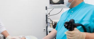
Modern Japanese gastroscopes, which our department has, are a flexible, elastic tube with a controlled end, where a video camera, light guides are located, the water/air channel and the channel for instruments are opened. The presence of an instrumental channel in the endoscope provides the possibility of performing additional manipulations such as:
- Biopsy - using special forceps, a piece of mucous membrane is taken for morphological examination, which allows a diagnosis to be made with high accuracy. This manipulation is absolutely painless.
- Express diagnosis of H. Pylori is also done using a biopsy, but a fragment of the mucous membrane is placed in a special medium that changes its color if the microbe is present.
- pH measurement is performed by touching the mucous membrane with a special probe that reacts to the acidity of the environment.
Thus, to summarize the above, gastroscopy is performed for:
- making a diagnosis
- confirmation of diagnosis (visual and morphological)
- clarification of process localization
- determining the prevalence of the process
- study of the secretory topography of the stomach
- determination of HP
- assessment of the effectiveness of conservative and surgical treatment
- carrying out medical procedures through an endoscope.
Reasons for performing gastroscopy may be:
- pain and heaviness in the abdomen after eating or on an empty stomach
- heartburn
- belching
- weight loss or lack of appetite
- nausea and vomiting
Contraindications to gastroscopy are:
- myocardial infarction in the acute stage
- acute stroke
- cardiovascular and cardiopulmonary failure of the 3rd degree.
- blood coagulation disorders
- stricture of the upper third of the esophagus
- extremely serious condition of the patient
No special preparation is required for gastroscopy - it is enough not to have breakfast and not to have a heavy or late dinner.
As mentioned above, gastroscopy, even with the use of additional techniques, is a painless procedure that requires the patient to be patient and strictly follow the instructions of the endoscopist. However, in some patients it causes pain that prevents them from deciding to undergo the study even with acute abdominal pain.
In this case, we recommend performing gastroscopy under medical sedation or “in your sleep.” Experienced anesthesiologists at our clinic provide anesthesia for the duration of the study, after which the patient is in the recovery room under medical supervision.
Additional features
Both methods of studying the digestive system are used not only for diagnostic purposes. They allow for therapeutic intervention and, if necessary, make it possible to apply tamponade and clips.
In addition, with their help it is possible:
- remove polyps;
- do a biopsy, collect material for histology;
- expand the area of the esophagus affected by stenosis. This procedure is called bougienage;
- take tissue to diagnose the presence of Helicobacter bacteria;
- apply the medicine to the pathological area, for example, perform chemical coagulation of the affected vessel.
During EGDS, as well as FGDS, it is possible to diagnose neoplasms and determine their nature.
Where to perform gastroscopy?
The effectiveness of treatment depends on the accuracy of diagnosis. Doctor Nearby clinics use the latest Pentax equipment. The study can be recorded on any electronic medium. The risk of infection is completely eliminated, since diagnostic instruments are disinfected using a special method in a washing and disinfection machine, which eliminates the “human factor”. The clinic employs doctors of the highest category and has anesthesiologists on staff, which allows the procedure to be carried out without pain or discomfort .
There are no age restrictions; the necessary medical procedures are performed in a complex. You can undergo the examination at any time convenient for you.
Preparation
Preparation for these research methods does not have any differences, which is associated with the diagnosis according to the same scheme. For 3 days before the procedure, the patient should adhere to a special diet, which is aimed at eliminating the consumption of fried, fatty, smoked, spicy, and canned foods. It is important to focus on liquid, well-digested, crushed food. There is also an emphasis on small but frequent meals, in small portions at least 5 times a day. This will help improve the digestibility of foods.
If a patient has a digestive disorder, he is prescribed enzyme agents during the preparatory period. Because during the procedure the stomach must be empty. You must come for the examination on an empty stomach. The last meal should end at 19:00 with a light dinner. It is also forbidden to drink water in the morning. This will help avoid the appearance of a gag reflex during diagnosis.
Side effects after endoscopy
Sometimes the patient complains that after the examination he is bothered by pain and a feeling of numbness in the throat, discomfort when swallowing, and his voice becomes hoarse. These phenomena are associated with the passage of a gastroscope; they go away on their own. There is no need to worry about this. To relieve discomfort and pain, your doctor may recommend using soothing lozenges.
What to do if your stomach hurts after endoscopy?
The pain may be caused by air being pumped into the stomach to smooth out the lining. It usually does not require treatment and goes away on its own.
How the research is carried out
Carrying out EGDS and FGDS is identical. Before the procedure, the throat is numbed. To avoid any unpleasant consequences, it is important to strictly follow all the recommendations of the doctor performing the procedure. The patient is placed on the couch on his left side. Next, a gastroscope with a microcamera is inserted, allowing visualization of internal organs. These procedures are quite unpleasant, cause discomfort, and can cause vomiting.
To make them more comfortable, you should try to relax, restore your breathing and strictly follow the doctor’s recommendations. During the study, you should not touch the probe with your hands or talk. There is a risk of damage to the mucous membrane, which can lead to internal bleeding. For the subject, the difference between EGDS and FGDS is only informative. There are no differences in the procedure and preparation.