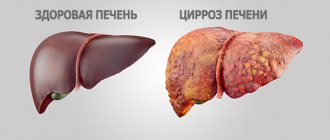
Cirrhosis of the liver (LC) is the final stage of many liver diseases, including chronic hepatitis, alcoholic and non-alcoholic fatty liver disease.1 This is the process of diffuse formation of connective tissue, which leads to dysfunction of the organ. Liver cirrhosis is irreversible; drug and non-drug treatment of liver cirrhosis is usually aimed at preventing the progression of the fibrosis process and preventing the development of complications.1 The prevalence of chronic hepatitis and fatty disease is growing, especially among people of working age - cirrhosis becomes not only medical, but also social and economically significant problem. The prevalence of the disease at the moment is more than 20 million.2 In the Russian Federation, about 200 thousand new cases of liver cirrhosis are registered annually. 2
Causes of cirrhosis
According to statistics, about 50% of liver cirrhosis is associated with alcohol abuse and the development of alcoholic liver disease (ALD). 1Moreover, 25% of them had a history of viral hepatitis.1
Other causes of cirrhosis include2:
- metabolic diseases associated with excessive accumulation of iron, copper and other elements;
- Autoimmune lesions;
- Primary and secondary biliary cirrhosis;
- Diseases of the vascular system (Budd-Chiari disease);
- Long-term use of medications that have a toxic effect on the liver;
- non-alcoholic fatty liver disease (NAFLD), which is associated with the accumulation of large amounts of free fatty acids in hepatocytes (liver cells), is characterized by a chronic course and potential progression from stage to stage: steatosis - steatohepatitis - fibrosis - cirrhosis (same as alcoholic disease liver, but the likelihood of developing cirrhosis in AFLLD is 10 times higher).5
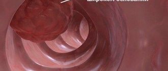
Regardless of the cause, cirrhosis develops in the same way: the lobular structure of the liver is disrupted with the formation of nodes in place of destroyed hepatocytes—the so-called false lobules.
- Small nodular - the size of fibrous nodes is no more than 3 mm;
- Large nodular - nodes 3-5 mm;
- Mixed.
Immunological analysis
A variety of diseases can cause fibrotic degeneration of the liver. The lion's share of these diseases are autoimmune processes.
These include:
- various types of autoaggressive hepatitis;
- cell damage in autoimmune polyendocrine syndrome;
- overlap syndrome;
- primary biliary cirrhosis;
- sclerosing cholangitis;
- lupus hepatitis (with systemic lupus erythematosus);
- inflammation in sarcoidosis.
Laboratory markers that can be used to identify autoimmune diseases:
- AMA (anti-mitochondrial antibodies);
- ANA (antinuclear);
- SMA (AT to smooth muscle cells);
- autoantibodies to kidney and liver microsomes.
Normally, the titer of AMA, SMA and autoantibodies to microsomes should be 1:40, and the ANA titer should be up to 1:160.
An immunological blood test can determine which autoimmune process is destroying the liver.
Symptoms of cirrhosis
The clinical picture of cirrhosis can be very different - from the complete absence of symptoms to the appearance of signs of liver failure, when liver function is completely disrupted1.
Most often, patients are concerned about:
Reduced ability to work; discomfort in the abdomen, mainly in the right hypochondrium
General weakness and increased fatigue
Dyspepsia (indigestion symptoms)
In the future, other symptoms appear, the diagnosis of which is carried out exclusively by a doctor.
May also be observed:
- Itching and yellowing of the skin
- Menstrual irregularities in women, impotence in men.
In the future, other symptoms appear, the diagnosis of which is carried out by a doctor.
What tests are performed?
Cirrhosis is a serious disease that results in destruction of the organ leading to death. The main causes of liver pathology include alcohol dependence, viral hepatitis, parasitic diseases, autoimmune disorders, and the use of drugs and substances of hepatotoxic properties.
If suspected, the doctor prescribes tests, especially the determination of blood counts when the patient complains of pain or discomfort in the right side, discoloration of the stool, darkening of the urine, and an unreasonable increase in temperature to the level of low-grade fever.
Blood tests for liver cirrhosis are divided into the following groups:
- Biochemical screening. It is important to determine the value of AST, ALT, bilirubin. They deviate from the norm even against the background of the early stage of cirrhotic processes.
- Enzyme research. It helps to identify the inflammatory reaction in the gland.
- Additional examinations (fibroscanning, ultrasound, etc.).
The diagnosis of cirrhosis is made based on a combination of test results and instrumental diagnostic methods. It is important to establish the degree of liver damage, the stage of the disease (initial or late), to differentiate the type of pathology - primary biliary, alcoholic, viral, etc.
Diagnostics
Diagnosis of liver cirrhosis begins with a clinical examination, which includes collecting complaints and examination.
During examination, the doctor may detect external signs of CP1:
- Redness on the palms and soles (palmaral erythema)
- Spider veins
- Reduction of hair in the armpits
- White nails
- Men with cirrhosis of the liver may experience gynecomastia, an enlargement of the mammary glands. The reasons for this are considered to be a reduced level of testosterone and an increased level of estradiol (the female sex hormone) in the blood due to various mechanisms: increased processes of aromatization of testosterone into estradiol, as well as increased production of globulin that binds sex steroids, and thus a decrease in the level of free testosterone.6 .7
Later, edema may appear, especially in the lower extremities, jaundice, and the development of ascites - accumulation of free fluid in the abdominal cavity.
When palpating the abdomen, an enlarged liver is detected; it is often possible to palpate the nodal surface of the organ, the pointed edge. However, in the final stage of the disease, the size of the liver may decrease, and the spleen may increase.1 To confirm the diagnosis and establish the stage of the disease, laboratory and instrumental studies are carried out, and calculated indices are also used.
Laboratory methods
- Standard biochemical tests that determine the level of bilirubin, total protein, prothrombin index, serum iron, ferritin and enzyme activity (AST, ALT, alkaline phosphatase). These tests are nonspecific, their deviations can be observed in other diseases, but they help assess the degree of liver dysfunction.
- General clinical blood tests can reveal the presence of anemia, a decrease in the number of leukocytes, platelets, which can also be observed with cirrhosis.
- Tests for hepatitis viruses and other infectious agents may be ordered to determine the potential cause of the disease.
Calculation indices:
The use of test panels, such as FibroTest, FibroIndex, Hepascore, can be used to assess the stage of fibrosis. For example, the use of the Fibrotest score avoided biopsy in 50% of patients.2
To assess the severity of cirrhosis and predict survival, the Child-Pugh classification is most often used.2 The scale, which takes into account the results of laboratory tests and clinical manifestations, allows one to assess the degree of functional impairment and determine the stage of the disease. The higher the score, the worse the prognosis.2
Instrumental studies
In addition to needle biopsy, which is recognized as the “gold standard” for diagnosing fibrosis and cirrhosis, liver imaging methods play an important role in diagnosis. These include:
- Ultrasound of the liver, abdominal organs and retroperitoneal space. The most accessible and fairly informative method. Allows you to evaluate the anatomical and structural features, shape, size, vascular pattern. CP can be judged by an increase in the size of the liver and by its heterogeneous structure and uneven tuberous contour. Also, with liver cirrhosis, ultrasound may show signs of portal hypertension: ascites, dilation of portal veins, enlarged spleen.1
- CT, MRI (computer and magnetic resonance imaging of the liver) - by performing a large number of thin sections, it provides comprehensive information about the structure of the organ, the presence of fibrous nodes, false lobules, additional vessels, etc.
- Ultrasound elastometry (FibroScan device) can serve as an alternative to liver tissue biopsy, as it makes it possible to assess the density of the organ, which increases as the percentage of connective tissue increases.2
Only a comprehensive diagnosis can give a complete picture of the disease, so a combination of various methods is used, which sometimes have to be repeated several times.
General blood analysis
In order to diagnose liver cirrhosis, when typical symptoms appear, the doctor prescribes a general blood test for the patient. This test will identify or confirm the presence of pathology. For OAK, blood is taken from a finger. The collection is carried out in the morning, on an empty stomach.
With cirrhosis of the liver, certain changes occur in the composition of a person’s blood, which allow the doctor to draw specific conclusions:
- There is a decrease in blood hemoglobin levels. The norm for women is not less than 120 g/l, for men - not less than 130 g/l.
- An increase in the level of leukocytes is recorded. The leukocyte norm in a healthy person is 4–9*10⁹/l.
- Against the background of liver damage, an increase in the erythrocyte sedimentation rate is observed: high ESR levels are a sign of an inflammatory process in the body. In men, the ESR rate exceeds 10 mm/h, in the female population – 15 mm/h.
- Changes in the protein composition of the blood are also detected - a decrease in albumin levels is observed.
A complete blood count helps in making a diagnosis
The data obtained make it possible to diagnose liver cirrhosis. To clarify the current stage of the disease and the extent of organ damage, a biochemical blood test is prescribed.
Complications of liver cirrhosis
Complications from cirrhosis can pose a threat to the patient’s life, therefore their timely diagnosis is one of the primary tasks of the doctor.
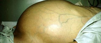
Ascites
The most common complication occurs within 10 years in almost 60% of patients3. Its main causes are increased pressure in the portal vein system (portal hypertension) and insufficient excretion of sodium in the urine, which leads to fluid retention. The development of ascites in a patient with cirrhosis is associated with an unfavorable prognosis and a decrease in the quality of life of patients. There are 3 degrees of severity of this condition: from minor, which is detected only by ultrasound, to intense, in which ascites is noticeable to the naked eye3 (the abdomen greatly increases in size, the vessels dilate, the skin becomes tense, becomes dry, and flakes off).
Ascites can trigger the development of other complications.
Spontaneous bacterial peritonitis
With ascites, infection of the fluid in the abdominal cavity can occur, and the source and route of infection usually cannot be determined. Additional factors are defects in the immune system and the penetration of microorganisms from the intestines into the ascitic fluid.
Symptoms of peritonitis: sharp abdominal pain, tension in the anterior muscle wall, general signs of inflammation, shock, renal failure, gastrointestinal bleeding. However, in every fifth patient it can be asymptomatic. The prognosis is unfavorable.3
Hepatorenal syndrome
Hepatorenal syndrome (HRS) is characterized by the development of renal failure against the background of severe stages of cirrhosis. Hepatorenal syndrome is a diagnosis of exclusion, that is, it is established after excluding other diseases that could cause kidney damage. There are 2 types of hepatorenal syndrome: rapidly progressive, which is usually associated with severe alcoholic hepatitis or the development of spontaneous bacterial peritonitis, and slowly progressive. The prognosis is unfavorable for the development of any type of HRS3.
Hepatic encephalopathy
This is a whole complex of psycho-neurological disorders, which, if the cause can be eliminated, can be reversible. 80% of patients with cirrhosis show signs of encephalopathy3. Symptoms: loss of attention, slow reactions, which is especially dangerous for those who drive vehicles. Treatment of HE is effective if started in a timely manner, that is, at the stage of minimal hepatic encephalopathy. According to the West Haven classification, there are 4 stages: from absent-mindedness and inability to concentrate to severe disorientation and coma.3
Portal hypertension and bleeding from dilated veins of the esophagus and stomach
The mechanism of development of the complication: a constant increase in pressure in the portal vein system leads to varicose veins of the esophagus and stomach with or without bleeding and ascites.
Diagnosed in half of patients with cirrhosis. The higher the severity of cirrhosis, the greater the risk of developing portal hypertension. Bleeding poses the greatest threat, as it causes death in 15-20% of patients.
Portal hypertension can be:
- suprahepatic,
- Intrahepatic,
- Subhepatic
Treatment approaches depend on its type. If hypertension is detected, all patients are given bleeding prophylaxis.3
Liver assessment
The diagnosis in the early stages of liver cirrhosis is difficult to establish, since there are often no pronounced changes in the liver.
First of all, an ultrasound examination of the liver (ultrasound diagnostics) is performed, which makes it possible to identify diffuse changes in the liver tissue and an increase in its size (although this does not always happen). It is advisable to carry out Dopplerography of the vessels of the abdominal cavity to determine the width of the lumen of the vessels and the speed of blood flow. This allows us to determine the presence of signs of portal hypertension. Important for characterizing the structural and functional state of liver cells is a biochemical blood test (ALT, AST, GGT, bilirubin, alkaline phosphatase, protein fractions), as well as a clinical blood test and coagulogram - blood clotting.
To accurately diagnose the degree of fibrosis, modern non-invasive (replacing biopsy) examination methods are used: elastometry (elastography) of the liver using the Fibroscan, Fibrotes, FibroMax apparatus. Liver damage is characterized by degrees from 0 to 4; 0 – healthy liver, 4 – cirrhosis.
Treatment of cirrhosis
Treatment of cirrhosis involves:
- Treatment of the underlying disease that led to the development of cirrhosis;
- Specific therapy for complications, including medications: for example, the prescription of diuretics for ascites, the prescription of antibiotics for spontaneous bacterial peritonitis, etc.
- Surgical treatment of ascites and bleeding from dilated veins of the esophagus and stomach1
Important components are diet, exclusion of harmful effects on the liver, such as alcohol, hepatotoxic drugs, treatment of concomitant chronic diseases.4 If the measures taken are ineffective, organ transplantation is indicated1.
Diagnosis by history
How to diagnose liver cirrhosis? Unfortunately, identifying the disease in the initial stages of development is quite difficult. There are no pain receptors in hepatocytes, so in the first few days, degenerative changes in the liver are practically asymptomatic. Over time, it ceases to cope with its functions, as evidenced by the appearance of dyspeptic symptoms.
Patients suffering from cirrhosis may present various complaints. The clinical manifestations of the pathology are determined by the degree of damage to the digestive gland. Cirrhosis can be identified by the following symptoms:
| CPU stage | Features of development | Clinical manifestations |
| compensated | Most of the hepatocytes function without failures, so the manifestations of pathology are weakly expressed | discomfort and heaviness in the abdomen, weakness and loss of appetite, flatulence and nausea, fatigue, low temperature (up to 37.2 ⁰C) |
| subcompensated | the vast majority of hepatocytes cease to function, causing irreversible changes in the body | yellowing of the skin, sleep disturbance, dull pain in the hypochondrium, itching, bowel dysfunction (diarrhea, constipation), increased gas formation |
| decompensated | the digestive gland is almost completely covered with connective tissue scars, as a result of which it ceases to function | low-grade fever (temperature 37.5°C), nosebleeds, muscle atrophy, internal bleeding, abdominal enlargement (abdominal hydrops), confusion (a sign of encephalopathy) |
Decreased appetite, chronic fatigue, heaviness in the abdomen and flatulence are symptoms that may indicate the development of cirrhosis.
Non-alcoholic forms of this disease are provoked by exogenous and endogenous causes, which can be learned from the patient himself. An unbalanced diet and previous diseases (hepatitis, cholangitis, cholelithiasis) negatively affect the functioning of the digestive gland and can cause irreversible processes in it. Classification of liver cirrhosis according to the type of morphological changes allows us to predict the course of the disease and create the most appropriate treatment regimen. Undesirable processes in the organ can be suspended at the stages of compensation and subcompensation.
Laboratory research
Diagnosis of liver cirrhosis involves several types of laboratory tests. Dynamic monitoring of biochemical blood parameters allows us to assess the course of the disease and the rate of its progression. If the results of the examination reveal that the patient has developed anemia, leukocytosis and microhematuria, he is sent for additional hardware examination.
Clinical blood test
If cirrhosis is suspected, a clinical blood test is performed, during which the number of platelets and formed elements - red blood cells and leukocytes - is calculated. When liver tissue is replaced by fibrous adhesions, the number of formed elements in the blood decreases. The formation of scars in the digestive gland leads to stagnation of venous blood, resulting in the development of hypersplenism syndrome - hypertrophy (enlargement) of the spleen, accompanied by a decrease in the number of leukocytes and red blood cells in the blood.
Hypertrophy of the liver and spleen manifests itself in the subcompensated and decompensated stages of cirrhosis.
The spleen destroys aged blood cells, but with an increase in its activity, the concentration of formed elements sharply decreases. As a result, this leads to the development of thromocytopenia, anemia and leukopenia. An increase in erythrocyte sedimentation rate (ESR) signals the presence of low-grade inflammation in the body. In addition, ESR can be triggered by a change in the amount of protein components in the blood.
Biochemical indicators
The liver is the organ in which most of the proteins and enzymes are produced. If during laboratory tests a change in its biochemical status is detected in the blood, this indicates a dysfunction of hepatocytes. If hepatic cirrhosis is suspected, special attention is paid to studying the following indicators:
- bilirubin is a toxic substance that is inactivated by the digestive gland; an increase in its concentration signals a malfunction of the organ (the range from 8.5-20.5 µmol/l bilirubin in the blood is considered normal);
- alanine aminotransferase (AlT) is an enzyme whose maximum concentration is concentrated in liver cells; a decrease in the amount of enzyme indicates the destruction of hepatocytes;
- alkaline phosphatase is an enzyme that is localized in the cells of the bile ducts; an increase in the amount of phosphatase in the blood signals the development of cholestasis, in which biliary cirrhosis is most often diagnosed (normal alkaline phosphatase is 80-306 U/l);
- albumin is a protein substance that is produced in the digestive gland; with the development of cirrhosis, the amount of albumin in the bloodstream decreases sharply, as a result of which intercellular fluid leaks into the surrounding cavities and tissues (the normal level of albumin is 35-50 g/l).
Sometimes it is necessary to differentiate liver cirrhosis from alveolar echinococcosis, hepatocellular carcinoma (liver cancer) and other pathologies. To ensure that the diagnosis is correct, a number of additional biochemical tests are performed, during which attention is paid to the concentration of serum iron and gamma globulins in the blood.
Urine and stool analysis
If CP is suspected, urine and stool tests are no less indicative. If bilirubin, red blood cells, platelets and protein substances are detected in the chemical composition of urine, in most cases this indicates the progression of cirrhosis. In a healthy person, these substances are practically absent in the urine.
Stool analysis can provide valuable information about the rate of development of cirrhotic processes. Even a visual examination of the biomaterial can indirectly indicate dysfunction of the digestive gland. Discoloration of stool or the appearance of a clay tint is explained by the lack of stercobilin in the body, an enzyme that colors stool brown.
Blood clots in the stool indicate bleeding hemorrhoidal veins. The appearance of the symptom is associated with varicose veins, which often accompanies cirrhosis. Stool instability, frequent constipation or diarrhea are indirect evidence of disturbances in the functioning of the digestive gland.
Blood clots in the stool are a symptom indicating the development of portal hypertension, which causes severe complications - ascites, splenomegaly, internal bleeding.