- Chronic hepatitis and liver cirrhosis;
- Hepatitis of various etiologies and other liver diseases;
- Cholelithiasis;
- Chronic cholecystitis;
- Dyskinesia of the gallbladder;
- Gallbladder cholesterosis.
Symptoms Classification Diagnostics Treatment Information
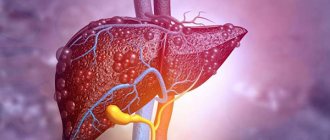
The liver is involved in all metabolic processes, serves as a source of bile formation, and performs a filtration function.
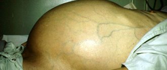
The peculiarity of the course of liver diseases is that there are no pain receptors in the organ. The disease occurs without severe pain. The patient feels pain only when the liver has already significantly increased in size and caused stretching of the liver capsule.
Attention! As a result of asymptomatic progression, the disease often becomes chronic.
THE INSTITUTE OF ALLERGOLOGY AND CLINICAL IMMUNOLOGY has:
- range of services for accurate diagnosis of diseases
- special treatment programs
- consultations are carried out by doctors with academic degrees.
When identifying concomitant diseases, consultations are held with specialists in related fields (otorhinolaryngologists, endocrinologists, neurologists, gastroenterologists, dermatovenerologists), consultations with Doctors of Medical Sciences.
How does the liver work?
The liver is the largest gland in humans. It is located in the upper floor of the abdominal cavity on the right. It consists of two main lobes - right and left, which in turn are divided into 8 segments.
In an adult, it weighs about 1/40 of the total body weight (approximately 1.6 kg in men and 1.2 kg in women). The liver has a dual blood supply: approximately 80% of the blood entering the liver comes from the portal vein (venous blood collected from all unpaired abdominal organs), the rest (oxygenated arterial blood) comes from the hepatic artery. Having entered the portal of the liver, both vessels give rise to multiple branching smaller vessels. At the exit from the liver, 3-4 hepatic veins are formed, which flow into the inferior vena cava.
The main structural unit of the liver is the hepatic lobule, which consists of liver cells - hepatocytes. This cell is one of the most important biochemical laboratories of the body. The hepatocyte takes part in the metabolism of proteins, carbohydrates, fats, bile acids, and vitamins. Neutralizes toxic substances coming from the intestines, followed by the release of associated products into the intestinal lumen. An important function of the hepatocyte is the synthesis of bile, which is involved in digestion.
Treatment of CKD
A common consequence of all forms of cholestasis is the retention of fatty acids in hepatocytes. Elevated FA levels lead to apoptosis or necrosis of hepatocytes and ultimately to chronic CKD [13]. In some cholestatic disorders, FAs also enter the peribiliary space, causing portal vein inflammation and fibrosis through the induction of chemokines and cytokines.
In this regard, the following pharmacological targets are identified for the treatment of intrahepatic cholestasis (Fig. 3) [13]:
stimulation of orthograde secretion of the biliary tract and retrograde secretion of FA and other toxic cholephiles into the systemic circulation for excretion by the kidneys;
stimulation of the metabolism of hydrophobic FAs and other toxic compounds to more hydrophilic but less toxic metabolites;
protection of affected cholangiocytes from the toxic effects of bile; inhibition of apoptosis caused by increased levels of cytotoxic FAs;
inhibition of fibrosis caused by FA leakage into the peribiliary space.
Drug therapy for CKD is presented in Table. 1. In table. Tables 2 and 3 present the main symptoms that occur with CKD and medications to relieve them.
The only generally accepted drug for medical treatment of most chronic CKD is ursodeoxycholic acid (UDCA) [12, 15–19]. The ability to treat cholestasis is considered one of the most important and most valuable properties of this drug. In table Figure 4 presents the main properties of UDCA, which, in the author’s opinion, are most fully disclosed in articles [15, 16].
The positive effect of UDCA in such cholestatic diseases as PBC has been most convincingly proven. In a combined analysis of French, Canadian and North American cohorts of patients with 2-4 years of follow-up, a decrease in mortality and the need for liver transplantation was noted in groups with moderate and severe disease. The Barcelona study of 192 patients treated with UDCA for 1.5 to 14 years showed that survival in patients who responded to UDCA therapy (response assessed by the level of reduction in ALP) was higher than predicted by the Mayo prognostic model and consistent with the population [ 16].
A dose of UDCA of 13–15 mg/kg/day for most cholestatic diseases has advantages in biochemical response and cost compared with low and high doses. An exception is cystic fibrosis, where doses of 20–30 mg/kg/day are recommended [16]. For PSC, recommended doses have not been determined.
There is evidence of a positive effect of UDCA on drug-induced cholestasis, including that caused by one of the most frequently causing hepatotoxicity drugs, amoxicillin/clavulanate [16].
The European Association for the Study of the Liver, 2009 [20] and the Russian Gastroenterological Association [1] recommend mandatory administration of UDCA as basic therapy for a number of CKD: primary biliary cirrhosis, PSC, cystic fibrosis, progressive familial cholestasis type 3 (Progressive Familial Intrahepatic Cholestasis 3), intrahepatic cholestasis of pregnant women, and also discuss its use in drug-induced cholestasis and benign familial cholestasis.
The concept of structured lifelong therapy for PBC is based on 3 main elements: 1) risk stratification and matching therapy; 2) determination of the stage and observation in accordance with it; 3) active management. Assistance is always tailored to the individual characteristics of the patient and the health care capabilities of the country, but its effectiveness is ensured by these 3 supporting elements [21].
As of today, the original UDCA drug Urso (Japan) is not registered in Russia. In this situation, when choosing generic drugs that are widely represented on the domestic pharmaceutical market, one should be guided, first of all, by the price-quality ratio. The domestic UDCA drug Urdoxa®, produced by JSC FP Obolenskoye, which appeared in recent years, is not inferior to the generic forms previously registered in our country [16]. This domestic drug has characteristics that allow it to be considered the drug of choice at any stage of medical care for patients with a wide range of nosologies, including chronic CKD. Urdoxa® is produced from a European substance (Italy, Industria Chimica Emiliana) at a Russian enterprise according to the international standard GMP (Good Manufacturing Practice). The high safety profile allows the drug to be used in children over 3 years of age [22].
The drug has successfully passed all registration procedures required by law, which allows us to speak of its bioequivalence to reference drugs containing UDCA [23, 24].
Confirmation of the pharmaceutical equivalence of UDCA preparations is not only the same amount of active substance and excipients in 1 capsule of the drug, but also the presence of identical infrared spectra obtained by infrared spectroscopy of the finished dosage forms of the compared UDCA preparations [25–27].
The reliability of the dynamics of standard biochemical indicators of liver function during therapy - a simple and informative diagnostic tool - has been confirmed by the last 10 years of its widespread use. Response to UDCA therapy can be assessed using a discrete binary variable model or a scoring system based on the calculation of continuous variables [21]. So, according to the Paris criteria, a good biochemical response after 12 months. For UDCA therapy, a serum bilirubin level ≤17 µmol/L (1 mg/dL), an ALP level ≤3 upper limit of normal, and an aspartate aminotransferase level ≤2 upper limit of normal are considered.
What is bile and why is it needed?
Bile is a secretion produced by hepatocytes. The color of bile ranges from light yellow to dark green. Bile contains bile pigments - bilirubin and biliverdin, cholesterol, fatty acids, lecithin, mucin.
Bile is the main participant in digestion. It neutralizes hydrochloric acid that enters the duodenum from the stomach, thereby creating favorable conditions for the action of pancreatic juice and parietal digestion in the jejunum.
Bile acids emulsify fats, allowing them to be absorbed in the intestines.
Bile has a bactericidal effect that prevents the proliferation of pathogenic microflora.
How are the bile ducts arranged and how do they work?
The bile ducts are a system of ducts that originate from hepatocytes and gradually gather into interlobular, segmental and right and left lobar ducts, which unite into the common hepatic duct.
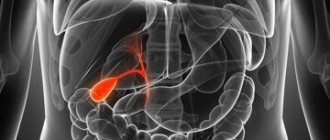
One of the stages of bile removal is its accumulation in the reservoir - the gallbladder. The main function of the gallbladder is to concentrate bile and release it into the lumen of the duodenum during digestion. The volume of this organ is about 80 ml.
The gallbladder is located on the lower surface of the liver. It is a hollow muscular organ, externally covered with a serous membrane and internally lined with mucous membrane. The structure of the gallbladder is divided into the bottom, body and neck of the bladder, which passes into the cystic duct.
Why do gallstones form?
The exact cause of cholelithiasis has not yet been fully elucidated. Only a few factors are known that increase the likelihood of its occurrence.
- Genetic: family predisposition increases the risk of cholelithiasis by 4-5 times;
- Congenital anomalies of the biliary tract;
- Gender - the risk of developing gallstones in women is approximately 2-3 times higher, which is associated with the influence of estrogens on the ability to form gallstones;
- Age - the maximum frequency of clinical manifestations of cholelithiasis is recorded at the age of 40-69 years;
- Dietary (high-calorie diet, poor in plant fiber, as well as with an excess of simple carbohydrates, animal proteins, foods containing cholesterol);
- Use of medications (oral contraceptives, fibrates, ceftriaxone, somatostatin, nicotinic acid);
- Concomitant diseases and conditions (obesity - a tendency to form gallstones in persons with a body mass index of more than 30 is 3 times higher, type 2 diabetes mellitus, hypothyroidism, cirrhosis of the liver, etc.);
- Pregnancy - the risk of developing cholelithiasis increases during pregnancy, especially with repeated pregnancies (the likelihood of stone formation increases by 10-11 times). During pregnancy, bile heterogeneity, or the so-called biliary sludge, develops in 20-30% of patients, stones - in 5-12% of cases.
There is a so-called 5F rule that describes patients at greatest risk of developing stones:
1. Female (female);
2. Fat (overweight/obesity);
3. Forty (age 40 years or more);
4. Fertile (fertile women - that is, pregnant and giving birth);
5. Fair (light-skinned blondes).
CHOLESTATIC (subhepatic) JAUNDICE
Cholestatic jaundice develops when there is an obstruction to the flow of bile at any level of the biliary system. Obstructive or subhepatic (obstructive) jaundice develops when bile flow is obstructed at the level of the extrahepatic bile ducts (choledochal stones, pancreatic cancer, chronic pancreatitis, post-traumatic narrowing of the common bile duct, pancreatic pseudocysts, etc.). 70% of cases are associated with stones in the common bile duct and cancer of the head of the pancreas. Intrahepatic cholestasis is most often observed in acute drug-induced hepatitis and primary cirrhosis, less often in viral, alcoholic hepatitis, cholangitis, liver cirrhosis, metastases, etc. Jaundice develops slowly, often preceded by itchy skin. It is typical for both extrahepatic and intrahepatic cholestasis. Diagnostic criteria:
- light-colored (non-pigmented) stools
- dark brown urine
- traces of scratching on the skin,
- yellowish gloss of nails,
- xanthelasma (cholesterol deposits on the eyelids),
- xanthoma (cholesterol deposits on the palmar folds and tendons),
- hepatomegaly – enlargement of the liver.
When blocked by a stone, jaundice develops within 24 hours after the onset of pain, and fever occurs due to developing cholangitis. Itchy skin does not occur in all patients. Unlike a tumor, long-term stone blockage is rarely complete. Diagnostics: Laboratory tests of blood, feces, urine. Instrumental diagnostics: Ultrasound, CT, MRI diagnostics with contrast, Endoscopic diagnostics.
How are gallstones formed?
The process of stone formation is quite well studied. The key links are:
- An increase in the lithogenicity of bile (the ability to form stones) means an increase in the content of cholesterol and bilirubin in it.
- Impaired contractile function of the gallbladder.
- Increased pressure in the bile duct system.
Mucin is a natural gel, constantly secreted by the wall of the gallbladder; when lithogenicity increases, it begins to “absorb” cholesterol crystals, thereby forming a “fossilization nucleus.” The formed suspension, with impaired contractile function, continues to attach salt crystals encrusted with calcium salts. Thus, gallstones can grow up to 5-7 mm per year.
How does gallstone disease occur?
The main factors influencing the course of the disease are the nature of nutrition and the size of the stones. Gallstone disease can occur both in the form of chronic stone carriage and in acute, and sometimes in complicated, form.
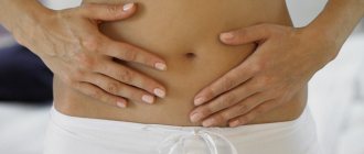
Where does pain come from with gallstone disease?
By themselves, gallstones do not cause any pain and can be an accidental finding during medical examination. When food enters the duodenum, the mechanoreceptors are irritated and cause contraction of the gallbladder. The stones wedge into the cervix, causing a prolonged spasm, which manifests itself as pain. When taking antispasmodic drugs, the calculus can “move” back from the cervix and the attack of hepatic colic is stopped. If the stone remains in the cervix, then inflammation begins to develop against the background of obstruction.
Chronic calculous cholecystitis is an asymptomatic stone carrier. The size of the stones can vary from 2 – 3 mm to 5 cm. This stage is characterized by an almost complete absence of clinical manifestations of the disease. Stones can remain in the lumen of the gallbladder for years without causing any symptoms. However, if there is an error, discomfort may occur in the right hypochondrium, which can be relieved independently or while taking antispasmodic drugs.
Hydrocele of the gallbladder - occurs against the background of obstruction of the neck of the gallbladder by a calculus, cutting off the latter from the entry of bile. The bladder is filled with mucus secreted by the mucous membrane and is not involved in digestion.
Acute calculous cholecystitis - occurs with prolonged obstruction of the neck of the gallbladder by a calculus. The pain syndrome persists against the background of ineffective conservative treatment. The patient is bothered by vomiting, which does not bring relief; pain can be felt behind the sternum - cholecystocardial syndrome, in the right shoulder blade. Body temperature rises and chills appear. Inflammatory changes begin to develop in the wall of the gallbladder, which progress from catarrhal, phlegmonous and gangrenous forms. Against the background of the latter, perforation of the gallbladder or abscess may develop.
Choledocholithiasis and acute biliary pancreatitis. Choledocholithiasis develops as a result of migration of biliary sludge or small stones from the gallbladder into the common bile duct. Once in the common bile duct, small stones freely descend to the sphincter of Oddi, which is a release mechanism - a circular muscle cuff. Sludge and stones “wedge” into the sphincter, causing its long-term spasm, causing a violation of the outflow of bile - obstructive jaundice. The patient's skin and sclera become a rich yellow color. This condition requires emergency decompression of the bile ducts - restoration of bile outflow.
An increase in pressure in the common bile duct promotes the reflux of bile into the main pancreatic duct and intraductal activation of pancreatic juice enzymes - a trigger for the development of acute biliary pancreatitis.
Mirizzi syndrome . A fairly rare complication of gallstone disease. Develops as a result of prolonged stone bearing. First, a stricture forms, then a bedsore between the gallbladder and the common bile duct.
Mirizzi syndrome presents significant difficulties for surgical treatment. This is due to the fact that in the human body there is no plastic material to close the defect of the common bile duct. Surgical treatment of Mirizzi syndrome is often accompanied by the formation of an anastomosis between the small intestine and the common bile duct. When a stone migrates into the small intestine, acute intestinal obstruction may develop, requiring emergency surgical treatment.
In the photographs, in the lumen of the resected section of the small intestine there is a calculus that caused intestinal obstruction.
CHRONIC AALCTIC CHOLECYSTITIS (CAC)
It occurs much less frequently than is diagnosed. If there is a clinical picture of chronic cholecystitis, it is necessary to exclude, first of all, cholelithiasis and parasitic infestations. CBC can be caused by microbial flora. The infection can enter the gallbladder from the gastrointestinal tract through the common bile and cystic ducts (ascending infection). Downward spread of infection from the intrahepatic bile ducts is also possible. The development of the inflammatory process in the gallbladder is promoted by stagnation of bile. CBC is characterized by a long progressive course with periods of remissions and exacerbations, aching pain in the right hypochondrium, less often in the epigastric region, lasting for many hours, days, sometimes weeks. The occurrence or intensification of pain is associated with the consumption of fatty and fried foods, eggs, cold and carbonated drinks, wine, beer, and spicy snacks. The pain may be combined with nausea, belching, bloating, and fever. There is a constant feeling of heaviness in the upper abdomen. Treatment. Diet, fractional meals, pain relief, anti-inflammatory and antiparasitic therapy, taking enzymes.
How to diagnose gallstone disease?
The gold standard for diagnosing gallstone disease is ultrasound examination of the abdominal cavity. This method allows you to identify stones in the gallbladder. Assess their size, shape and location. An important indicator is the thickness of the gallbladder wall. Normally, it does not exceed 2-3 mm. Thickening of the wall is an absolute sign of inflammatory changes - acute cholecystitis. An important criterion is the diameter of the common bile duct. If there are stones, its diameter increases from 3 – 6 to 20 mm.
Magnetic resonance imaging allows you to see the intra- and extrahepatic bile ducts, as well as the main pancreatic duct. This diagnostic method is used when there is a suspicion of the presence of stones in the common bile duct. This is necessary for treatment planning.
Multislice computed tomography is indicated for the development of acute biliary pancreatitis. This diagnostic method allows you to identify inflammatory changes in the pancreas and surrounding tissues.
Gastroduodenoscopy - this diagnostic method is especially valuable for complicated cholelithiasis, as it allows not only to evaluate the stomach, duodenum, but the flow of bile through the major duodenal papilla. If necessary, gastroduodenoscopy can easily be transformed from a diagnostic procedure into a therapeutic one. Using special instruments, you can “paint” the bile ducts and remove stones.
In the figure, the arrow indicates a stone in the common bile duct. Using a duodenoscope, a dissection of the sphincter of Oddi was performed in the duodenum. A radiopaque substance was introduced to allow visualization of the calculus. Using a special basket, the stone is captured and removed.
POSTCHOLECYSTECTOMIC SYNDROME
In 15% , pain and dyspeptic disorders remain or recur These symptoms are associated with impaired motility of the sphincter of Oddi, dyskinesia of the extrahepatic bile ducts and duodenum . The main features are:
- recurrent colicky pain in the right hypochondrium,
- fat intolerance,
- diarrhea,
- bloating.
Patients often have duodenogastric reflux, antral gastritis, duodenitis, causing a feeling of heaviness and pain in the epigastrium, nausea, bitterness in the mouth, flatulence, unstable stool, etc. Inflammation of the gastroduodenal mucosa is usually associated with a microbial factor and the damaging effect of bile acids
Treatment of gallstone disease
Treatment of gallstone disease is only surgical. Conservative treatment, taking choleretic drugs promotes the migration of stones from the gallbladder into the common bile duct and can cause acute biliary pancreatitis and obstructive jaundice.
Depending on the access and equipment used, cholecystectomy can be performed openly, laparoscopically through punctures in the anterior abdominal wall, and robotically using the DaVinci apparatus. Each method has its own indications and limitations.
Open cholecystectomy, this type of surgical intervention is indicated if there are contraindications for laparoscopic cholecystectomy. Such as severe cardiovascular or respiratory failure, abdominal adhesive disease, the presence of internal biliodigestive fistulas, Mirizzi syndrome.
Laparoscopic cholecystectomy is the gold standard in the treatment of gallstone disease. This method is low-traumatic and provides excellent visualization and functionality. Modern high-tech equipment reduces the risk of intraoperative complications.
One of the options for laparoscopic surgery is robotic cholecystectomy using the DaVinci device. Allows you to isolate extrahepatic bile ducts with pinpoint accuracy, thereby protecting the patient from a dangerous complication - intersection of the common bile duct.
Special categories of patients
Elderly and senile patients require special attention. Many concomitant diseases can cause refusal of planned surgical treatment in favor of surgical treatment “for health reasons” in other clinics. Our clinic has accumulated extensive experience in providing care to patients of the older age group. After preoperative evaluation and preparation, older patients can expect to undergo surgical intervention.
Patients suffering from diabetes with a high body mass index require increased attention. Against the background of diabetes mellitus, the body's reparative capabilities are significantly reduced, which, along with severe pain and prolonged bed rest, can lead to a number of undesirable complications.
In both groups of patients, laparoscopic cholecystectomy is the operation of choice, because minimally invasive access can significantly reduce surgical trauma and significantly speed up the process of patient activation. As a rule, you can get out of bed 3-4 hours after surgery. Thanks to early activation, the risk of developing cardiovascular complications and abdominal adhesive disease is significantly reduced. The use of special extended instruments allows laparoscopic surgery to be performed on patients with a body mass index of more than 40 kg/m2.
How is laparoscopic cholecystectomy performed?
The photographs show in detail the stages of laparoscopic cholecystectomy.
The picture shows the stage of mobilization of the neck of the gallbladder, its isolation from the adhesions with the duodenum.
In this image, using a laparoscopic instrument, the peritoneum in the area of the neck of the gallbladder is opened and the cystic duct is mobilized.
In this image, the cystic duct is highlighted and a clip is applied to it.
After applying two clips to the remaining part of the cystic duct and one to the outgoing part, it can be crossed.
After crossing the cystic duct, the cystic artery is isolated and clipped.
Now that the cystic artery has been crossed, all that remains is to separate the gallbladder from the bed and remove it from the abdominal cavity.
This image shows the separation of the gallbladder from its bed in the cervical region. Seemingly simple to perform, it requires very careful work to avoid liver damage.
The final stage of the operation. The gallbladder is separated from the bed and removed from the abdominal cavity.
What do gallbladder stones look like?
The photographs show removed gallbladders with extracted stones. The appearance of stones is varied. They differ in shape, size, appearance and structure.
Common pathogenic mechanisms of CKD
Impaired bile outflow can be caused by several factors (Fig. 1), the main ones being antigenic stimuli, exotoxins, endotoxins, xenobiotics and microorganisms. These external factors cause an inflammatory response of cholangiocytes, which develops into a cholestatic state [6]. Obstruction of bile transport is another predisposing factor. Intrahepatic and extrahepatic obstruction may occur due to external benign compression (cystic diseases), the effect of a malignant tumor (cholangiocarcinoma), or due to the formation or migration of gallstones along the biliary tract. Conditions that slow the flow of bile promote a cholestatic state with increased concentrations of bile acids. Sepsis, hyperestrogenic conditions (pregnancy), chronic heart failure and dysfunction of bile acid transporter genes can change the basic characteristics of bile, activating a more cytotoxic component of bile acid.
The primary protective reaction of cholangiocytes allows the damage to be stopped. However, persistent disruption of proinflammatory genetic and/or epigenetic regulatory mechanisms can cause persistent dysfunctional disorder, ultimately leading to a fibrogenic state with biliary and periportal fibrosis, loss of tissue homeostasis, and autocrine and paracrine remodeling. Proliferation can cause cell cycle changes, senescence, apoptosis, ductopenia, mesenchymal infiltration, and sometimes malignant transformation.
Currently, there are several concepts of the occurrence of cholestasis.
Ductular (tubular) reaction -
first major concept of cholestasis
.
Intrahepatic and extrahepatic bile ducts of varying sizes are lined with cholangiocytes, which regulate and change the volume and composition of bile. They vary in size, metabolic rate, and ability to proliferate and be plastic. The ductular response is part of the liver cell response to injury in CKD [7]. The response of cholangiocytes to inflammation can lead to a reduction in damage, and in the case of persistent inflammatory effects leads to fibrosis of the bile ducts [8].
FA cytotoxicity and mitochondrial dysfunction
is the second fundamental basis of the pathogenesis of CKD, described in several studies in extrahepatic cholestasis [9].
Influence of immunogenetic and epigenetic factors on the immunoinflammatory response
is the third fundamental aspect of the pathogenesis of CKD. Patients with CKD have a variety of genetic changes that account for different elements of each CKD. However, some of these genes may be directly involved in the progression of the cholestatic phenotype. Therefore, they may become potential targets for new therapeutic agents, or their transcriptional activators may indirectly serve as modulatory targets. This modulation represents a type of epigenetic control of gene expression as a pathogenic mechanism of cholestasis [2].
Clinical spectrum of CKD [2]:
Idiopathic:
primary biliary cholangitis, primary sclerosing cholangitis, autoimmune cholangitis, IgG4-associated cholangitis, idiopathic ductopenia, biliary atresia.
Secondary sclerosing cholangitis
: choledocholithiasis, drug-induced (toxic) cholangitis, portal hypertensive biliopathy, HIV-associated cholangitis, abdominal trauma, ABCB4 deficiency, iatrogenic biliary strictures, vascular/ischemic cholangitis, sickle cell disease, recurrent pyogenic cholangitis.
Genetic
: cystic fibrosis, Caroli syndrome, Alagille syndrome, autosomal dominant polycystic kidney disease, autosomal recessive polycystic kidney disease, autosomal dominant polycystic liver disease, cholestasis during pregnancy.
Oncological
: cholangiocarcinoma.
Dysfunctional matrix rearrangements and fibrogenesis
- the fourth concept of the pathogenesis of CKD. Fibrogenesis is a complex dynamic process associated with the interaction of immunoinflammatory mechanisms, changes in the secretion of tissue metalloproteinases, cytokine networks and disruption of mesenchymal cell infiltration with the final loss of supporting tissue homeostasis. Fibrogenic processes affect damaged and undamaged bile ducts, as well as the periportal sinusoidal system, leading to progressive cholestasis [10].
Complications after laparoscopic cholecystectomy
The most common intraoperative complications are the intersection of the cystic artery and the common bile duct.
Bleeding from the transected cystic artery is stopped by clipping and does not cause technical difficulties for an experienced surgeon.
Damage to the common bile duct requires reconstructive intervention, which is associated with significant difficulties. In the long term, such a surgical injury can lead to a significant decrease in quality of life and disability.
Performing cholecystectomy using minimally invasive and high-tech equipment can significantly reduce the risks of intraoperative complications.
Postoperative period
In case of an uncomplicated course of the postoperative period, early activation of the patient is practiced, 3-4 hours after the operation. Eating is allowed on the second day. Discharge from the hospital occurs within 3-4 days, and depends on the individual threshold of pain sensitivity. Removal of sutures is not required, as skin wounds are sutured with cosmetic sutures and absorbable sutures.
A return to normal lifestyle and physical activity is possible within 10–14 days, after complete healing of skin wounds.
Appearance after laparoscopic cholecystectomy:
What questions can you ask the doctor at your appointment?
- Do I need surgery?
- Is it possible to dissolve stones?
- Why is it better not to dissolve stones?
- Is it possible to simply remove the stones and leave the bubble?
- How will my life change after cholecystectomy?
- Will there be dietary restrictions?
- Will it hurt after the surgery?
- What kind of anesthesia will it be?
- When can I eat fatty heavy foods and alcohol?
- Are dressings necessary after discharge from the hospital?
You can contact the clinic of coloproctology and minimally invasive surgery for diagnosis and treatment of gallstone disease. Thanks to modern operating rooms and highly qualified staff, the clinic provides all types of treatment for gallstone disease, including robotic surgeries.
Treatment under the compulsory medical insurance policy is possible.
Sign up for a consultation and receive quality treatment as soon as possible.
Liver and gallbladder diseases
Clinical guidelines from the European Society for the Study of the Liver (EASL) and the European Society of Clinical Microbiology and Infectious Diseases (ESCMID) for the management of patients with liver disease during the COVID19 pandemic 2020
There is currently limited data on the impact of pre-existing liver disease on the course of SARS-CoV-2 infection, and many open questions remain. However, patients with severe liver disease and liver transplant recipients are high-risk groups and are likely to be at increased risk of contracting infection and/or severe COVID-19. In addition, the COVID-19 pandemic is increasing the burden on healthcare systems around the world, which may negatively impact the treatment of patients with chronic liver disease who require ongoing medical care.
Translation into Russian of EASL and ESCMID clinical guidelines for the management of patients with liver diseases during the COVID19 pandemic 2022
The translation was carried out by doctors of the Gastroenterological Center Expert Yulia Rinatovna Kulikova, Maria Vladimirovna Ukhova and Svetlana Igorevna Kovaleva.
European Society for the Study of the Liver Clinical Guidelines on Drug-Drug Liver Injury (DILI) 2019
A large proportion of drug-induced hepatotoxicity occurs as a result of prescribing drugs according to recommendations, which is quite specific. As a consequence, the prevalence and incidence of most drug side effects, such as hepatotoxicity, are still only partially known.
Translation into Russian of EASL clinical guidelines on drug-induced liver injury 2022
The translation was carried out by doctors of the Gastroenterological Center Expert Yulia Rinatovna Kulikova, Maria Vladimirovna Ukhova and Svetlana Igorevna Kovaleva.
European Society for the Study of the Liver clinical guidelines for the treatment of hepatitis C 2022
Hepatitis C virus (HCV) infection is the leading cause of chronic liver disease in approximately 71 million infected people worldwide. Clinical care for patients with chronic hepatitis C has improved significantly due to greater understanding of the pathophysiology of the disease, as well as changes in diagnosis and significant improvements in therapy and prevention. These recommendations describe optimal therapy for patients with acute and chronic HCV infections.
European Society for the Study of the Liver Clinical Guidelines for the Treatment of Alcoholic Liver Disease 2018
Alcohol abuse is responsible for approximately 3.3 million deaths annually, corresponding to almost 6% of all deaths worldwide. Therefore, effective treatment of alcoholic liver disease is a public health issue. The following clinical practice guidelines review the latest evidence for the treatment of alcohol-related liver disease.
(English)
American College of Gastroenterology (ACG) Clinical Guidelines for the Treatment of Alcoholic Liver Disease 2018
Alcoholic liver disease includes fatty liver disease, alcoholic hepatitis and cirrhosis of the liver with its complications. Alcohol abuse is a leading cause of chronic liver disease worldwide and accounts for up to 48% of deaths from cirrhosis in the United States. Alcohol is also a common factor in the progression of other liver diseases, such as hepatitis C, where it accelerates the development of fibrosis. People who abuse alcohol for a long time are at risk of developing alcoholic steatohepatitis, cirrhosis and hepatocellular carcinoma. Currently, the most effective therapy for reducing the symptoms of alcoholic liver disease and restoring the liver is long-term abstinence from alcohol.
(English)
Clinical recommendations of the Russian Society for the Study of the Liver for the diagnosis and treatment of adult patients with hepatitis C 2022
Authors : V.T. Ivashkin, N.D. Yushchuk, E.A. Klimova, M.V. Mayevskaya, O.O. Znoiko, I.V. Shestakova, Ch.S. Pavlov, V.P. Chulanov, V.E. Syutkin, I.G. Nikitin The need to publish the 3rd edition of the Recommendations for the diagnosis and treatment of adult patients with hepatitis C is primarily due to the registration in Russia of new antiviral drugs for the treatment of viral hepatitis C: a combination drug containing dasabuvir, ombitasvir, paritaprevir, ritonavir, narlaprevir, asunaprevir, daclatasvir and sofosbuvir , used in the complex therapy of viral hepatitis C as part of various regimens.
Clinical recommendations of the Russian Society for the Study of the Liver for the management of adult patients with alcoholic liver disease 2022
Authors : V.T. Ivashkin, M.V. Mayevskaya, Ch.S. Pavlov, Yu.P. Sivolap, V.D. Lunkov, M.S. Zharkova, R.V. Maslennikov The problem of alcohol consumption with harmful consequences for health is extremely relevant for Russia. Alcoholic liver disease (ALD) is a clinical and morphological concept that includes several types of damage to the liver parenchyma due to alcohol abuse - from steatosis to alcoholic hepatitis (steatohepatitis), leading to the development of successive stages - fibrosis, liver cirrhosis and hepatocellular carcinoma.
European Society for the Study of the Liver clinical guidelines for the diagnosis and treatment of primary biliary cholangitis 2022
Primary biliary cholangitis is a chronic inflammatory autoimmune cholestatic liver disease that, without proper treatment, leads to biliary cirrhosis. This is a rare disease that occurs more often in women; it was previously called primary biliary cirrhosis. The diagnosis is usually made on the basis of biochemical blood tests confirming the presence of cholestatic hepatitis and determination of antimitochondrial antibodies (AMA) and specific antinuclear antibodies (ANA) in the blood serum. Early biochemical markers include elevated serum ALP and GGT levels, followed by hyperbilirubinemia in more advanced stages.
Clinical guidelines of the Russian Gastroenterological Association for the diagnosis and treatment of cholelithiasis 2016
Authors : V.T. Ivashkin, I.V. Maev, E.K. Baranskaya, A.V. Okhlobystin, Yu.O. Shulpekova, A.S. Trukhmanov, A.A. Sheptulin, T.L. Lapina cholelithiasis is characterized by a fairly high prevalence in countries with a Western lifestyle (Europe, North America, Russia): this disease is registered with a frequency of approximately 10–15%. Such a high frequency, in addition to the contribution of genetic factors, is explained by dietary features - consumption of increased amounts of simple carbohydrates. In Africa, Asian countries and Japan, the prevalence of cholelithiasis is 3.5–5%.
European Society for the Study of the Liver Clinical Guidelines for the Prevention, Diagnosis and Treatment of Non-Alcoholic Fatty Liver Disease 2016
Clinical guidelines are intended for practical use and serve as a vector for the development of scientific research to gain knowledge about NAFLD in adults; In addition, NAFLD in children is further discussed here. The ultimate goal of the guidelines is to help raise awareness of the importance of NAFLD, improve the quality of care, and optimize the process of treatment selection.
Draft clinical guidelines of the Russian Gastroenterological Association for the diagnosis and treatment of patients with gallbladder sludge 2016
Authors : Ivashkin V.T., Okhlobystin A.V., Bordin D.S., Osipenko M.F., Selezneva E.Ya., Shulpekova Yu.O. If gallbladder sludge is detected in a patient with complaints of pain in the right hypochondrium, it is necessary to conduct a general clinical examination to exclude other clinically significant diseases (tumors, acute cholecystitis, pancreatitis). In the absence of other changes other than sludge, an initial course of therapy with hymecromone in combination with the drug ursodeoxycholic acid (UDCA) is indicated.
Clinical recommendations of the Russian Society for the Study of the Liver and the Russian Gastroenterological Association for the diagnosis and treatment of non-alcoholic fatty liver disease 2016
Authors : V.T. Ivashkin, M.V. Mayevskaya, Ch.S. Pavlov, I.N. Tikhonov, E.N. Shirokova, A.O. Bueverov, O.M. Drapkina, Yu.O. Shulpekova, V.V. Tsukanov, S.N. Mammaev, I.V. Maev, L.K. Palgova. Non-alcoholic fatty liver disease (NAFLD) includes steatosis, non-alcoholic steatohepatitis (NASH) and liver cirrhosis (LC). An important criterion for distinguishing NAFLD from alcoholic liver disease (ALD) is the absence of alcohol consumption by patients in hepatotoxic doses, i.e. more than 40 g of pure ethanol per day for men and more than 20 g for women. In most patients, NAFLD is associated with metabolic syndrome (MS). Some authors propose to consider NAFLD as one of the components of MS.
Clinical guidelines of the European Society for the Study of the Liver for the prevention, diagnosis and treatment of cholelithiasis 2016
Gallstone disease is a major health problem in Europe and other developed countries, affecting up to 20% of the population. Over the past decades, due to the growing understanding of pathophysiological mechanisms and the availability of technical developments in the field of endoscopic surgical treatment, the medical care of patients with cholelithiasis has improved significantly. In contrast, primary prevention of this disease is still in its infancy.
Clinical recommendations of the Russian Gastroenterological Association and the Russian Society for the Study of the Liver for the diagnosis and treatment of cholestasis 2015
Authors : V.T. Ivashkin, E.N. Shirokova, M.V. Mayevskaya, Ch.S. Pavlov, O.S. Shifrin, I.V. Maev, A.S. Trukhmanov Cholestasis is a disorder of the synthesis, secretion and outflow of bile. Clinical signs of the disease are weakness, itching and, in some cases, jaundice. Cholestasis is usually divided into intra- and extrahepatic. Differential diagnosis for cholestasis includes a fairly wide range of different diseases. Cholestasis is considered chronic if it lasts more than 6 months.
Clinical guidelines of the Russian Gastroenterological Association for the diagnosis and treatment of cholelithiasis 2015
Authors : V.T. Ivashkin, E.K. Baranskaya, A.V. Okhlobystin, Yu.O. Shulpekova The evidence base for the recommendations is publications included in the Cochrane Library, EMBASE and MEDLINE databases. The search depth was 15 years. Gallstone disease (GSD, syn. cholelithiasis) is a chronic disease with a genetic predisposition, in which the formation of stones in the bile ducts is observed. GSD is characterized by a fairly high prevalence in countries with a Western lifestyle (Europe, North America, Russia): this disease is registered with a frequency of ≈ 10-15%. Such a high frequency, in addition to the contribution of genetic factors, is explained by dietary patterns and consumption of increased amounts of simple carbohydrates.
Clinical guidelines for the non-invasive diagnosis of liver diseases of the European Society for the Study of the Liver (EASL) 2015
The Journal of Hepatology, published by the European Society for the Study of the Liver, has published updated recommendations for the non-invasive diagnosis of chronic liver diseases. The list of literature used in preparing the recommendations included work carried out under the leadership of the President of ROPIP, Academician V.T. Ivashkin. .
European Association for the Study of the Liver (EASL) Clinical Guidelines: treatment of patients with hepatitis C 2015
Hepatitis C virus (HCV) is one of the leading causes of chronic liver disease worldwide. The effects of long-term exposure to hepatitis C virus vary from minimal histological changes to extensive fibrosis and cirrhosis with or without hepatocellular carcinoma (HCC). The number of chronically infected people in the world is about 160 million, but most of them are unaware of their infection. These EASL guidelines for the treatment of hepatitis C are intended for the treatment of patients with acute and chronic HCV infection.
Clinical recommendations of the Russian Society for the Study of the Liver for the diagnosis and treatment of non-alcoholic fatty liver disease (NAFLD) 2015
Authors : V.T. Ivashkin, V.Kh. Vasilenko, M.V. Mayevskaya, Ch.S. Pavlov, E.N. Shirokova, A.O. Bueverov, O.M. Yu.O.Shulpekova, V.V.Tsukanov, S.N.Mammaev, L.K. Palgova, I. N. Tikhonov.
NAFLD is a very heterogeneous group of diseases that differ significantly in the combination of etiopathogenetic factors, rate of progression and prognosis. While in most patients steatosis remains steatosis throughout life, in some of them the disease is characterized by a steadily progressive course with the development of cirrhosis, and in some cases, hepatocellular carcinoma. The heterogeneity of NAFLD causes the absence of a single generally accepted standard of treatment for such patients.
Clinical recommendations of the Russian Society for the Study of the Liver for the treatment of complications of liver cirrhosis 2015
Authors : V.T. Ivashkin, M.V. Mayevskaya, Ch.S. Pavlov, E.A. Fedosina, E.N. Bessonova, I.Yu. Pirogova, D.V. Garbuzenko
Recommendations for doctors on the diagnosis and principles of treatment of the main complications of liver cirrhosis: hepatic encephalopathy, ascites, hepatorenal syndrome, spontaneous bacterial peritonitis, variceal bleeding, dilution hyponatremia, compiled taking into account modern research.
Clinical recommendations of the Russian Gastroenterological Association and the Russian Society for the Study of the Liver for the diagnosis and treatment of adult patients with hepatitis B 2014
Authors : V. T. Ivashkin, N. D. Yushchuk, M. V. Mayevskaya, O. O. Znoiko, K. R. Dudina, G. N. Karetkina, E. A. Klimova, S. L. Maksimov, Yu. V. Martynov, I. V. Maev, Ch. S. Pavlov, E. A. Fedosina, A. O. Bueverov, D. T. Abdurakhmanov, N. A. Malyshev, I. G. Nikitin, Ya. G. Moysyuk, T. L. Lapina, A. S. Trukhmanov, G. M. Kozhevnikova, K. V. Zhdanov, A. G. Rakhmanova, V. P. Chulanov, I. V. Shakhgildyan, V. E. Syutkin, P. O. Bogomolov.
Approximately one third of the world's population has markers of previous hepatitis B virus (HBV) infection and 350 million people have markers of current chronic infection, characterized by a wide range of clinical variants and disease outcomes - from inactive HBV carriers with low levels of viremia to chronic hepatitis B (CHB) ) with pronounced activity and the possibility of developing unfavorable outcomes - liver cirrhosis (LC) and hepatocellular carcinoma (HCC).
Clinical recommendations of the Russian Gastroenterological Association for the diagnosis and treatment of autoimmune hepatitis 2013
Authors : V.T. Ivashkin, A.O. Bueverov, M.V. Mayevskaya, D.I. Abdulganieva.
Autoimmune hepatitis (AIH) is a chronic liver disease of unknown etiology, characterized by periportal or more extensive inflammation, and occurring with significant hypergammaglobulinemia and the appearance of a wide range of autoantibodies in the serum.