For a long time, Soviet medicine considered the appendix to be a kind of rudiment, an obsolete organ that we inherited from herbivorous monkeys. Such conclusions were made on the basis that predator animals do not have an appendix at all, while herbivores, for example, cows, have an extremely developed appendix. This attitude towards the small appendage of the cecum persisted for more than 100 years. There have been cases where the appendix was cut out at birth to avoid further unpleasant consequences. But the human body is a single, interconnected system in which there is nothing superfluous. The removal or failure of one organ is compensated by an increased load on other organs and on the entire body as a whole. And although the appendix is supposedly part of the digestive system, it does not take part in this process. This small ten-centimeter process has a different function. Why is an appendix needed?
What is the appendix and what is its role in the body
Clear localization of pain in appendicitis
The appendix is part of the lymphatic system, and is directly involved in the functioning of the immune system, that is, the system that resists various diseases. Observations revealed that those children whose appendix was cut out in early childhood were significantly behind their peers in mental and physical development. And most importantly, people with a removed appendix get sick much more often than those who have this organ functioning well. American researchers from Duke University also came to the conclusion that the appendix is a kind of farm for the reproduction of beneficial microorganisms for the gastrointestinal tract.
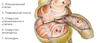
The appendix is inserted into the cecum, through a small lumen microorganisms enter the gastrointestinal tract, but the intestinal contents cannot penetrate from the gastrointestinal tract into the appendix, due to which the cavity of the lymphatic organ remains free. The appendage produces amylase and lipase, enzymes involved in digestion, the breakdown of fats, and the hormone serotonin, which is called the hormone of happiness. Serotonin, along with other functions, is involved in the work of sphincters and intestinal motility.
Etiology of appendicitis
There are several theories about the occurrence of appendicitis. The mechanical theory believes that the cause of the inflammatory process is blockage of the lumen caused by fecal stones, and increased reproduction in the cavity of the appendix of pathogenic microorganisms that entered it before this blockage occurred. The second reason is the pathological proliferation of lymphoid follicle cells. Much less often, the lumen in the appendix is blocked by a foreign body, tumor, or accumulation of parasitic microorganisms. As a result, the mucous membrane becomes inflamed and the walls of the appendix die. It has been noticed that in people suffering from lazy bowels and constipation, appendicitis occurs more often. At the beginning of the last century, there were reports that inflammation of the appendix was found in patients suffering from ascariasis.
Proponents of the infectious theory believe that appendicitis can be provoked by infectious diseases such as tuberculosis, typhus, amoebiasis and other parasitic infections. But this theory does not find scientific confirmation, since the parasites that provoke this pathology have not been identified. The vascular theory sees the etiology of appendicitis in vasculitis, in other words, inflammation of the blood and lymphatic vessels that penetrate the body of the appendix. There is also an endocrine theory, the proponents of which believe that the provoking factor is serotonin, a hormone produced in the mucous membrane of the appendix.
The first, mechanical theory, with all the variety of factors, is more confirmed than others by research and data from postoperative analyzes. But, despite the fact that other theories are poorly supported, they once again prove that the appendix is important in the body.
How does the appendix work?
The appendix has its own mesentery in the shape of a triangle between the cecum and ileum. It contains adipose tissue, vessels and nerve branches. At the base of the process, the peritoneum forms folded pockets. They are important in terms of limiting the inflammatory process.
The wall of the appendix is formed by three layers or membranes:
- serous - representing the continuation of a single layer of peritoneum with the ileum and cecum;
- subserous - consists of adipose tissue, it contains a nerve plexus;
- muscular;
- mucous membrane.
The muscular layer, in turn, consists of:
- from the outer layer with the longitudinal direction of the fibers;
- internal - the muscles move circularly.
The submucosal layer is formed by cruciform elastic and collagen fibers and lymphatic follicles. In an adult, there are up to 80 follicles per cm2 of area with a diameter of 0.5 to 1.5 mm. The mucous membrane forms folds and outgrowths (crypts).
In the depths there are secreting Kulchitsky cells that produce serotonin. The epithelium is prismatic single-row in structure. Between it are goblet cells that secrete mucus.
The appendix communicates with the lumen of the cecum through its orifice. Here it is covered by Gerlach's own valve, formed by a fold of mucous membrane. It is well expressed only by the age of nine.
Inflammation of the appendix and its symptoms
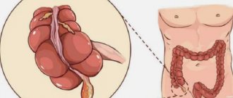
Appendix: schematic representation
Inflammation of the appendix can be recognized by the following signs:
- The pain first appears in the upper abdomen (at the level of the stomach), or near the navel. Sometimes it spreads throughout the entire abdominal cavity. And after a few hours the pain moves down to the right.
- For some time, the pain is moderately constant, but at some point it may stop due to the necrosis of the nerve fibers. The pain may intensify while walking, coughing, or sudden movements.
- In acute appendicitis, appetite disappears, nausea and vomiting appear, which is reflexive in nature, and body temperature rises to 37-38oC. If you measure the temperature in the right and left armpits, then on the right it will be higher.
Location of the appendix
The study of topographic anatomy obliges doctors to know not only on which side the appendix is located, but also to provide options for its normal location.
There are 8 main positions of the appendix:
- pelvic or descending (half of the cases according to the frequency of detection) - the freely hanging end reaches the pelvic organs, in women it can be “soldered” to the right ovary, in men it is in contact with the ureter (64%);
- ascending (subhepatic) - rare;
- anterior in the iliac fossa on the right is a rare occurrence;
- median (0.5%) - the apex is retracted into the sacral area;
- lateral (1%) - outside the cecum;
- intraperitoneal or retroperitoneal - the process is located posterior to the cecum (another name is retrocecal, observed in 32% of cases);
- extraperitoneal or retroperitoneal (2%);
- intramural - the process is fused with the posterior wall of the cecum and can be located in its layers.
So, to the questions “which side is the appendix on” and “in which side to look for the appendix” we will answer with a high degree of probability - on the right. Because the left-sided position of the process is very rare.
Mobility and movement of the free end are accompanied by pain of various types in appendicitis. In 70% of cases, the appendix is freed from adhesions along its entire length. But in 30% of people it is fixed by various adhesions.
Positions are determined by the deviation of the process body
Diagnostics
Appendicitis, or inflammation of the appendix, occurs, as a rule, at an active age - 20-40 years. Less often - in children. Women get sick much more often than men, and this is probably why in the Middle Ages doctors mistook inflammation of the appendix for uterine abscesses. The incidence of the disease is 4-5 people per 1000 per year. A doctor can detect appendicitis by palpating (feeling) the right lower abdomen. There is pain here, the muscles are overly tense. There is a feeling of fullness and pain in the stomach, radiating to the right iliac or left hypochondrium if you press at McBurney's point (in the middle between the navel and the ilium on the right). Laboratory diagnosis of appendicitis is carried out only after surgery; it allows us to understand the morphological nature of the disease. There are 3 main forms known:
- Catarrhal
- Phlegmonous
- Gangrenous
Postoperative diagnosis is necessary to prevent subsequent postoperative complications. Today, the only, and perhaps the most effective method of treating acute appendicitis is appendectomy, that is, a surgical operation to remove the inflamed organ.
How to find the appendix?
The vermiform appendix extends from the lower part of the cecum 2–3 cm below the junction of three longitudinal muscle bundles (ribbons). The appendix normally looks like a pink shiny cord. It has a tubular structure. The length of the appendix ranges from 2 to 25 cm, and the thickness is 0.4–0.8 cm.
Types of discharge from the cecum:
- the intestine narrows funnel-shaped and smoothly passes into the appendix;
- the intestine sharply narrows and bends before the transition;
- the process extends from the dome of the intestine, although its base moves posteriorly;
- extends back and down from the confluence of the ileum.
The base, body and apex of the process are distinguished. The shape of the process can be:
- embryonic - the continuation of the cecum is emphasized;
- stem-shaped - has the same thickness along the entire length;
- cone-shaped - the diameter at the base is wider than at the top.
The greatest difficulty in diagnosing appendicitis is associated with the varied location of the body and apex of the appendix. This feature causes diagnostic errors and makes it possible for inflammation to masquerade as symptoms of other diseases of neighboring organs.
In addition to the McBurney point, there are many recommendations from different authors that surgeons can use
For doctors, the McBurney point is a reference point on the human abdomen. It can be determined by mentally drawing a straight line from the navel to the superior process of the ilium on the right (or on the left in case of a rare feature - mirror image of organs). Next, the distance must be divided into 3 equal parts.
The desired projection point for the base of the appendix can be found at the junction of the outer and middle parts. This is only a single example of the projection of the appendix.
Acute appendicitis is a disease that is not to be trifled with
Scar after appedixectomy
It is important to know that at the first sign of illness you should immediately go to the hospital. Inflammation of the appendix develops quickly. Therefore, the phrase “delay is like death” is precisely about appendicitis. Self-medication in this case is unacceptable. Traditional medicine also does not know methods of treatment for inflammatory processes of the appendix. Sometimes two days are enough for a patient who is not provided with timely assistance to die. The reason for such a rapid development of the disease is that the pus produced in the inflamed organ does not find a way out and bursts the walls, causing perforation of the tissue. People say that the appendix burst.
Pus leaks into the abdominal cavity, causing infection of the abdominal tissue and blood. True, traditional medicine has had its say about the prevention of the disease, and traditional medicine agrees with it in that the diet of each of us must necessarily contain fiber, which gently cleanses the intestines of fecal stones. Therefore, we should eat more plant foods in the form of greens, vegetables and fruits.