In the laboratory of the CMD Central Research Institute of Epidemiology, you can take a stool test for the following types of research:
- General analysis of stool - coprogram
- Fecal Occult Blood Test
- Feces for occult blood (Colon View Hb/Hp) (Colon View hemoglobin (Hb) and hemoglobin/haptoglobin complex (Hb/Hp))
- Fecal analysis for helminth eggs
- Analysis of stool for helminth eggs and protozoan cysts using a Parasep concentrator
- Stool analysis for carbohydrates
Coprogram
A coprogram is a general clinical study that includes the study of physicochemical properties and microscopic examination of feces.
The coprogram allows you to evaluate the function of the stomach and pancreas, liver dysfunction, the speed of food passage through the digestive tract, the presence of inflammatory, destructive changes, and absorption processes in the intestine. A general stool analysis is also informative in the diagnosis of colitis, intestinal infections, and dysbacteriosis.
During the study, it is possible to detect pathological impurities that indicate the presence of a pathological process in the organs of the gastrointestinal tract: ulcerative defects, helminthic infestations, intestinal tuberculosis, disintegrating tumors, etc.
Stool analysis includes assessment of physical properties, chemical analysis and microscopy.
- Physical properties: consistency, shape, color and smell of feces, macroscopic mucus impurities, blood, remains of undigested food.
- Chemical analysis: blood content, bilirubin, stercobilin, leukocytes, pH reaction.
- Microscopy: food residues, epithelium, mucus, crystals, erythrocytes, leukocytes, protozoa, yeast and other indicators.
Types of stool diagnostics
There are the following types of stool analysis:
- general research;
- examination for occult blood;
- analysis for the presence of helminths;
- bacteriological analysis.
A general stool analysis is also called a coprogram. The study covers macroscopic indicators that can be determined visually - color, shape, consistency, visible impurities. These characteristics make it possible to assess the condition of the digestive tract. The presence of various inclusions may indicate a lack of a certain enzyme. Changes in color or consistency also provide information about the disease state. A general examination using a microscope helps to assess the level of substances that are normally found in the stool. Acidity, the presence of various chemicals, admixtures of blood, pus or mucus are assessed.
The results of the coprogram play an important role in making a diagnosis and determining treatment tactics. In addition, stool analysis must be done over time. This will allow you to assess the adequacy of treatment and its effectiveness, and will also indicate the need for correction of treatment measures.
Laboratory diagnosis of stool for occult blood is an indispensable diagnostic technique in surgery. It is also used in other medical fields where it is necessary to exclude or confirm bleeding in the digestive tract. Even a small number of blood cells can be determined using the test. The study is carried out with drugs that are highly sensitive to blood components and react to their presence in the stool.
Analysis of feces for the presence of helminths allows us to detect elements or traces of the activity of various parasites that live in the human body. If eggs or cysts of worms are found in the stool, this is a criterion for diagnosing the disease. Unfortunately, the absence of a positive result does not always make it possible to exclude pathology. It is advisable to repeat the analysis several times if clinical characteristics indicate the presence of invasion, but this fact has not been confirmed in the laboratory.
Bacteriological research allows you to assess the state of the microflora that is present in the digestive tract. The digestive system is a continuation of the external environment into the internal one. This factor, as well as the need for certain biological processes, determines the fact that a significant number of bacteria are present on the walls of organs, from the oral cavity to the rectum. Normally, they do not cause harm to the body and, on the contrary, participate in physiological processes. Deviations from the norm cause their uncontrolled growth or contribute to the addition of other, pathological flora. Determining the type of bacteria in feces (for example, dysentery), their quantity and sensitivity to antibacterial therapy is an important element in the diagnosis of infectious pathology, dysbacteriosis, as well as a determining factor in the choice of therapy.
go to analyzes
Fecal occult blood test
A fecal occult blood test is intended to diagnose hidden bleeding from the gastrointestinal tract (GIT).
The cause of bleeding in the gastrointestinal tract can be erosive or ulcerative damage to organs, varicose veins, helminthic infestations, tumors, diverticula, polyps, hemorrhoids, etc. The intensity of bleeding varies significantly. The most difficult thing to diagnose is small chronic bleeding, which does not manifest itself clinically for a long time.
A fecal occult blood test is also included in the medical examination program and is carried out once every 2 years, starting at the age of 49, for early diagnosis of colorectal cancer.
The CMD laboratory has two options for testing stool for occult blood:
- A fecal occult blood test is used to diagnose hidden bleeding from the lower gastrointestinal tract. The test is based on the detection of red blood cell hemoglobin in the stool.
- Colon View Hb/Hp is a new generation test based on the detection of hemoglobin and hemoglobin/haptoglobin complex (Hb/Hp). The hemoglobin molecule breaks down as it passes through the gastrointestinal tract, and the hemoglobin-haptoglobin complex is a stable form of hemoglobin, so it can be detected even after a fairly long passage through the intestines. Thus, this test is used for screening examination of all parts of the gastrointestinal tract.
What diseases can be diagnosed using a stool test?
Let us consider the diseases indicated by changes in various parameters of this study.
- An increased amount of feces is observed with enteritis, pathology of the hepatobiliary zone, with diarrhea and pancreatitis. A reduced amount is observed with constipation.
- Stool compaction accompanies constipation, stenosis and intestinal spasms. A more liquid consistency is observed during fermentation processes, during diarrhea, pancreatitis, cholecystitis, and infectious processes.
- The color of feces can change with putrefactive dyspepsia, bleeding, colitis, constipation, the presence of ulcers, impaired pancreatic function, hepatitis, and pathology of the hepatobiliary area.
- Mucus in stool will help in the diagnosis of infectious processes, polypous growth, malabsorption, hemorrhoids, cystic fibrosis, constipation, and intestinal obstruction.
- The presence of blood is observed in hemorrhoids, proctitis, ulcerative colitis, infectious process, the presence of diverticula, cirrhosis, and oncological process.
- Soluble protein in feces accompanies ulcerative colitis, bleeding, dyspepsia, pancreatitis and other inflammatory diseases.
- Stercobilin in feces is observed with pathological hemolysis of red blood cells and increased bile production.
- Discoloration of stool accompanies obstructive jaundice, pancreatitis, hepatitis, cholangitis, cholelithiasis.
- The presence of inclusions indicates a violation of enzymatic function. Fatty elements are observed with insufficiency of bile, starch appears with insufficiency of pancreatic enzymes. Fiber and protein fibers may also appear, which also indicates decreased digestion.
Analysis of stool for helminth eggs and protozoan cysts
Helminths (worms) are parasitic worms that live in the body of humans and animals and cause diseases called helminthiasis (helminthic infestations). The disease in humans is most often caused by roundworms (nematodes) and flatworms (platytodes), which are divided into tapeworms - cestodes and flukes - trematodes. Helminths change their host in the process of life development, and the range of hosts, routes and mechanisms of transmission are individual.
Clinical manifestations are largely determined by the localization of the parasite, its number and feeding habits. Helminths, parasitizing in the human body, can have a mechanical effect, damaging the tissues and organs of the host, toxic and allergic phenomena, metabolic disorders, closing the lumen of the intestines, excretory ducts of the liver, gall bladder, pancreas, disrupting intestinal motility, irritating the receptors of the intestinal wall.
Protozoa are single-celled organisms. Like helminths, during the development process protozoa go through several stages and change hosts. Many protozoa have two forms of existence - vegetative and cystic. The cyst has a durable shell and ensures the survival of the protozoan in unfavorable, and even extreme, conditions. It is cysts that usually infect humans.
A stool test for helminth eggs and protozoan cysts must be taken if a parasitic infection is suspected, to monitor the effectiveness of antiparasitic treatment, before hospitalization, when preparing a medical record, or certificates. For example, for children before visiting the pool.
In the CMD laboratory, stool analysis for helminth eggs and protozoan cysts is carried out using two different methods:
- Feces for helminth eggs and protozoan cysts: microscopic method; "thick smear" method and Kato staining
- Modified sedimentation method using PARASEP concentrators (enrichment method). Analysis of feces for helminth eggs and protozoan cysts using a Parasep concentrator: the use of PARASEP concentrators makes it possible to identify helminths and protozoa even in small quantities.
Rules for storing stool before analysis
Before performing the test, you should check with your doctor about how to properly store the container with feces. Recommendations may vary depending on the initial diagnostic goals. It is often possible to collect material in the evening.
If a coprogram is required, you should have a bowel movement in the morning and immediately take the feces for examination. You can also do this in the evening by placing the container with the material in the refrigerator on the main shelf. If stored on the door or in the freezer, the result will be distorted. The maximum allowable time between defecation and transportation is 9 hours.
If testing for helminth eggs is carried out, feces can be stored in the refrigerator. It will be necessary to additionally use preservative components. They must be taken from the laboratory assistant in advance. It is better to deliver the material as soon as possible. Otherwise, the enzymes present in the excrement will destroy the helminths.
Stool analysis for carbohydrates
Stool analysis for carbohydrates is used to diagnose congenital or acquired deficiency of the lactase enzyme in children of the first year of life.
Lactase is an enzyme that is produced by intestinal epithelial cells (enterocytes) and participates in the biochemical reaction of the breakdown of lactose, otherwise known as milk sugar. When consuming milk and dairy products, if there is not enough lactase, digestion is disrupted and unpleasant symptoms appear: diarrhea, flatulence, cramps, etc.
Causes of lactase deficiency:
- decrease in enzyme production by intact enterocytes due to genetic defects or physiological immaturity of enzyme systems in children of the first year of life - primary, or congenital, lactase deficiency;
- a decrease in enzyme production due to damage to enterocytes during an inflammatory or other (food allergy, tumor) process - secondary, or acquired, lactase deficiency.
Indications for taking a stool test for carbohydrates:
- the presence in children of symptoms such as flatulence, frequent regurgitation, diarrhea, cramps, abdominal pain;
- control of correct diet selection.
Dysbacteriosis
Intestinal dysbiosis (dysbiosis) is a disorder in the gastrointestinal tract of the relationships between microorganisms. This disorder causes changes in vital processes in the body: from digestion to immune changes, accompanied by manifestations of allergies or signs of chronic inflammation.
Recently, the term “intestinal dysbiosis” has been widely used, created from the Latin words “dis” - difficulty, disturbance, disorder, and “bios” - life.
Dysbiosis is a disruption of the functioning and mechanisms of interaction of the human body, its microflora and the environment. According to the Russian Academy of Medical Sciences, almost 90% of the Russian population experience various pathological changes in the microflora, indicating the presence of intestinal dysbiosis (dysbiosis).
The main causes of dysbiosis:
- uncontrolled use of antibiotics;
- malnutrition;
- disturbed ecology;
- state of chronic stress;
- existing functional or inflammatory diseases of the gastrointestinal tract;
- cancer therapy;
- immunity disorders.
The main manifestations of dysbiosis:
- diarrhea (or constipation);
- flatulence;
- bloating;
- stomach ache.
In most cases, patients experience intolerance to certain foods and experience general allergic reactions in the form of skin itching, urticaria, various skin rashes, and prolonged cough. Characteristic symptoms of intoxication (general malaise, lack of appetite, headaches). Patients with dysbiosis often get sick and often have recurrent upper respiratory tract infections.
The importance of normal flora for humans
Each person has an individual composition of normal flora, but the functions are always the same. The total number of microorganisms on the mucous membranes of an adult is 10 times greater than the number of his own cells.
This symbiosis of flora protects the mucous membranes from the invasion of pathogenic microbes, fungi, allergens and harmful substances through the walls of the mucous membranes into the human blood, thereby maintaining the constancy of its internal environment (homeostasis).
Normal flora forms bonds with secretory immunoglobulins (protective antibodies) and forms biofilms on the surface of mucous membranes.
Normoflora is involved in digestion and produces B vitamins and vitamin K.
The biomass of microorganisms populating the intestines of an adult healthy person is 2.5–3 kg (approximately 5% of its total weight) and includes up to 450–500 different types of microorganisms.
Age-related features of intestinal microflora
In a healthy child, from the moment of birth, the intestines are rapidly colonized by bacteria. Normal microflora should populate the newborn’s intestines on the 3rd–5th day of life. In children who are breastfed, by the 10th–21st day of life, a steady growth of lactobacteria, bifidobacteria, and lactic acid streptococcus is observed in the intestines.
Human milk contains a number of substances, so-called bifidus factors, which contribute to the colonization of the intestines by these types of microorganisms in adequate quantities.
The formation of normal microbial flora in a child may be disrupted if:
- use of antibacterial drugs during pregnancy and after childbirth;
- feeding with formula rather than breast milk;
- neurological disorders in the baby, in particular with perinatal damage to the child’s central nervous system.
Under the influence of all these factors, the timing of the formation of normal intestinal microflora, as well as its qualitative and quantitative composition, changes.
In a healthy child of the first year of life, 90–98% of the entire colon microbiocenosis should be bifid flora, which provides protection for the body from pathogenic microbes at this age.
In children older than one year, when their nutritional pattern approaches the usual diet of adults, the composition of the intestinal flora stabilizes, its quantitative indicators approach the norms of adults.
In elderly people, the composition of intestinal microbes also changes: the content of bifidobacteria and lactobacilli is reduced, the content of E. coli is increased, with a change in their enzymatic activity.
Laboratory diagnostic methods Laboratory methods can be direct (isolation of living microflora from a material) and indirect (determination of products associated with the vital activity of flora).
- Direct methods include stool cultures - Dysbacteriosis with determination of sensitivity to bacteriophages or Dysbacteriosis with determination of sensitivity to antibiotics and bacteriophages
- An indirect method is: biochemical analysis of feces - Biochemical study of the metabolic activity of intestinal microflora
When can dysbacteriosis be confirmed by laboratory analysis:
- Reduction or disappearance of bifidobacteria.
- Reducing the content of complete E. coli.
- Increased content of hemolytic Escherichia coli strains.
- Change in total E. coli count.
- Presence of opportunistic enterobacteria.
- Change in the number of enterococci.
Doctors assess the degree of imbalance of intestinal flora as follows:
First degree
Latent phase of dysbiosis: manifests itself only in a decrease by 1–2 orders of magnitude in the amount of protective microflora - bifidobacteria, lactobacilli, as well as full-fledged E. coli - up to 80% of the total amount. The remaining indicators correspond to the physiological norm (eubiosis). As a rule, the initial phase does not cause intestinal dysfunction and occurs as a reaction of the body of a practically healthy person to the influence of unfavorable factors, such as, for example, a violation of the diet and a change in the climatic zone (season of the year). In this phase, it is possible for a small number of individual representatives of the opportunistic flora to grow in the intestines. There are no clinical manifestations of dysbacteriosis in this phase.
Second degree
The starting phase of more serious disorders is characterized by a pronounced deficiency of bifidobacteria against the background of a normal or reduced number of lactobacilli or their reduced acid-forming activity, an imbalance in the quantity and quality of E. coli, among which the proportion of lactose-negative or citrate-assimilating variants is increasing. At the same time, against the background of a deficiency of protective components of the intestinal microbiocenosis, the proliferation of either plasma-coagulating staphylococci, or Proteus, or fungi of the genus Candida occurs. Vegetation in the intestines of Proteus or plasma-coagulating staphylococci in this phase of the development of dysbacteriosis is often transient rather than permanent. Functional digestive disorders are not clearly expressed - sporadically loose stools of a greenish color with an unpleasant odor, with a shift in pH to the alkaline side, sometimes, on the contrary, stool retention, sometimes nausea is noted.
Third degree
The phase of aggression of the aerobic flora is characterized by a clear increase in the content of aggressive microorganisms - at the same time, Staphylococcus aureus and Proteus, hemolytic enterococci multiply up to tens of millions in association, replacement of full-fledged Escherichia with bacteria of the genera Klebsiella, Enterobacter, etc. is observed. This phase of dysbiosis is manifested by intestinal dysfunctions with disorders of motility, secretion enzymes and absorption. Patients experience frequent, loose stools, often green in color, decreased appetite, deterioration in health, children become lethargic and moody.
Fourth degree
The phase of associative dysbiosis is characterized by a deep imbalance of the intestinal microbiocenosis with a change in the quantitative ratios of the main groups of microorganisms, a change in their biological properties, and the accumulation of toxic metabolites. Vegetation of enteropathogenic serotypes of E.coli, Salmonella, Shigella and other pathogens of acute intestinal infections is characteristic. Clostridia may multiply. This phase of dysbiosis is characterized by functional disorders of the digestive system and disturbances in general nutritional status, body weight deficiency, pale skin, decreased appetite, frequent stools mixed with mucus, greens, and sometimes blood, with a sharp putrid or sour odor.
According to I. B. Kuvaeva and K. S. Ladodo (1991)
If the study for dysbacteriosis does not reveal pathogens of dangerous intestinal infections, then to restore the normal balance of microflora, bacteriophages can be used, which are destructive to microbes, but safe for humans. The result form, which indicates the sensitivity of the identified pathogenic microorganism to bacteriophages, indicates the manufacturer of the drug and the batch.
Using these data, you can buy exactly the bacteriophage with which the testing was carried out.
Good health to you! Your KDL laboratory
How to prepare for a stool examination?
In agreement with the attending physician, 2 days before the study, medications that affect intestinal motility (laxatives, enzymes, sympathomimetics) are discontinued; drugs that affect the blood coagulation system (acetylsalicylic acid, warfarin, non-steroidal anti-inflammatory drugs); rectal suppositories and medications, the impurities of which in stool change its color.
The study is carried out no earlier than 2 days after an X-ray examination of the stomach and intestines using contrast agents. It is prohibited to collect stool from diapers, after an enema, during menstruation or within 3 days before or after it, in the presence of bleeding hemorrhoids, bleeding with prolonged constipation, with impurities of urine or discharge from the genitals, with pieces of undigested food.
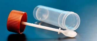
Biomaterial for analysis is accepted in a sterile plastic container, which can be obtained free of charge at any CMD office.
After natural bowel movements, feces are collected in a container with a spoon and a screw cap in the amount of 1/3 of the container’s volume from 3-5 different places. Avoid contact of the biomaterial with the surface of the toilet bowl. Delivered to the laboratory in a primary container. Screw the container lid tightly.
To analyze stool for carbohydrates : stool from a pre-disinfected pot or bedpan (you can use a plastic bag or oilcloth) is collected in a sterile disposable container. Take a small amount (4-5) from several places with a disposable spatula attached to the lid of the container. If the stool is liquid, then a small amount, no more than 5 ml, should be poured into a container. Close the container lid tightly. Delivery to the laboratory on the day of collection of the biomaterial.
General analysis (coprogram)
The coprogram helps to assess the functioning of the gastrointestinal tract, identify inflammation, colitis of various origins and other pathologies.
Preparation for the analysis involves avoiding taking medications and using enemas. You also need to adhere to a special diet for 4-5 days. It involves avoiding exotic foods, fruits and vegetables with coloring pigments. These are tomatoes, beets, blueberries.
It is better to collect feces after a night's sleep. If this is not possible, the procedure should be performed in advance. The maximum allowable time before transportation to the laboratory is 8 hours.
In this case, the container with feces must be stored in the refrigerator. You must first urinate and perform genital hygiene. It is recommended to collect feces from different places, avoiding particles of undigested food.


