Lymphoid hyperplasia
Lymphoid hyperplasia in the terminal ileum is a common finding in childhood and adolescence.
In this case, against the background of the absence of an inflammatory process, symmetrically located nodules with clear contours are visualized. Differential diagnosis between Crohn's ileitis and lymphosarcoma can sometimes be difficult. Narrowing of the lumen against the background of thickening and smoothing of the folds of the mucous membrane is not a manifestation of benign lymphoid hyperplasia, in which there is also no ulceration of the mucous membrane. Lymphoid hyperplasia can occur in combination with familial colon polyposis [19]. The ability to distinguish lymphoid hyperplasia from adenomatosis is extremely important to prevent unnecessary surgical treatment.
Lymphoid hyperplasia of the colon can also occur in newborns and children [8,15,21]. Small, homogeneous polypoid lesions are found throughout the entire colon or are limited to a segment of it. The lesions are relatively homogeneous, spherical in shape, with clear contours, their size varies from 1 to 3 mm in diameter [21].
On histological examination, the lesions are solitary lymphoid follicles of the submucosal layer and the lamina propria protruding into the intestinal lumen. They are absolutely benign.
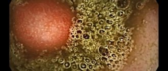
Rice. 18–9. Lymphoid hyperplasia. The granular mucosa contains hyperplastic lymphoid follicles.
Follicular hyperplasia in most cases is transient and may soon disappear. The true mechanism of intestinal lymphoid hyperplasia remains unknown, but it has been established that follicular hyperplasia may be a morphological reaction of functionally inadequate lymphoid tissue in response to various stimuli.
Patients may suffer from diarrhea, abdominal pain, and stool mixed with mucus and blood. Using double-contrast irrigoscopy, it is easy to detect multiple small-focal lesions.
The endoscopic picture of each individual lesion is as follows: a spherical nodule, with a smooth surface, covered with an intact or hyperemic mucous membrane (Fig. 18–9). A central umbilical retraction on the surface of a small formation can be detected during irrigoscopy in the form of a barium speck or directly visualized during endoscopy. The nodules are localized against the background of intact mucosa. The diagnosis is easily made based on obtaining lymphoid tissue from a biopsy.
Lymphoid hyperplasia should be differentiated from many diseases - cystic fibrosis, polypoid gangliofibromatosis, Gardner's syndrome, trichocephalosis, Peutz-Jeghers syndrome, giardiasis with hypogammaglobulinemia and polyposis with Hirschsprung's disease [21]. However, the most important differential diagnoses are SATC and ulcerative colitis.
Morphometric characteristics of lymphoid nodules (Peyer's patches) of the small intestine in ontogenesis
ANNOTATION
The interconnectedness in time and space of the development and formation of the structural and functional units of the small intestine, the lymphoid nodules of the feather plaque, is a morphological reflection of the integration of its hydrolytic-transport immune functions.
ABSTRACT
Time and space interconnectedness of development ormation of structurally functional units of the small intestine, lymphoid nodules plaque morphology is a reflection of the integration of its hydrolytically transport and immune function.
Key words: small intestine, lymphoid nodules, integration, morphology, Peyre's patches, structure, function, structure, ontogenesis, immunity, follicles, cell.
Keywords: small, intestine, lymphoid, nodules, integration, morphology, peyrlovoy, plaque, function, structure, ontogenesis, immunity, follicles, cell.
Among the immune formations of the digestive system, the lymphoid nodules (Peyer's patches) of the small intestine play an exceptional role. They, like the thymus, tonsil, and appendix of mammals, belong to the lymphoepithelial organs in which lymphopoiesis occurs and are in close interaction with reticular tissue and epithelium. In birds, the thymus and bursa of Fabricius are the primary organs of lymphopoiesis, in which T- and B-lymphocytes are differentiated, respectively. In mammals, an analogue of the bursa of Fabricius has not been found, although some authors consider the appendix and Peyer's patch as such. The basis for such assumptions may be their topographic location - the digestive tract, as well as their location on the antimesenteric side, in contact with the epithelium [1,2,3,4,5,6,7,8,9,10,11.].
Small intestine of white outbred rats at the ages of 1, 3, 7, 14, 21, 30 and 90 days after birth. was secreted from the pyloric part of the stomach to the cecum. Macroscopically, under a stereoscopic microscope MBR-9, areas of lymphoid nodules were identified. After determining its parameters in two mutually perpendicular planes, the distances between them, the number of nodules in each cluster, and the total mass after their careful isolation were measured. On semi-thin sections in each cluster nodule, structural and functional zones, lymphocytes, lymphoblasts, reticulocytes, plasmacides, macrophages, and mitotic dividing cells were determined on an area of 15,000 μm2.
Lymphoid plaques of the small intestine are formed on the 19th day of embryonic development in the caudal part of the duodenum on the antimesenteric side. In newborns, they have the appearance of an oval or round formation, visible from the side of the serous membrane. As with the development and formation of the small intestine at all stages, the development of Peyer's patch can be divided into stage I - 1-3 days; II - 7-14; III - 15-21; IV - 22-90 days. Each of these stages is characterized by certain changes in number, structure and cellular composition. The most distal plaque is formed on the first day after birth of the rat near the ileocecal region, also on the free edge of the small intestine. No other patterns were found in the topography of lymphoid nodules at birth.
During this period, lymphocytes and blast cells are located diffusely, nodules are not detected. Plasmocides are not detected; macrophages, both light optical and electron microscopic, are found in isolated cases. Among the accumulation of blast and stromal cells, mitotic cells are found. It must be assumed that in lymph nodes the number of cells increases from term to term due to the division of diffusely located cells, as well as their migration from the circulating blood.
After 7 days along the small intestine, the number of lymph nodes increases by 3 times on average. Linear dimensions increase (Table 1) almost 10 times compared to 1 day after birth. In the diffuse state of lymphoid nodules, the absolute number of lymphocytes and blast cells increases, respectively, by an average of 2 and 1.65 times (Table 1). On the same area, while maintaining the number of stromal (reticular) cells, mitotic dividing lymphoblasts increase significantly - up to 0.8 ± 0.03. For the first time, single macrophages and plasmacides are detected among the accumulation of lymphoid cells. If macrophages in the cytoplasm contain moderate amounts of polymorphic secondary lysosomes, then plasmocids are not functionally active: the profiles of the granular endoplasmic reticulum are few, the cisterns are not expanded, the Golgi complex is moderately developed.
Table 1.
Dynamics of morphometric characteristics of lymphoid nodules of the small intestine of white rats (M ± m , n = 6)
| Age, days after birth | Number of nodules along the small intestine | Nodule sizes cm x 101 | Number of follicles in individual nodules |
| Newborns | 2,3±0,5 | (1.0±0.8)x(2.0±0.1) | Not determined |
| 7 | 7,5±2,5 | (2.2±0.4)x(2.5±0.5) | Not determined |
| 14 | 10,8±2,0 | (4.3±0.8)x(3.5±1) | 5,6±2,0 |
| 21 | 14,5±2,5 | (5±1)х(5±1) | 12,0±3,0 |
| 30 | 14,5±2,8 | (5.6±1.4)x(6.5±1) | 14,0±2,5 |
| 90 | 2,8±2,4 | (6.1±1.8)x(6.5±1) | 13,5±2,0 |
Figure 1. Dome-shaped bulging lymphoid nodule of Peyer's patch of the small intestine 14 days after birth in rats. Staining: hematoxylin-eosin. OK. 10, vol. 40
14 days after birth along the small intestine the number of lymphoid nodules reaches 10.8±2.0. the size of each of them increases on average 10 times. If in the previous periods of the study, accumulations of lymphocytes were diffuse tissue located in the lamina propria under the developing villi and crypts and slightly protruded into the intestinal lumen, then by the end of the second week after the birth of rats, the localization zone of the lymphoid nodule begins to bulge dome-shaped into the intestinal lumen and does not contain villi. and crypts (Fig. 1). The epithelium lining this area of the mucous membrane contains single goblet cells and is infiltrated with a relatively large number of lymphocytes (compared to the epithelium of the villi or crypts). It should also be noted that in the absence of zones, individual nodules first appear among the accumulation of lymphoid cells. They differ more clearly in the proximal part of the small intestine, although a similar trend appears in the distal (ileal) part of the small intestine. Simultaneously with the increase in lymphoid plaques, the boundaries between individual nodules and zones do not differ. However, this reveals an increase in the absolute number of lymphocytes (110±3.0) while maintaining the absolute number of blast and reticular cells per unit area. Plasmocids and macrophages are still rare. The number of mitotic dividing cells increases on average 2.5 times and becomes equal to 2.0±0.3 (Table 1).
After 3 weeks, the rats switch to definitive feeding. By this time, the total number of Peyre's plaques increases by an average of 134% compared to the previous period and becomes equal to 14.5 ± 2.5, it should only be noted that there is a significant variability in their number - min 11, max - 18. Such a significant difference in their number along It is still difficult to explain the organ in animals of the same litter.
It is by this time that they increase significantly - (5±1x5±1cm x10-1) - in size, and also increase quantitatively: in each of the plaques the number of follicles increases almost 2 times over the last week under study. Histologically, it should be noted the appearance in the follicles of the nodule of zones characteristic of an adult animal: germinative, follicular (Fig. 2), parafollicular and dome, lined on the outside with single-layer epithelium and infiltrated with numerous lymphocytes (Fig. 3). Counting individual types of cells in different zones of the follicles indicates that most mitotic divisions (3.1 ± 0.2) are in the germinal zone; in other zones they are rare. Medium and small lymphocytes are insignificant in the germinal center (70±1.5) and are on average 1.7 times larger in other zones. Lymphoblasts are naturally numerous in the center of the follicle (38.5 ± 1.0) and relatively few in all other zones. There are relatively fewer reticulocytes in the parafollicular and follicular zones, more and, on average, in almost equal numbers in the germinal zone and dome (9.0-10.0).
The relatively sharp increase in the number of macrophages in the dome of each nodule is noteworthy. Plasmicides are rare in all zones. The epithelium associated with the dome zone contains numerous medium and small lymphocytes. They are located at different levels of the epithelial layer. The basement membrane separating the epithelium and the dome of the follicle is thin, but discontinuous. Lymphocytes pass through it in both directions.
90 days after the birth of rats, macroscopically, from 17 to 28 lymphoid nodes are detected on the free edge of the small intestine, located singly or in groups in the lamina propria of the mucous membrane. The distance between them ranges from 50 to 180 mm (on average 55.1 ± 2.2 mm). They, as we noted earlier, are located between the distal part of the duodenum and the terminal part of the ileum. If this segment is divided into 3 parts, then in the proximal and distal third of the small intestine the proportion of lymphoid nodules is 26.6±3.9% and 33.4±3.1%, respectively. The rest of them are concentrated in the middle third of the organ (40.2±4.3%, P<0.001). It must be assumed that the relatively larger number of lymphoid formations in the middle part of the small intestine is associated with a high concentration of peptides that undergo final hydrolysis and absorption.
Figure 2. Germinal and follicular zones of the lymph node of Peyer's patch of the ileum of 3-week-old rats. Staining: hematoxylin-eosin. OK. 10, vol. 20
Figure 3. Epithelium over the dome of the lymph node of Peyer's patch 3 months. rats is infiltrated with numerous lymphocytes. Staining: hematoxylin-eosin. OK. 10, vol. 100
Having isolated visible lymphoid nodules and determined their weight, we were able to establish that in mature animals it ranges from 3 to 52 μg (on average 29.6 ± 1.5 μg). The total mass of tissue aggregated into lymphoid nodules averages 704.5 ± 15 μg.
The dimensions of the nodules are (6.1±1.8x6.5±1) cm x 10-1. In each nodule the number of follicles is 13.5±2.0. The nodules protrude dome-shaped into the intestinal lumen and are surrounded at the periphery by crypts and villi. The epithelium lining the dome continues at the base into the crypt. In this part, the epithelium contains mitoses and single goblet cells containing a PAS-positive secretory product. Most of the epithelial lining does not contain goblet cells.
The prismatic epithelial cells lining the dome of the lymph node do not have a glycocalyx on the surface of the microvilli. Their nuclei are at different levels due to interepithelial lymphocytes located in the basal or middle part of the layer. The basement membrane is about 100 µm thick, separating the epithelium from the underlying dome of the lymphoid nodule, but is discontinuous due to the migration of lymphocytes in both directions.
The lymph node, regardless of the number of follicles composing it, is located mainly in the lamina propria and captures part of the submucosa, thinning the muscular layer, spreading to the serous membrane. In this regard, they are distinguished under the serous membrane along the free edge of the small intestine as an independent small-tuberous formation.
Regardless of the number of follicles that make up a lymph node, each of them has a round or oval shape, separated from the adjacent one by a parafollicular zone. Along the periphery, each lymphatic follicle, without sharp boundaries, passes into the surrounding loose connective tissue of the lamina propria or submucosa.
Lymphoid follicles of lymph nodes are characterized by a layer-by-layer arrangement of lymphoid cells in it. The separation of one zone from another is carried out by reticular cells (Fig. 4). In the germinal center, blast cells are loosely located. The surrounding follicular zone consists mainly of small lymphocytes (120±2.0). Other cells are in relatively small numbers; the number of mitotic dividing cells is almost the same as in the germinal center.
Lymphoblasts are large round cells with a relatively large round nucleus. Less commonly, the core may have a bay-shaped depression. In the cytoplasm, the Golgi complex consists of 1-2 short flattened cisterns and single vesicles. Mitochondria are small, single. Ribosomes are numerous, evenly distributed along a narrow rim of the cytoplasm. Accumulating in groups of 3-4, they form groups separated by long-processed reticular cells.
In the follicular and parafollicular zones, small and medium-sized lymphocytes are located more densely, in groups of 5-8 cells, surrounded by the same anastomosing reticular cells. The dome of the follicle is significantly different from other zones. In this zone, large blast cells are not visible, however, small and medium-sized lymphocytes, macrophages, single plasmablasts, tissue basophils and eosinophils are detected in groups in this zone.
Figure 4. Reticular cells of the lymph node of Peyer's patch and layer-by-layer arrangement of lymphoid cells. Semi-thin slice. Color: basic fuchsin-methylene blue. OK. 10, vol.20.
Macrophages in the cytoplasm have secondary lysosomes and myelin-like formations of various sizes and shapes.
Compared to other zones, the dome contains relatively more lymphatic and blood capillaries lined with flattened endothelium.
Thus, lymph nodes and the follicles of which they consist develop mainly after birth and their formation, the formation of zones occurs by the time of transition to definitive nutrition. The final formation of each zone and the perfect integration of the cells that make up each zone occurs by the time of puberty.
The immunocompetent cells of the lymphoid nodules of the gastrointestinal tract, in contrast to similar other organs of the immune system not related to the gastrointestinal tract, are distinguished by the highest ability to migrate, tens of times greater than in other organs. The antigen is transported from the intestinal lumen through M cells to the dome area of the Peyer's patch. There, with the help of a macrophage, it is presented to T- and B-lymphocytes. Activated, they are delivered through the lymphatic tract to the mesenteric lymph nodes and spleen. Subsequently, T- and B-lymphocytes enter the lamina propria of the mucous membranes of the gastrointestinal tract, respiratory and genitourinary systems, lacrimal, salivary, and mammary glands. T-lymphocytes are predominantly found between epithelial cells, B-lymphocytes differentiate predominantly (80%) JgA - secreting plasmocides. On this basis, grouped lymph nodes should be considered as the main activator of the immune properties of both the gastrointestinal tract and the lung and urogenital tracts. Stimulation of the immune system of the small intestine by normal microflora leads to an increase in the level of sJgA in the secretions of the bronchopulmonary tract, cervix, elimination of bacterial vaginosis, and remission of bronchopulmonary diseases [7,8, 9, 10, 11].
The predominant settlement of activated B-lymphocytes in the mucous membranes and differentiation into JgA-producers occurs under the influence of Th-2 lymphocytes producing IL-4, IL-5 [10,11].
Under the condition of full activation in the dome zone, T helper cells are activated and proliferate under the influence of the IL-4 they secrete, and then differentiate into Th1 and Th2. Unbalanced activation of these cells is fraught with the development of autoimmune cell-type processes. Th2 secretes IL-4 and IL-5, which determine the development of a humoral response, as well as antiparasitic defense with the participation of eosinophils. Excessive predominance of Th2-dependent processes leads to the development of allergies.
Cytotoxic CD8+ T cells migrate to the lamina propria and interepithelial areas. Being between the enterocytes, CD8+ cells under the influence of bacterial antigens penetrating here, they are activated and participate in the presentation of the antigen and the production of cytokines. It is not excluded that they simultaneously induce apoptosis of enterocytes that allow antigens to pass through.
Thus, grouped lymphoid nodules are an important tool for the dialogue of the macroorganism with antigens of microorganisms and food components. Developing and being activated under their influence, they provide an optimal relationship between the central and peripheral organs of the immune system, a barrier function against the introduction of foreign antigens by activating its humoral and cellular components, and the development of tolerance.
References: 1. Azizova M.A., Akhmedova Kh.Yu., Yuldashev M.A. Kinetics of lymphocyte populations in the lymph node of the ileum of rats // Ros. morphol. Gazette.- 2001.-No. 1-2.-S. 121-122. 2. Aminova G.G. Cytoarchitecture of lymphoid tissue associated with the wall of the cecum in humans during adolescence // Morphology. - 2002. - No. 4. - P. 53-55. 3. Afanasyev Yu.I., Nozdri V.I., Subbotin S.M. Lymphatic nodule of the appendix //Arch.anat.-1983.-T.85, No. 8.-S. 73-82. 4. Afanasyev Yu.I., Subbotin S.M., Nozdri V.I. Grouped lymph node // Advances in modern biology. - 1997.-T.104, No. 1(4).-P. 79-88. 5. Age-related features of the structure of lymphoid nodules of the small intestine A.Yu. Yuldashev, Z.A. Kakharov, M.A. Yuldashev, N.U. Abdukarimova //Uzbekistan Tibbiyot Journal.-2006.-No. 1.-B.72-77. 6. Grigorenko D.E. Lymphoid structures of the human duodenum in adolescence and youth // Morphology.-2002.-No. 5.-P.63-65. 7. Ulanova M.A. The system of local immunity of the digestive tract and its features in childhood // Advances of modern times. biol. and medical - 1988.-T.105, No. 2.-S. 202-216. 8. Bjenenstock J. The derivation, distribution and function of intestinal mucosae lymphocytes //Sumposium: Endocytosis and exocytosis in host devense Basel ea-2001.-Pf.1.-P.362-369. 9. Bjenen Stock J., Befus D. Gut- and bronchus – associated lymphoid tissue //Amer. J. Anat.-1994.-Vol. 80, N 3.-P.437-455. 10. Chin K., Hudson G. Ultrastructure of Peyer's patches in the normal mouse //Acta anat.- 2001.-Vol. 78, N 2.-P. 306-310. 11. Fink R., Dancygier Das Immunsystem des gastrointestinal Traktes //Leber, Magen, Daim – ROOG. -Vol. 16, N 12.-P. 93-103.
Amyloidosis
Amyloidosis is a pathological condition characterized by the accumulation of glycoproteins in connective tissue.
Primary and secondary types of amyloidosis can be distinguished. The latter develops against the background of inflammatory and neoplastic processes. Amyloid can be deposited in various parts of the body, including the gastrointestinal tract. Damage to the gastrointestinal tract can be localized anywhere from the oral cavity to the rectum. Colon involvement was noted in 44% of patients with amyloidosis [37].
Rice. 18–10 a, b. Endoscopic picture of amyloidosis. The appearance of the mucous membrane resembles ulcerative colitis in the healing stage (swelling and mild hyperemia). Small ulcers may be visible (b).
Amyloidosis of the colon can be either primary or secondary. Symptoms, if any, include constipation or diarrhea. Amyloidosis of the colon does not have pathognomonic morphological signs.
The endoscopic picture is normal in most cases, even if biopsy specimens reveal amyloid deposition. However, there are also cases with the presence of pathological changes in the mucous membrane.
Mucosal loosening and ulceration are sometimes observed and have been reported [16] to mimic ulcerative colitis (Fig. 18–10).
Thickening and smoothing of the folds of the mucous membrane may be observed, up to complete loss of haustration. Zones of narrowing and foci of ulceration may form, which must be taken into account when carrying out differential diagnosis with ischemic colitis (ischemic zones) [11].