Reflux esophagitis (GERD)
After conducting a comprehensive diagnosis, the gastroenterologist comprehensively assesses the patient’s health status, analyzes the severity and nature of the disease, and selects an individual treatment regimen.
The goal of treatment of gastroesophageal reflux disease is to relieve its symptoms, treat esophagitis, prevent or eliminate complications of the disease, and improve the patient’s quality of life. Treatment of GERD can be conservative or surgical.
Conservative treatment of gastroesophageal reflux disease (GERD)
Conservative treatment is indicated for mild to moderate reflux disease.
Properly selected antireflux therapy can reduce reflux, reduce the damaging properties of refluxate (gastric contents), improve esophageal clearance and protection of the esophageal mucosa.
Effective anti-reflux treatment is based on lifestyle changes, in particular it is necessary to normalize body weight, seriously adjust the diet, volume and time of food intake (avoid eating fatty, sour, foods that increase gas formation, as well as chocolate, coffee, carbonated drinks). It is very important to avoid smoking and drinking alcohol, and you should refrain from taking drugs that inhibit the function of the lower esophageal sphincter. Patients should avoid overeating, as well as stress on the abdominal muscles.
Antireflux drug therapy includes regular intake of antacids and alginic acid derivatives, prokinetics and antisecretory drugs that reduce the acidity of gastric juice, protect the esophageal mucosa, activate peristalsis, increase the activity of the esophageal sphincters and improve the motility of the esophagus and stomach as a whole.
With complex drug therapy for moderate reflux esophagitis, most patients experience a significant reduction in symptoms and an improvement in quality of life.
The basic course of treatment should be at least one month, and then the patient should receive maintenance treatment for 6-12 months. Without maintenance treatment, there is a high probability of relapse of erosive esophagitis - it reaches 90% within a year.
It should be borne in mind that many antacid drugs contain a large amount of aluminum, and accordingly, long-term, unsystematic use of them leads to its accumulation in the body, which in old age increases the risk of developing Alzheimer's disease.
Pathogenesis and diagnosis of bile reflux gastropathy
Authors: Rushandi Martamala, Dharmika Djoningrat, Chudahman Manan, A. Aziz Rani Department of Gastroenterology, Department of Internal Medicine, Faculty of Medicine, University of Indonesia, Jakarta
lane from English N.D. Firsova
Introduction
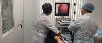
Bile reflux gastropathy is a clinical condition that is relatively unrecognized in everyday practice. This is due to the fact that there is no accurate, yet simple, accessible and practical method for diagnosing this condition. Diagnosis of bile reflux gastropathy requires upper endoscopy of the gastrointestinal tract, and its clinical complaints are difficult to identify.
Duodenogastric reflux (bile reflux) is a pathological condition that is a reverse flow of fluid (reflux) from the duodenum, consisting of bile, pancreatic juice and secretions of the intestinal mucosa. This condition may be asymptomatic, but often manifests as serious clinical conditions.
Pathogenesis of bile reflux gastropathy
There are external physical and chemical factors that can potentially damage the stomach: radiation, medications, alcoholic beverages and psychological stress. Physical stress consists of severe illness and duodenogastric reflux. In healthy people, duodenogastric reflux occurs on an empty stomach or after overeating. In some gastric pathologies, such as peptic ulcers, cholelithiasis or conditions after gastric surgery, prolonged reflux is observed at a higher rate than in healthy individuals.
The mechanism of duodenogastric reflux is still unclear. Some researchers have shown that in patients with peptic ulcers, the mobility of the antrum (antrum is the pyloric part of the stomach, the place where the stomach connects to the duodenum) is significantly reduced, and most of them have a high concentration of bile in the gastric juice both on an empty stomach and after eating . Poor antrum mobility, even in cases where the duodenum is normal, will lead to duodenogastric reflux. Many patients with chronic antrum gastritis maintain normal antrum mobility, and yet many of them have a high concentration of bile in the gastric juice. This is due to pyloric sphincter incontinence.
Other researchers have associated duodenogastric reflux with abnormalities in pressure fluctuations during peristalsis, which leads to uncoordinated antroduodenal motility, creating the opportunity for reflux of duodenal contents into the stomach. This may also occur in patients who have had a partial gastrectomy, since the absence of the pyloric sphincter facilitates duodenogastric reflux.
In addition to food, the duodenum also contains bile salts and pancreatic juice in high concentrations. If reflux occurs, all these substances enter the stomach, creating a pathological condition in the form of gastritis with corresponding clinical manifestations. This condition is known as bile reflux gastropathy (bile reflux gastropathy). It does not always manifest itself clinically. The clinical condition depends on the amount of fluid, the concentration of components in the reflux fluid, the duration of its contact with the gastric mucosa and the degree of sensitivity of the gastric mucosa.
Of all the contents of the duodenum, bile and pancreatic juice are potentially the most destructive to the gastric mucosa. The concentration of these two fluids also determines the severity of symptoms. Chenodeoxycholic (dihydroxycholic) acid is more toxic than trihydroxycholic acid. This is probably due to its greater lipophilicity, which allows it to penetrate deeper into the lipid structure of the cell membrane.
A study has shown that both bile and pancreatic juice can lead to chronic gastritis if one of them enters the stomach over a long period of time (200 days); the condition worsens if both are present at the same time. The mechanism is still unclear. It is possible that bile and pancreatic juice destroy the mucus barrier on the mucous membrane and then the underlying layers, causing inflammation.
In an experiment, rats that were given bilious dishes for three months developed erosive gastritis, while in another experiment, rats that were given bilious dishes for 200 days developed atrophic gastritis. Both groups suffered from lesions of the stomach, especially the pyloric region.
Bile has also been shown to cause a decrease in the transmembrane potential of the gastric mucosa, an increase in the re-diffusion of H+ ions and an increase in the concentration of sodium ions in the gastric lumen. These effects increase at high bile concentrations (20 mM) and low pH (pH = 2).
It is also believed that bile causes destruction of the stomach lining through mast cell degeneration and the subsequent release of large amounts of histamine. One study showed that bile causes instability of the mucosal cell lysosome. This disruption of lysosomes leads to the efflux of proteolytic lysosomal enzymes into the cell cytoplasm, which causes degeneration and disruption of cellular metabolism. In addition, the pores of the mucous membrane also expand. These two events influence mucosal permeability. As mucus breaks down, the permeability of the membrane against H+ ions increases, facilitating the movement of H+ ions into the mucosa. In the mucosa, H+ ions activate pepsinogens, which are then converted to pepsin, which further destroys the mucosa. H+ ions that enter the muscle layer also cause degranulation of mast cells, which then release histamine into the surrounding tissue. Histamine promotes vasodilation of mucosal capillaries, increases their permeability and causes swelling of the mucous membrane. The released histamine also stimulates the parietal cells to secrete more HCl into the gastric lumen, further increasing the concentration of H+ ions, which undergo re-diffusion, thereby starting an endless chain of reactions.
In addition to these developments, several researchers have also discovered through animal testing that the presence of bile in the stomach can increase the secretion of the hormone gastrin, which stimulates the secretion of HCl and reduces blood circulation in the mucosa, thereby impairing tissue regeneration.
It is important to note that bile in the gastric fluid causes a decrease in the secretion of PGE2 (prostaglandin E2) by the epithelium of the gastric mucosa, which reduces its cytoprotective function. Unfortunately, the mechanism by which PGE2 secretion is reduced remains unclear.
It is known that phospholipase A and lysolecithin contained in pancreatic juice can directly destroy the mucosal barrier. Phospholipase A is a pancreatic enzyme, while lysolecithin is the product of lecithin after dehydrolysis by phospholipase A. After destruction of the mucous membrane, the subsequent events are similar to those that occur in the presence of bile, re-diffusion of H+ ions and all those consequences that lead to damage to the gastric mucosa .
Clinical symptoms
Clinical symptoms of bile reflux gastropathy are nonspecific. In general, the manifestations resemble gastritis of any other etiology. There is often no connection between the extent of gastropathy and its symptoms.
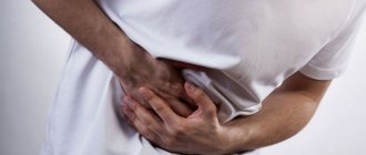
Patients complain of discomfort in the upper gastrointestinal tract, gas in the stomach, nausea and heartburn associated with meals. They usually feel pain while eating, relief comes soon after, and then the discomfort returns about 3 hours later.
Nausea and vomiting with quite severe colic in the form of heartburn are often observed. If vomiting occurs, yellowish-green bile is found in the vomit, and the patient experiences relief.
Diagnosis
Clinical diagnosis is based on medical history, physical examination, endoscopy and histopathology.
Pathogenetically, bile reflux gastropathy is defined as a chronic condition in which intestinal contents are thrown into the stomach both through an intact pylorus with a dysfunctional sphincter, and through a passage formed after gastric surgery.
The main problems that cause bile reflux gastropathy in a patient who has undergone gastric surgery are dysfunction of the pyloric sphincter and abnormal motility of the duodenum. The diagnosis of bile reflux gastropathy is based on history, clinical manifestations, measurement of bile fluid in the stomach and proximal intestine, X-ray results, endoscopy and histological analysis of the gastric mucosa. Reflux, which does not manifest itself clinically, differs from reflux with biliary gastropathy in frequency, number of episodes and duration of the disease.
Endoscopically, with reflux gastropathy, bile is detected in the stomach, which adheres to the gastric mucosa in the form of crusts, and also leads to its changes. The mucous membrane becomes hyperemic, fragile and erosive. Clinically, the patient complains of a feeling of fat in the throat, milk intolerance, and vomiting with bile.
Histopathological examination demonstrates an inflammatory response, foveal hyperplasia, edema, smooth muscle hypertrophy of the propria, and dilation and congestion of blood vessels. These results are identical to those of gastropathy due to NSAIDs. There are no specific pathological or pathognomonic data to confirm this diagnosis.
Conclusion
- Cases of bile reflux gastropathy are rarely reported due to the special procedures required to confirm the diagnosis.
- The pathophysiology of bile reflux gastropathy is well established, although further research is still needed to further understand this condition.
- Diagnosis is based on clinical findings confirmed by endoscopic and pathological findings.
- There is no pathognomonic pathological sign that would confirm bile reflux gastropathy.