Platyphyllin
Diseases of the cardiovascular system in which an increase in heart rate (HR) may be undesirable: atrial fibrillation, tachycardia, chronic heart failure, coronary heart disease, mitral stenosis, arterial hypertension, acute bleeding.
Thyrotoxicosis (possibly increased tachycardia).
Increased body temperature (may further increase due to suppression of the activity of the sebaceous glands).
Reflux esophagitis, hiatal hernia, combined with reflux esophagitis (decreased motility of the esophagus and stomach and relaxation of the lower esophageal sphincter can slow gastric emptying and increase gastroesophageal reflux through the sphincter with impaired function).
Diseases of the gastrointestinal tract accompanied by obstruction: achalasia and pyloric stenosis (possibly decreased motility and tone, leading to obstruction and retention of gastric contents).
Intestinal atony in elderly or debilitated patients and paralytic intestinal obstruction (possible development of obstruction).
Diseases with increased intraocular pressure: closed-angle glaucoma (mydriatic effect, leading to an increase in intraocular pressure, can cause an acute attack), open-angle glaucoma (mydriatic effect can cause a slight increase in intraocular pressure; therapy adjustment may be required), age over 40 years (risk of undiagnosed glaucoma).
Nonspecific ulcerative colitis (high doses can inhibit intestinal motility, increasing the likelihood of paralytic ileus; a complication such as megacolon may occur or worsen).
Dry mouth (long-term use may cause further worsening of xerostomia).
Liver failure (decreased metabolism), renal failure (risk of side effects due to decreased excretion).
Chronic lung diseases, especially in young children and weakened patients (a decrease in bronchial secretion can lead to thickening of secretions and the formation of plugs in the bronchi).
Myasthenia gravis (condition may worsen due to acetylcholine inhibition).
Autonomic (autonomic) neuropathy (urinary retention and paralysis of accommodation may increase), prostatic hypertrophy without urinary tract obstruction, urinary retention or predisposition to it, or diseases accompanied by urinary tract obstruction (including bladder neck due to prostatic hypertrophy).
Preeclampsia (possibly increased arterial hypertension).
Brain damage in children (central nervous system effects may be enhanced).
Down's disease (possibly unusual pupil dilation and increased heart rate).
Central paralysis in children (response to anticholinergics may be more pronounced)
Pregnancy, lactation period.
Features of the impact
Antispasmodics relieve pain.
The effect of antispasmodics directly depends on the form of the disease for which these drugs are used.
With the help of medications, the organs of the digestive tract are relaxed, spasms of which can manifest themselves in the head, heart, abdominal cavity, etc.
With the help of traditional drugs, the activity of the heart is stimulated, blood vessels and bronchi dilate, and blood pressure increases. Also, the action of antispasmodics is aimed at eliminating:
- Renal and intestinal colic;
- Fever;
- Heat;
- Inflammation;
- Stomach pain.
Each drug in this group has a specific mechanism of action. Due to the local effect of myotropic compositions, the transport of nerve impulses is blocked. When taking these medications, the secretion of hydrochloric acid decreases and the excretory activity of the glandular organs decreases.
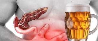
The chronic form of pancreatitis is in most cases accompanied by dull or aching pain. After eating, increased pain is observed. In most cases, patients complain of nausea and vomiting.
Patients also say that their stomach rumbles quite often. During the period of taking medications, spasms from the sphincter muscles are relieved. Thanks to long-term use of antispasmodics, this form of the disease relieves obstruction and pain.
The acute form of pancreatitis is characterized by a rather sharp manifestation after eating. Inflammation of the pancreas is very often accompanied by acute intense pain that has a bursting character.
Types of drugs
Methocinium - acts on nerve impulses and nerves.
According to the mechanism of action, traditional medicines are divided into several groups. They can be:
- Neurotropic. They act on the transmission of nerve impulses to nerves. With the help of these medications, the smooth muscles of certain internal organs are stimulated. This group of medications includes Platiphylline, Metocinium, Arpenal, Scopolamine, etc.
- Myotromic. These traditional medications have a positive effect on smooth muscle cells. With their help, a change in biochemical processes in the human body is carried out. Medicines in this group are represented by Halidor, Mebeverine, Nitroglycerin, etc.
- Natural. These are traditional medicines used to eliminate spasms. They are prepared on the basis of common tansy, May lily of the valley, belladonna, pharmaceutical lovage, etc. Traditional medicines may differ in the form of release. Most often, the production of antispasmodics is carried out in the form of capsules, which are represented by Duspatalin,
- Sparex. Tablet medications in the form of No-shpa, Halidor, Papaverine, Dicetel, Spazoverine, etc. are quite convenient to use. Treatment of spasms in pancreatitis can be carried out using antispasmodics in ampoules - Trigana,
- Diabazol, Droverina. The use of these medications is carried out for intramuscular injections. Peppermint tincture is often used to relieve pain. Antispasmodics can be produced in the form of suppositories - Papaverine, Buscopan, belladonna extract. These medications require rectal administration. Pain in pancreatitis can be treated with drops of Zelenin and Valorserdin. These medications are taken orally. You can also use granules
- Plantacida, Plantaglucida. They are used to prepare a solution for oral administration.
Thanks to the various types of the drug, it is possible to choose the most convenient option for the patient.
Instructions for use (Method and dosage)
Used orally, parenterally (subcutaneously, intravenously), rectally or topically.
Platiphylline Hydrotartrate is used intramuscularly if necessary - prescribed and adjusted by the attending physician, who is responsible for this prescription. The dose is determined depending on the symptoms and degree of the disease, route of administration and age of the patient.
Platyphylline Hydrotartrate is usually prescribed for the treatment of:
- Relief of acute ulcerative pain , intestinal, hepatic or renal colic, prolonged attacks of bronchial asthma , cerebral and peripheral vasospasms for adults - 1-2 ml 1 or 2 times a day.
- As an antispasmodic for adults - 1 ml daily for one month. The maximum single dose for an adult subcutaneously is 10 mg, no more than 30 mg per day.
Interaction
- Platiphylline Hydrotartrate is able to prolong the hypnotic effect of phenobarbital , sodium etaminal , magnesium sulfate ;
- capable of blocking proserin ;
- may increase the effect of H2-histamine blockers , Digoxin and Riboflavin ;
- may enhance mydriasis with adrenergic agonists and nitrates , increasing intraocular pressure;
- compatible with caffeine for cerebral vascular spasms;
- compatible with Cordiamin and Diafillin ;
- with Morphine eliminates bradycardia , nausea, vomiting;
- with Verapamil eliminates bradycardia .
Pharmacodynamics and pharmacokinetics
Platyphylline Hydrotartrate is characterized by a cholinolytic effect: by blocking M-choline receptors , it converts them into forms insensitive to acetylcholine . Due to the direct myotropic antispasmodic effect on the smooth muscles of the intestines, bile ducts, bronchi and eyes, it is considered a papaverine-like agent . In addition, it has a mild direct inhibitory effect on vascular smooth muscle, a calming effect on the central nervous system, especially on the vasomotor center, providing vasodilatory and hypotonic effects.
Platyphylline Hydrotartrate is well absorbed from the subcutaneous layer of the epidermis . However, the administration of large doses causes its accumulation in the tissues of the central nervous system in significant concentrations. It is excreted through the urinary as well as the digestive system.
Shcherbakov P.L. On the issue of pancreatitis // Attending Physician. Gastroenterology. 2011. No. 7.
On the issue of pancreatitis
P.L. Shcherbakov
Diseases of the pancreas are one of the most common lesions of the digestive organs. Women predominate among the patients. This is apparently due to a higher incidence of cholelithiasis (GSD) and lipid metabolism disorders in them. A significant proportion of patients are elderly and elderly people, but recently lesions of the pancreas are increasingly common in children of all ages. Currently, there are various recommendations and standards dedicated to the management of patients with pancreatitis. The best known are the 1992 Atlanta criteria [1] and the 1999 Santroni consensus [2], revised in 2003.
Among the inflammatory diseases of the pancreas, acute and chronic pancreatitis are distinguished. The causes of damage to the pancreas are varied. In particular, these include: obstruction of the pancreatic duct, exposure to drugs or toxic poisoning with various substances, metabolic disorders of both the pancreas itself and other organs and systems, infectious and parasitic diseases, vascular disorders and injuries.
Acute pancreatitis is a polyetiological disease. Acute pancreatitis is based on enzymatic damage to the pancreas, which is autocatalytic in nature. There are two groups of causes of the disease. The first includes factors that cause difficulty in the outflow of pancreatic juice through the pancreatic ducts and, as a result, lead to an acute increase in pressure in them, with the development of the hypertensive ductal form of acute pancreatitis. The second group consists of factors leading to primary damage to acinar cells with the development of the primary acinar form of the disease.
Acute damage to the pancreas can occur when taking various drugs, such as methyldopa, 5-aminosalicylates, azathioprine, cimetidine, furosemide, metronidazole, tetracyclines, or when the pancreatic parenchyma is overfilled with radiopaque agents during instrumental endoscopic examinations. In addition, the causes of the development of acute pancreatitis in both adults and children can be metabolic disorders, in particular hypertriglyceridemia - hyperlipidemia type I, IV or V.
Of the infectious diseases, the most common causes of pancreatitis are: viral diseases (cytomegaly, herpes, hepatitis A, B, C), bacterial (mycobacteriosis, leptospirosis), fungal infections (cryptococci, candida), as well as parasitic infestations (roundworms that clog the lumen of the excretory pancreatic duct, or pneumocystis).
Chronic pancreatitis is a long-term, usually progressive, inflammatory disease of the pancreas. In this case, focal or diffuse destruction of gland tissue develops with its gradual replacement by connective tissue. The main etiological factors for the development of chronic pancreatitis are the same as for acute pancreatitis, which directly damage the acinar elements or contribute to an increase in pressure in the pancreatic ducts, but act longer and less intensely.
According to the Atlanta criteria, a correct diagnosis of acute pancreatitis should be made in all patients within 48 hours of admission (grade C recommendation). The etiology of acute pancreatitis should be determined in at least 80% of cases and in no more than 20% should be classified as idiopathic (grade B recommendation) [3].
If the cause of the disease cannot be established, we should talk about idiopathic chronic pancreatitis.
According to most studies, the cause of approximately half of cases of acute pancreatitis is cholelithiasis, 20–25% are associated with alcohol abuse. The “idiopathic” group includes patients in whom no obvious cause for the development of this condition has been identified [4]. The diagnosis of idiopathic pancreatitis cannot be established without a targeted search for gallstones. Ultrasound examinations must be performed at least twice. After one negative ultrasound, the most sensitive diagnostic test for gallstones is a repeat ultrasound, which identifies stones that may have been missed [5]. The advent of endoscopic ultrasound (EUS) and magnetic resonance cholangiopancreatography (MRCP) has expanded the range of tests available to determine the cause of acute pancreatitis. In such situations, EUS can detect microlithiasis in the gallbladder or common bile duct, and MRCP can detect most stones in the duct, as well as abnormalities of the ducts themselves, such as bifurcation of the pancreas. EUS is also accurate and safer than endoscopic retrograde cholangiopancreatography (ERCP) for detecting common bile duct stones. Bile testing may be the only way to identify patients with recurrent acute pancreatitis due to microlithiasis. Ductal manometry
(used to identify sphincter of Oddi dysfunction) may have a significant risk of exacerbation of acute pancreatitis and should only be performed in specialized units. Careful selection of patients for manometry is necessary [6]. To determine the nature of the development of pancreatitis, it is necessary to conduct studies on the content and level of plasma lipids and the concentration of calcium in the blood. Early and convalescent titers of antibodies to viruses (mumps, Coxsackie B4 and others) can also identify a possible cause of acute pancreatitis, although no specific therapy is prescribed. It is necessary to consider the possibility of the existence of a concomitant neoplasm or chronic pancreatitis and examine the patient accordingly.
The studies necessary to determine the etiological factors that caused acute pancreatitis, depending on the stage of the disease, as well as anamnestic data to exclude other causes of acute pancreatitis, are presented in the table.
Long-term dysfunction of the pancreas can lead to the development of chronic changes manifested as chronic calcific pancreatitis, chronic inflammatory pancreatitis or chronic obstructive pancreatitis.
Congenital disorders of the pancreas, which may later manifest as an inflammatory response, are associated with autosomal dominant abnormalities of the gene or chromosome 7G associated with cancer of the head of the pancreas.
A disruption of the pancreas, which is not usually accompanied by an inflammatory reaction, but has certain clinical symptoms, is called pancreatic insufficiency.
There are primary and secondary pancreatic insufficiency. Primary pancreatic insufficiency develops due to the influence of so-called non-modifiable factors, which a person (the patient himself or the doctor) cannot influence and influence. These include diseases such as cystic fibrosis of the pancreas, congenital obstruction of the pancreatic duct, Shwachman syndrome, isolated lipase deficiency, isolated trypsin deficiency, hereditary recurrent pancreatitis.
In clinical practice, secondary or relative pancreatic insufficiency (pancreatopathy) is more common, usually caused by the intake of unusual food, its excess amount or temporary disorders of the functioning of the pancreas. Secondary deficiency can accompany various inflammatory diseases of the upper digestive tract (UPDT).
Clinical manifestations of pancreatic insufficiency are signs of disruption of the pancreas - abdominal pain, changes in appetite (decrease or complete disappearance), nausea, rumbling in the abdomen, flatulence and flatulence, steatorrhea. The intensity and severity of these signs depends on the degree of damage to the pancreas.
Diagnosis of secondary pancreatic insufficiency can often present significant difficulties due to the vagueness of clinical symptoms and minor changes in instrumental research methods. Therefore, for correct diagnosis and timely prescription of adequate treatment, the doctor must use the entire arsenal of tools at his disposal. With pancreatic insufficiency, pain is localized in the epigastrium, left hypochondrium, or can be encircling, radiating to the left hypochondrium, under the left shoulder blade, or in the back. The pain can be paroxysmal and constant; it intensifies after overeating, consuming fatty, spicy and fried foods, and alcohol. Heat increases the pain, the use of cold reduces it somewhat. The pain is difficult to relieve with medications. The pain eases somewhat when the patient is forced into a knee-elbow position, sitting, bent forward, lying on his side with his knees pulled to his chest. The pain syndrome is accompanied by symptoms of intestinal dyspepsia and stool disorders, while patients complain of bloating and rumbling in the abdomen, and there may be constipation and diarrhea. The stool during diarrhea is copious, liquid, foamy, light yellow in color due to the large amount of fat. Signs of gastric dyspepsia are also characteristic - nausea and vomiting, which does not bring relief.
For pancreatic insufficiency, various medications containing enzymes are used. Traditionally, pancreatin is used for this, a drug prepared from animal pancreas [8]. However, under conditions of intense acid formation in the stomach, its partial inactivation occurred, and the drug did not have the expected therapeutic effect. Subsequently, with the development of the pharmaceutical industry and knowledge about the mechanism of digestive processes, new forms of drugs appeared containing pancreatin in the form of tablets, dragees, granules with a protective shell and microspheres placed in a capsule. Currently, enzyme preparations used in clinical practice must meet certain requirements: 1) non-toxicity; 2) good tolerability; 3) absence of significant adverse reactions; 4) optimal action in the pH range 5–7; 5) resistance to the action of hydrochloric acid, pepsins and other proteases; 6) content of a sufficient amount of active digestive enzymes; 7) have a long shelf life [9].
Depending on the phase of development of the pathological process, four forms of acute pancreatitis can be distinguished: acute interstitial, corresponding to the phase of edema (serous, hemorrhagic, serous-hemorrhagic), acute necrotic, expressing the phase of necrosis formation (with or without a hemorrhagic component); infiltrative-necrotic and purulent-necrotic, corresponding to the phase of melting and sequestration of necrotic foci.
To facilitate the choice of treatment tactics, the volume of infusion therapy and the correct interpretation of the form of pancreatitis, mild, moderate and severe degrees of intoxication are distinguished.
A mild degree (usually found with serous edema of the pancreas) is characterized by a satisfactory general condition of the patient, moderate epigastric pain, nausea, single vomiting, absence of symptoms of peritoneal irritation, unchanged skin color, pulse rate within 88–90 beats per minute, normal or slightly elevated blood pressure (BP), high uroamylase levels, low trypsin and lipase activity, preserved or slightly reduced circulating blood volume (CBV) (deficiency 7–15%), moderate leukocytosis, low-grade fever.
The average degree of intoxication (observed with small focal necrosis of the gland) is manifested by persistent pain in the epigastrium, which does not disappear with the use of antispasmodics and analgesics, pallor and cyanotic skin, repeated vomiting, muscle tension in the epigastric region, pulse rate up to 100–110 beats/min. , a drop in blood pressure below the initial level, an increase in peripheral and a decrease in central venous pressure (CVP), high uroamylase levels, early high activity of trypsin and lipase, a decrease in Ca levels and an increased content of sialic acids, a decrease in BCC (deficiency 16–35%), decreased diuresis , a rise in body temperature to 38 °C.
A severe degree of intoxication (occurs with widespread necrosis of the gland) is characterized by a severe general condition of the patient, severe pain in the epigastric region, painful vomiting, sharply pale or cyanotic skin, often jaundice, the appearance of symptoms of peritonitis, a pulse rate over 120 beats/min, a drop in blood level Blood pressure and central venous pressure, decreased uroamylase, trypsin and lipase activity, Ca level, low diuresis, up to complete anuria, a sharp decrease in blood volume (deficiency 36–50%), high temperature, significant dysfunction of the heart, liver, lungs, kidneys.
In the treatment of acute pancreatitis, from the very beginning, the surgical method proposed by the German surgeon Korbe in 1894 prevailed. However, the high mortality rate (90–100%) even at that time forced people to be cautious about this method [10]. At the 5th All-Russian Congress of Surgeons, Academician V.S. Savelyev (1978) emphasized that currently the predominantly conservative method of treating acute pancreatitis is generally accepted. However, treatment tactics are dictated mainly by the form of the disease. If for interstitial and necrotizing pancreatitis timely conservative therapy and, to a lesser extent, surgery are crucial, and for infiltrative-necrotizing pancreatitis only conservative treatment is crucial, then the purulent-necrotic form requires mandatory surgical intervention.
Modern conservative therapy of acute pancreatitis solves the following problems:
1) elimination of pain and spasm, improvement of microcirculation in the gland;
2) combating shock and restoring homeostasis;
3) suppression of exocrine secretion and activity of gland enzymes;
4) fight against toxemia;
5) normalization of the activity of the lungs, heart, kidneys, liver;
6) prevention and treatment of complications.
In the treatment of pancreatitis, the leading importance is to ensure a decrease in pancreatic secretion, since increased secretion of pancreatic juice rich in enzymes, especially with significant blockage of the duct, leads to progression of the process. Patients with severe acute pancreatitis should be treated in the intensive care unit.
For any variant of the course of acute pancreatitis, treatment begins with the appointment of fasting, which, depending on the severity of the disease, is prescribed for 3–5 days. With a mild process, after this period, patients are able to eat without pain, expand their diet, gradually recover and do not require further treatment. In case of a more severe disease, constant aspiration of gastric contents is carried out in the first days and the optimal tactics for managing the patient are quickly determined. It should be noted that with proper management, patients with acute pancreatitis of moderate severity without the development of pancreatic necrosis and parapancreatic phlegmon can be cured using conservative methods.
When choosing treatment tactics for patients with acute pancreatitis, it is most important to differentiate between edematous and interstitial forms of acute pancreatitis. Indicators of the onset of the necrotic process are changes in the blood serum concentration of C-reactive protein and elastase. Patients with an edematous form of pancreatitis require conservative therapy and dynamic observation, while patients with pancreatic necrosis require intensive care. The choice of patient management tactics is determined by the functional viability of the organs and the presence of concomitant pathology. Patients without concomitant organ pathology and with limited necrosis can be treated conservatively, whereas patients with multiple organ failure or progression of comorbidity are candidates for surgical treatment.
In order to suppress pancreatic secretion, proton pump inhibitors are currently used, which are advisable to be administered parenterally, in particular pantoprazole (Controloc®). This representative of proton pump inhibitors has the longest effect time (up to 48 hours), providing reliable blocking of hydrochloric acid production in the stomach and thereby suppressing all enzyme-forming reactions in the pancreas. Unlike other representatives of proton pump inhibitors, pantoprazole (Controloc®) after administration is not metabolized in the liver using cytochrome P450 and its coenzymes, therefore pantoprazole does not interact with drugs undergoing metabolism in the liver, does not compete with them and can be combined with other drugs for the treatment of pancreatitis in both acute and chronic stages. The initial daily dose of Controloc® is 80 mg. If necessary, the dose can be titrated, increasing or decreasing, depending on the levels of acid secretion in the stomach. For doses exceeding 80 mg per day, they must be divided into two doses. It is possible to temporarily increase the dose above 160 mg of pantoprazole, but the duration of use should be limited to only the period necessary to adequately control acid secretion. The powder is dissolved in 10 ml of physiological sodium chloride solution, which is added to the bottle. This solution can be administered directly or after mixing with 100 ml of saline sodium chloride solution, 5% or 10% glucose solution. Intravenous administration should be carried out over 2–15 minutes. These medications should be used in the above doses until the pain syndrome is relieved and the “evasion” of enzymes into the blood is eliminated, after which it is necessary to continue taking them at half the dose orally for another month or replace them with antacids, such as Almagel, Maalox, Phosphalugel, etc. 6–8 times a day.
The most important importance in the acute period of pancreatitis is given to the elimination of the leading pain syndrome, for which a combination of non-narcotic analgesics and antispasmodics is most often used.
- Metamizole sodium orally 250–300 mg 2–3 times a day
or
- 50% solution IM or IV 0.1–0.2 ml/10 kg until pain relief
or
- Paracetamol orally 0.5 g 2–3 times a day until pain relief
in combination with:
- Hyoscine butyl bromide orally 20 mg 3–4 times a day until pain relief
or
- Drotaverine orally 30–40 mg 3–4 times a day
or
- Papaverine orally or rectally 15–20 mg 3–4 times a day until pain relief
or
- Platiphylline orally or subcutaneously 3–4 mg 2–3 times a day until pain relief.
In case of severe pain, it is rational to use narcotic analgesics such as Promedol [11].
Enzyme replacement therapy for pancreatitis is aimed at eliminating disturbances in the digestion of fats, proteins and carbohydrates. Among the large number of pancreatic enzymes used in gastroenterology, preference is given to drugs that best meet modern requirements:
- resistance to hydrochloric acid, presence of an acid-resistant shell;
- lipase activity of at least 25,000 units per dose, optimal enzyme action in the pH range of 5–7;
- uniform and rapid mixing with food, microcapsule size no more than 2 mm;
- rapid release of enzymes in the duodenum.
Traditional pancreatic enzymes in the form of tablets or dragees are usually destroyed by hydrochloric acid of gastric juice, which requires an increase in the daily dose of drugs to correct exocrine insufficiency. Microgranulated enzymes have the best effect [12].
Diet for pancreatitis plays an important role at all stages of monitoring patients and is based on mechanical, thermal and chemical sparing of the pancreas, suppression of hyperfermentemia, reduction of stasis in the ducts and duodenum, and reduction of reflex excitability of the gallbladder.
Currently, a new concept of nutritional support for pancreatitis has been developed and the attitude to the duration of the “starvation” diet, parenteral and enteral nutrition has been revised. It has been proven that fasting increases the rate of lipolysis, causes the development of hypodysproteinemia, metabolic acidosis, and aggravates degenerative changes in the pancreas.
Nutritional support is complete nutrition through partial or complete parenteral and enteral nutrition. Its main goal is to provide the body with energy donors (carbohydrates, lipids), plastic material (amino acids); correction of metabolic disorders and restoration of the trophological status of the patient. Early parenteral and enteral nutrition accelerates reparative processes in the gastrointestinal tract.
The nutritional support algorithm is compiled taking into account the assessment of the patient’s condition and includes the stages of parenteral, enteral or mixed nutrition and dietary therapy itself (diet No. 5P). Medical nutrition is considered as pharmacotherapy for various metabolic disorders and is the main way to qualitatively provide the energy-plastic needs of the patient’s body.
The effectiveness of therapy for pancreatitis is assessed by the dynamics of pain and dyspeptic syndromes, normalization of the level of blood and urine enzymes, coprogram indicators, fecal elastase and the increase in the patient’s body weight.
Literature
- Bradley EL III. A clinically based classification system for acute pancreatitis. Summary of the International Symposium on Acute Pancreatitis, Atlanta, Ga, September 11 through 13, 1992 // Arch Surg. 1993; 128:586–590.
- Dervenis C., Johnson CD, Bassi C. et al. Diagnosis, objective assessment of severity, and management of acute pancreatitis. Santorini consensus conference // Int J Pancreatol. 1999; 25: 195–210.
- Tooli J., Brook-Smith M., Bassi C. et al. Guidelines for the management of acute pancreatitis // J Gastroenterol Hepatol. 2002; (Suppl 17): S15–39.
- Johnson CD, Abu-Hilal M. Persistent organ failure during the first week as a marker of fatal outcome in acute pancreatitis // Gut. 2004; 53:1340–1344.
- McKay AJ, Imrie CW, O'Neill J. et al. Is an early ultrasound scan of value in acute pancreatitis? // Br J Surg. 1982; 69: 369–372.
- Hogan WJ, Sherman S, Pasricha P et al. Sphincter of Oddi manometry // Gastrointest Endosc. 1997; 45: 342–48.
- Brown A, Baillargeon JD, Hughes MD et al. Can fluid resuscitation prevent pancreatic necrosis in severe acute pancreatitis? // Pancreatology. 2002; 2: 104–107.
- Graham DY Enzyme replacement therapy of exocrine insufficiency in man. Relation between in-vitro potency in commercial pancreatic extracts // N. Engl. J. Med. 1977. 296. P. 1314–1317.
- Zlatkina A. R., Belousova E. A., Nikitina N. V., Siliverstova T. R. Modern therapy of chronic pancreatitis. 2nd Gastr. a week. 1996. Special issue. 4 s.
- Li H., Qian Z., Liu Z., Liu X., Han X., Kang H. Risk factors and outcome of acute renal failure in patients with severe acute pancreatitis // J Crit Care. Sep 23, 2009.
- Minushkin O. N. Chronic pancreatitis: issues of pathogenesis, diagnosis and treatment // Difficult patient. M., 2003. No. 1. P. 26–30.
- Ivashkin I. T. Modern issues of clinical pancreatology // Bulletin of the Russian Academy of Medical Sciences. 1993. No. 4. pp. 29–34.


