- Causes of biliary colic
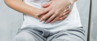
- Symptoms of an attack of gallbladder colic
- Algorithm for providing assistance for biliary colic
- What complications does biliary colic lead to?
- Diagnosis and treatment
Hepatic or biliary colic is pain of varying intensity that occurs during exacerbation of diseases of the liver and biliary tract. It is of a sharp, cramping nature, most strongly felt in the right hypochondrium and covers the abdomen with pain in the shoulder, neck, and shoulder blade. Gallbladder colic can last from 15 minutes to 5-6 hours. The condition is accompanied by vomiting and weakness.
What is hepatic colic?
The content of the article
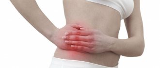
Hepatic colic, also known as biliary colic, is a condition characterized by severe abdominal pain in the right hypochondrium or middle epigastrium (upper abdomen).
Hepatic colic usually occurs when you eat heavy or fatty foods. The condition is very unpleasant, the pain can extend to the back, and associated symptoms may include gas, heartburn or vomiting.
Heartburn
The pain of hepatic colic comes on suddenly and disappears slowly, sometimes lasting up to several hours. Hepatic colic is not a disease in itself, but may indicate more serious diseases, such as the gallbladder. Ignoring the problem can lead to serious complications.
Classification
According to the type, pain due to biliary colic is classified as follows:
- cutting;
- piercing;
- tearing;
- constant;
- paroxysmal.
If you have these symptoms, you should consult a specialist to quickly determine their cause.
Clinical blood test (CBC/Diff - 5 leukocyte fractions) + ESR in Moscow from day 1
from 294 ₽
Sign up
Complete blood count (CBC/Diff - 5 fractions of leukocytes) in Moscow from 1 weekday
from 290 ₽
Sign up
Differential diagnosis of Crohn's disease and nonspecific ulcerative colitis - screening in Moscow from 2 to 5 days a week.
from 7990 ₽
Sign up
Hepatic (biliary) colic - causes
Eating large amounts of foods containing animal fats is associated with high cholesterol levels. This, in turn, causes the formation of deposits that accumulate in the gallbladder and interfere with its function. The function of the gallbladder is to push out the bile needed to digest food - if something interferes with the contraction of the gallbladder, it must contract more intensely. This is the direct cause of hepatic colic and the severe pain associated with it.
Colic occurs when accumulated bile has difficulty moving due to deposits in the gallbladder or bile ducts.
Cholelithiasis
Gallstones are nothing but the presence of deposits in the gallbladder or bile ducts. Gallstones are more likely to affect women, obese people, older adults, diabetics, and patients taking oral contraceptives or other estrogen medications.
Gallstones
Gallstones can occur at the same time as duct stones because deposits in the gallbladder are tiny and can travel with bile into the bile ducts, blocking their narrow lumen. In addition to hepatic colic, jaundice indicates urolithiasis, which is additionally accompanied by a change in the color of the stool and unpleasant itching of the skin.
Diagnosis of hepatic colic
As already mentioned, hepatic colic usually indicates gallstone disease, so this disease should not be ignored. In any case, you should visit a gastroenterologist who will perform a physical examination and imaging.
The most useful test for diagnosing hepatic colic (due to gallstones) is an abdominal ultrasound. This test can detect stones in the gallbladder and sometimes even in the bile ducts.
Abdominal ultrasound
However, endoscopic retrograde cholangiopancreatography or EUS (endoscopic ultrasound) is more sensitive. These two studies are performed when choledocholithiasis is suspected and allow visualization of the bile ducts from the inside and removal of deposits or dilation of the bile ducts to the duodenum.
Types of hardware research
At NEARMEDIC clinics, doctors take care of the health of patients with an attentive approach and accurate diagnosis. Modern, most advanced equipment allows you to avoid mistakes in making diagnoses, find even the smallest stones, accurately detect the place of their formation, and correctly prescribe treatment.
The following types of instrumental diagnostics are available at NEARMEDIC clinics:
- Ultrasound (ultrasound examination) allows you to detect the location of stones and determine their size;
- EGDS (Esophagogastroduodenoscopy) is necessary to exclude or confirm diseases of the digestive tract; a biopsy is performed during the study;
- Computed tomography allows for differential diagnosis.
Hepatic (biliary) colic - treatment
Treatment for hepatic colic is specialized because, as mentioned above, it is not a disease in itself, but a symptom of another ongoing disease. Colic goes away as soon as its cause is eliminated.
Thus, the treatment of this disease consists of the following measures:
- Taking painkillers such as non-steroidal anti-inflammatory drugs or paracetamol. Patients should not be given morphine as it may worsen symptoms;
- Administration of drugs that relax the smooth muscles of the gastrointestinal tract, such as papaverine, scopolamine or drotaverine;
- Through the procedure - gallbladder removal when we are dealing with symptomatic gallstones;
- The use of endoscopic retrograde cholangiopancreatography with prosthetics of the biliary tract or expansion of the outlet into the biliary tract.
Taking painkillers
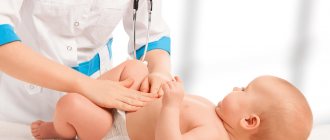
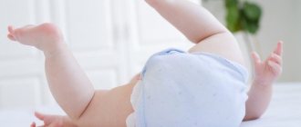
The main importance in stone formation in children is given to hereditary factors in combination with general metabolic disorders and abnormalities in the development of the biliary system. The formation of stones in children is not accompanied by an acute inflammatory process in the gallbladder. Functional disorders of the gastrointestinal tract are factors contributing to the development of cholelithiasis in children. The clinical picture of cholelithiasis in preschool children resembles an attack of hypertensive biliary dyskinesia, and in older children it occurs under the guise of esophagitis, chronic gastroduodenitis, and peptic ulcer.
Gallstone disease in children
Major importance in stone formation in children the hereditary factors in combination with common metabolic disorders and abnormalities of the biliary system are given. Forming concretions in children are not accompanied by an acute inflammation of the gall bladder. Functional disorders of the gastrointestinal tract are factors contributing to the development of cholelithiasis in children. The clinical picture of gallstone disease in preschool children recalls attack hypertonic biliary dyskinesia, and the older children takes place under the guise of esophagitis, chronic gastroduodenitis, peptic ulcer disease.
Gallstone disease (GSD, cholelithiasis, K80) is a multifactorial metabolic disease of the hepatobiliary system, characterized by the formation of stones in the hepatic bile ducts, common bile duct, and gallbladder [6]. Cholelithiasis is accompanied by a continuously recurrent inflammatory process, the outcome of which is sclerosis and dystrophy of the gallbladder. Currently, this pathology has been sufficiently fully studied in adults, however, direct copying of the mechanisms of formation of gallstones and associated clinical manifestations is completely unacceptable for pediatric practice [5].
Gallstone disease is quite widespread among the adult population of Russia (20-30%). At the same time, cholelithiasis is observed in women 5 times more often than in men. Among children with diseases of the gastrointestinal tract, cholelithiasis is detected in 0.1-1% of children. The ratio of boys and girls in the morbidity structure has age-related characteristics: up to 7 years of age, boys predominate, at 7-9 years of age the ratio is equalized, and by 10-12 years of age, cholelithiasis is 2 times more often detected in girls. In adolescence, the predominance of girls becomes clear and begins to approach the indicators in adults - 3:1 [11].
Etiology. Cholelithiasis is a multifactorial, polyetiological disease. In this case, the main importance in stone formation in children is given to hereditary factors in combination with general metabolic disorders and abnormalities in the development of the biliary system. 75-95% of children have a family history of gallstone disease among first-degree relatives. There is a known disturbance in the metabolism of phospholipids and lipoproteins. [3].
Nutritional characteristics play an important role in the etiology of cholelithiasis. Human milk is rich in taurine, which improves the absorption of lipids, and its conjugation with bile acids reduces toxicity and improves the absorption of the latter. Breastfeeding provides long-term protection against hyperlipidemia, hyperinsulinemia, obesity, etc. Children with cholelithiasis usually receive breast milk from 1 to 3 months, and a significant proportion receive formula immediately after birth. In addition, the diet of children with cholelithiasis is dominated by carbohydrates and fats, with a lack of consumption of fresh fruits and vegetables rich in macro- and microelements and dietary fiber [2, 15].
When the detoxification function of the microbial flora of the digestive tract decreases under the influence of food ingredients, xenobiotics, viruses, and bacterial pathogens, microecological disturbances occur in the intestine, which leads to metabolic (endotoxemia) and structural damage to hepatocytes. Microbial flora also plays an important role in maintaining stable cholesterol levels in the body [11]. The formation of gallstones is facilitated by physical inactivity, stress in the family and school, active and passive smoking, and in adolescents - alcohol and substance abuse.
The role of abnormalities in the development of the biliary system in the process of stone formation is high: more than half of children with cholelithiasis have abnormal gallbladder, common bile duct, or their combinations, which leads to the development of bile stasis both in the gallbladder and in the intrahepatic bile ducts [4]. Most children with cholelithiasis have various metabolic disorders: dysmetabolic nephropathy, nutritional-constitutional obesity of the 2nd–3rd degree, thyrotoxicosis, etc. [10].
In children under 12 years of age, bilirubin stones are more often detected, while in adolescence cholesterol lithiasis predominates [2, 14]. In children, unlike adults, inflammatory diseases of the gastrointestinal tract (gastroduodenitis, including those associated with Helicobacter pilory) and functional disorders of the gastrointestinal tract caused by autonomic dysfunction are factors contributing to the development of cholelithiasis, and not the result of the latter [6 ].
To date, the mechanisms of stone formation have not been precisely studied. In the pathogenesis of cholelithiasis, a large role is played by changes in the ratio of various bile acids and other components of bile. Bile is an isosomatic electrolyte solution, the main components of which are bile acids (67%), phospholipids (22%), proteins (4.5%), cholesterol (4%), bilirubin (0.3%). Lecithin, cholesterol and bile salts are amphiphilic compounds, so they form micelles in an aqueous environment. Human bile keeps cholesterol in a dissolved state. If the amount of bile acids and lecithin is insufficient to form micelles, insoluble cholesterol appears and the bile becomes supersaturated or lithogenic [9].
Currently, two types of lithogenesis are distinguished: cholesterol and bilirubin. Cholesterol lithogenesis is uncommon in young children and predominates in adolescents. In the mechanism of cholesterol lithogenesis, the main role is played by “vesical” factors against the background of intestinal dysfunction. In the distal intestine, anaerobic flora is activated, which promotes the deconjugation of bile acids, as a result of which they acquire increased water repellency. Deoxylate (secondary bile salt), when absorbed, returns to the liver, bile ducts and, together with water, is easily resorbed by the wall of the gallbladder. Hypersecretion of mucoid substances (mucin, glycoproteins) and a decrease in the evacuation function of the gallbladder contribute to the formation of the nucleus of the future calculus. In addition, a deficiency of essential microelements in the child’s body (selenium, zinc and iron) leads to the accumulation of lipid peroxidation products and disruption of enzymatic systems, as a result of which the pH of gallbladder bile shifts to the acidic side.
These factors cause damage to cell membranes and intracellular structures with the development of chronic inflammation in the wall of the gallbladder. The relationship between bile acids, phospholipids and cholesterol is disrupted, bile acquires lithogenic properties, and preconditions are created for the precipitation of insoluble salts (calcium and magnesium bilirubinate) and the formation of stones. In turn, lithogenic bile, having powerful abrasive properties, aggravates the course of chronic inflammation.
Bilirubin lithogenesis is most characteristic of childhood and is the leading one in most adults. With this mechanism of stone formation, the high concentration of the unconjugated free fraction of bilirubin in the bile and cholestatic processes in the liver and biliary tract are of primary importance. The gradual accumulation of copper and iron in bile contributes to the formation of pigmented gallstones. Both microelements form strong compounds with high-molecular proteins and free bilirubin [12].
There is no generally accepted classification of cholelithiasis in children. In practical healthcare, they use the classification adopted by the Congress of the Scientific Society of Gastroenterologists of Russia (2002). According to this classification, the initial or prestone stage is distinguished, characterized by thick heterogeneous bile, the formation of biliary sludge with microliths, putty-like bile, or a combination of these. Next comes the formation of gallstones, which can be localized in the gallbladder, common bile duct, and hepatic ducts; single or multiple stones; composition: cholesterol, pigment, mixed. The disease can occur latently or with clinical symptoms in the form of pain with typical biliary colic, dyspeptic form, or under the guise of other diseases. This is followed by the stage of chronic recurrent calculous cholecystitis, which is replaced by the stage of development of complications.
Clinical picture. Signs of cholelithiasis in childhood have their own characteristics and are not as typical as in adults. The presence of stones in the biliary tract in children is not accompanied by an acute inflammatory process in the gallbladder, which causes the classic symptoms of calculous cholecystitis/cholangitis. [7]. In the clinical course of cholelithiasis, three variants are distinguished: asymptomatic stone carriage (in about half of patients), a dyspeptic form and a painful form with typical biliary colic. In asymptomatic stone carriers, stones are discovered by chance. The children make no complaints.
Abdominal pain and dyspeptic disorders are among the main complaints when cholelithiasis can be suspected in children. The nature of the pain depends on the size of the stones. Multiple, small, easily moving stones cause acute, paroxysmal pain. Dull, drawing, vague pain is typical for patients with single stones. Children of preschool and primary school age equally often complain of both sharp and dull pain, localized throughout the abdomen or in the navel area. In patients of pre- and pubertal age, dull, aching, bursting pain in the abdomen predominates. In children aged 7-11 years, pain predominates in the epigastrium and pyloroduodenal zone, and in adolescents - in the right hypochondrium.
When stones are localized in the area of the bottom of the gallbladder (in a “silent”, weakly innervated zone), an asymptomatic course of the disease is more often observed, while in the presence of stones in the cervix (high pain sensitivity), acute early pain in the abdomen is observed, accompanied by nausea and vomiting. [7, 12, 15].
The pain syndrome has its own characteristics depending on the type of the autonomic nervous system: in vagotonic patients, paroxysmal acute pain predominates, the provoking factors of which are various psycho-emotional overloads and stress; in sympathotonic patients, on the contrary, dull, aching pain predominates. In such patients, the contractility of the gallbladder decreases, which leads to stagnation of bile and disruption of its passage into the duodenum. In addition, in children with cholelithiasis, you should pay attention to clinical signs characteristic of vago- or sympathicotonia: headache, marbling of the skin, pronounced vascular pattern, red diffuse or white dermographism, pasty tissue or predominance of pale and dry skin, increased sweating, sinus arrhythmia, tendency to tachycardia, palpitations, nausea.
The main complaints that patients present indicate digestive disorders in various parts of the gastrointestinal tract: heaviness in the stomach and right hypochondrium, a feeling of bitterness in the mouth, heartburn, belching of air, intolerance to fatty foods, unstable stools, flatulence, in preschoolers - during or immediately after eating, the urge to defecate (slipping syndrome). At the same time, children's appetite does not change; it may decrease only during periods of exacerbation.
In some children, the pain syndrome, by the nature of its clinical manifestations, such as “acute abdomen,” resembles biliary colic. Children complain of sharp pain in the right hypochondrium or in the stomach, sometimes radiating under the scapula or axillary region, lasting from 20 minutes to an hour. Colic may be accompanied by reflex vomiting, rarely - icterus of the sclera and skin, discolored stool. Jaundice coloration of the skin and visible mucous membranes is not typical for children with cholelithiasis. When it appears, we can assume a violation of the passage of bile, and with the simultaneous presence of acholic feces and dark urine - obstructive jaundice. [2, 6, 15].
Thus, the clinical picture of cholelithiasis in preschool children resembles an attack of hypertensive biliary dyskinesia. In older age groups, it occurs under the guise of esophagitis, chronic gastroduodenitis, peptic ulcer, etc. It is worth noting that during the period of remission, children do not present any complaints.
Diagnostics. In the clinical diagnosis of cholelithiasis, special attention should be paid to collecting an anamnesis with a search for burdened heredity, risk factors and manifestations of dyspeptic syndrome. During an objective examination, the symptoms of Grekov - Ortner, Kera, Mussi are rarely detected. The diagnostic value of “point” symptoms (Jonash, Riedel, Lyakhovitsky, Kharitonov, etc.) is small. Hepatomegaly is also uncharacteristic; a moderate increase in the size of the right lobe of the liver is possible if the outflow of bile is impaired.
There are no laboratory criteria for cholelithiasis in children. Indicators of cholestatic syndrome are alkaline phosphatase, gamma-glutamyl transpeptidase, leucine aminopeptidase, etc. When studying lipid metabolism, the level of total cholesterol in the blood of children with cholelithiasis is at the upper limit of age standards and only in rare cases slightly exceeds it. Among the indicators of the lipid complex, the content of triglycerides changes to a greater extent. In children with hypomotor dyskinesias and cholelithiasis, the concentration of total lipids decreases, and the concentration of triglycerides, which play an important role in the formation of micelles in bile, increases. An increase in the content of both total lipids and triglycerides is typical for children with cholelithiasis in combination with nutritional-constitutional obesity. A simultaneous increase in the level of triglycerides, non-esterified fatty acids and phospholipids is a sign of a severe disturbance in the metabolism of bile acids [1, 12, 13].
The screening method that has priority is ultrasound. This study allows us to determine 90-95% of stones, their number, location, mobility and size, identify anomalies in the shape and position of the gallbladder, septa and constrictions in the organ cavity, compaction and thickening of the gallbladder wall more than 2 mm, which is a sign of chronic cholecystitis. However, ultrasound makes it difficult to detect so-called impacted stones and stones in the bile ducts. “Young” loose cholesterol stones, polyps, cysts do not provide an acoustic shadow and are difficult to differentiate.
When performing a plain X-ray of the abdominal organs, only stones containing calcium can be detected. Cholecystography for the targeted detection of X-ray negative stones in the gall bladder is not very informative.
The information content of computed tomography (CT) in diagnosing gallbladder anomalies is lower than with ultrasound, and diagnosing a stone in the common bile duct is possible only if there is calcification in it. Nevertheless, CT is indispensable in the differential diagnosis of cholelithiasis with a tumor or abscess.
Magnetic resonance cholangiocreatography (MRCP) is a safe, highly effective diagnostic method used in children to detect bile duct stones, including intrahepatic ones, as well as biliary tract anomalies.
Endoscopic retrograde cholangiopancreatography (ERCP) is the “gold standard” for the X-ray diagnosis of cholelithiasis. The study allows us to identify stones in the gallbladder, cystic, hepatic and common bile ducts, as well as various developmental anomalies. In recent years, this method has been increasingly used in examining children. However, it should be remembered that the method is invasive, traumatic, and the development of reactive pancreatitis is possible [1, 2, 6].
Differential diagnosis. If the child does not have typical signs of hepatic colic, then the differential diagnosis of dyspeptic syndrome is carried out with almost all gastrointestinal diseases. In addition, diffuse abdominal pain requires the exclusion of acute appendicitis, gynecological diseases in girls, strangulated hernia, intestinal obstruction, pyelonephritis, cystitis, and urolithiasis. GSD in children must be differentiated from parasitic diseases of the biliary tract - ascariasis, echinococcosis, fascioliasis [7, 10].
Treatment. A child diagnosed with cholelithiasis must be provided with conditions to maintain an optimal daily routine with the limitation or exclusion of physical activity, which can lead to the movement of stones and the appearance of pain. At the same time, physical inactivity is considered as one of the unfavorable factors contributing to stone formation. Walking in the fresh air is acceptable. Food intake should be regulated. A calm, friendly environment should be created in the family; overload with audiovisual information is unacceptable.
In case of exacerbation of cholelithiasis, hospitalization is indicated. In a hospital setting, physical therapy is recommended: a gentle movement regime for 5-7 days, then a tonic regime (games - billiards, table tennis, walks), in the next 8-12 days - a training regime (mass games lasting up to 20 minutes, walks) [ 6].
Diet is a particularly important component of the treatment of patients with cholelithiasis. During an exacerbation, table No. 5 is prescribed. Moderate mechanical and chemical sparing of the gastrointestinal tract is provided. Recommended food products include beef, chicken, rabbit, turkey, boiled fish, cereals, vegetables, fruits, berries (excluding sharply sour and unripe ones), white and gray stale bread, dry cookies, pasta and vermicelli, vegetarian soups with vegetables and cereals, 30-40 g of butter and vegetable oil per day, sour cream only with food 2-3 teaspoons.
It is necessary to exclude egg yolks, fried, fatty foods, fresh baked goods, chocolate, legumes, sweet creams, cream, hot spicy, sharply sour and salty dishes and foods. Vegetables and fruits containing dietary fiber should be actively added to the diet. For asymptomatic stone carriers, it is enough to follow these dietary recommendations. [6].
To relieve pain, belladonna extract is prescribed in combination with drotaverine (no-shpa). Prescribing baralgin and spazgan is possible, but is not always effective. In case of a painful attack caused by spasm of the sphincter of Oddi, it is advisable to prescribe narcotic analgesics (Promedol). The use of morphine is not recommended due to its ability to intensify spasms. For severe pain, peripheral vasodilators (nitroxoline, validol) are effective.
The practice of prescribing choleretic drugs for cholelithiasis lacks clear scientific justification and often does more harm than good. This applies to drugs not only with kinetic, but also with choleretic activity. The administration of choleretics is contraindicated in the presence of stones in the common bile duct and any narrowing thereof.
As the main therapy for cholelithiasis, drugs that help dissolve cholesterol gallstones are currently used. Only “young” cholesterol stones up to 10 mm in diameter, occupying no more than ½ of the volume of the gallbladder, are subject to litholysis. At the same time, the contractility of the gallbladder and the patency of the ducts must be preserved. For children, ursodeoxycholic acid is prescribed at the rate of 10 mg/kg body weight per day. It is possible to use a combination of drugs urosodeoxycholic and chenodeoxycholic acids (both drugs at half the dose). The daily dose is divided into 2 doses: 1/3 of the daily dose in the morning and 2/3 of the daily dose in the evening due to increased cholesterol synthesis at night. The effectiveness of therapy is monitored by monthly monitoring of transaminase levels and ultrasound examination every 6 months. The duration of litholytic therapy with ursodeoxycholic acid drugs ranges from 6 months to 2 years. After the stone dissolves, it is necessary to continue taking the litholytic drug for another 3 months.
Litholytic therapy must be combined with hepatoprotectors (Essentiale, Hepatofalk). Contraindications can only be complete obstruction of the biliary tract and gross anomalies in the development of the gastrointestinal tract.
In addition to UDCA therapy, a mixture of terpenes is added, which are effective against stone formation in the common bile duct, but are ineffective against stones localized in the gallbladder [6, 8, 14]. In children with cholelithiasis, the use of medicinal herbs and herbs should be avoided, since they all have a powerful choleretic effect. This can lead to increased motor function of the biliary tract with the development of obstruction of the bile ducts by stones.
Dissolution of gallstones is a long process that requires especially conscientious attention of patients and their parents. It is this reason that often underlies unsuccessful treatment.
To improve the outflow of bile and liver function, it is acceptable to prescribe paraffin baths, inductothermy to the liver area, magnesium electrophoresis, papaverine hydrochloride solution, and platyphylline solution as physiotherapy. Fresh or pine baths with a temperature of 37-37.5°C, TES therapy, and the use of dry carbon dioxide (DC) have a general tonic effect. Sanatorium-resort treatment is important (Zheleznovodsk, Essentuki, Druskininkai, Lake Shira, Goryachiy Klyuch).
Surgical treatment is an alternative to conservative therapy. Children under 12 years of age are recommended for planned surgical intervention—laparoscopic cholecystectomy—followed by basic litholytic therapy. Removal of the gallbladder, greater functional and compensatory capabilities of the child’s body ensure the normalization of the rhythm of bile secretion and bile formation, which in turn allows us to restore the functional state of the liver and normalize digestive processes. Indications for surgical intervention in children with cholelithiasis are gross anomalies in the development of the gastrointestinal tract, disrupting the processes of bile secretion, as well as the dyspeptic form of cholelithiasis and repeated attacks of biliary colic.
You should refrain from surgery if the child has stones simultaneously in several parts of the gallbladder (gall bladder + intrahepatic bile ducts), and the outflow of bile is not impaired. At the age of up to 3 years, spontaneous or drug dissolution of gallstones is possible, but for recurrent pain, surgical intervention is indicated. For children 12-15 years old, basic conservative therapy is indicated; surgical intervention is performed only for emergency indications, because surgical intervention and the use of anesthesia can provoke the manifestation of hereditary diseases [13].
The problems of primary prevention include the formation of a healthy lifestyle, support for breastfeeding, and constant balanced nutrition. Secondary prevention includes regular ultrasound examinations of children at risk. Tertiary prevention is carried out for children with an established diagnosis of gallstone disease to prevent relapse of the disease.
Thus, cholelithiasis, being a typical representative of a group of multifactorial diseases, has etiological and clinical features characteristic of childhood, which requires individual approaches to the conservative and surgical treatment of these patients.
N.V. Pimenova, K.S. Kaznacheev, L.F. Kaznacheeva
Novosibirsk State Medical University
Pimenova Natalia Vladimirovna – Candidate of Medical Sciences, Associate Professor of the Department of Hospital Pediatrics
Literature:
1. Bogomaz L.V., Shcherbakov P.L., Tsarkova O.N. and others. Diagnostic algorithm for diseases of the biliary tract in children // Experiment. and wedge. gastroenterol. - 2010. - No. 1. - P. 8-14.
2. Bulatov V.P., Kamalova A.A., Khusnullina G.A. and others. Clinical, anamnestic, ultrasonographic and microecological features of cholelithiasis in childhood // Ros. Vestn. perinatol and pediatrician. - 2009. - No. 5. - P. 40-43.
3. Grigorieva I.N. Main risk factors for cholelithiasis // Ros. magazine gastroenterol., hepatol., coloproctol. - 2007. - No. 6. - P. 17-22.
4. Zaprudnov A.M. Diseases of the biliary tract in children: developmental anomalies, dysfunctional disorders // Ros. Vestn. perinatol and pediatrician. - 2005. - No. 5. - P. 36-42.
5. Zaprudnov A.M., Kharitonova L.A. Current aspects of biliary tract diseases in childhood // Experiment. and wedge. gastroenterol. - 2010. - No. 1. - P. 3-7.
6. Zaprudnov A.M., Kharitonova L.A. Biliary pathology in children. M.: Medical information agency. - 2008. - 376 p.
7. Zaprudnov A.M., Kharitonova L.A. Features of clinical manifestations of cholelithiasis in children // Ros. magazine gastroenterol., hepatol., coloproctol. - 1995. - No. 2. - P. 29.
8. Zaprudnov A.M., Kharitonova L.A. Treatment of children with cholelithiasis // Ros. Vestn. perinatol and pediatrician. - 2000. - No. 2. - P. 39.
9. Kovaleva L.P., Sizykh T.P. Modern theories of bile changes in cholelithiasis // Sibirsk. honey. magazine. - 2006. - No. 1. - P. 11-15.
10. Korovina N.A., Zakharova I.N. Cholepathies in children and adolescents. M.: Medpraktika-M, 2003.
11. Lupash N.G. Gallstone disease in young children (clinical, pathogenetic, epidemiological aspects): abstract. dis. ...cand. honey. Sci. - M., 2005. - 37 p.
12. Marakhovsky Yu.Kh. Gallstone disease: current state of the problem // Ros. magazine gastroenterol., hepatol., coloproctol. - 2003. - No. 1. - P. 81-92.
13. Semenova O.V. Gallstone disease in children is a problem for pediatricians and surgeons // News surgeon. - 2006. - No. 1. - P. 65-71.
14. Tsarkova O.N., Zaprudnov A.M., Kharitonova L.A. and others. Biliary sludge: clinical, diagnostic, treatment and prophylactic aspects // Ros. Vestn. perinatol and pediatrician. - 2009. - No. 6. - P. 38-42.
15. Sheina P.V., Cherednichenko A.M. Clinical and anamnestic characteristics of children with cholelithiasis // Uralsk. honey. magazine - 2007. - No. 5. - P. 15-19.
Hepatic colic and diet
A properly selected diet can not only cure hepatic colic, but also relieve the symptoms of cholelithiasis. The diet should be low-fat and easily digestible.
Patients should eliminate or limit the consumption of foods such as cheese, lard, mayonnaise and cream. In addition, frying should be avoided; it is better when you cook the dish in the oven or steam.
Patients who are obese should reduce their body weight, and those who additionally suffer from hypertriglyceridemia should reduce their levels of simple sugars and disaccharides.
Weight loss in obesity
Prevalence of the disease
Attacks of pain due to cholelithiasis can be very severe
According to statistics, in developed countries, 15% of the population suffers from this pathology. If we analyze the age groups of the sick, we can find a direct connection between the age, gender of the sick and their number. In particular, it is noted that women get sick twice as often as men.
If we consider women whose age has exceeded 40 years, then every fifth person will become ill. Men of the same age have one case in 10 people. The distribution of the number of cases by age group is as follows:
- 40 – 50 years – 11%;
- 50 – 69 years old – 23%;
- 70 years and older – 50%.
Complications of hepatic colic
Ignoring the disease of hepatic colic can lead to unpleasant consequences requiring hospitalization. Among the complications of untreated hepatic (biliary) colic, we note:
- acute pancreatitis;
- acute cholecystitis;
- inflammation of the bile ducts;
- in rare cases, secondary cirrhosis of the liver may occur;
- chronic inflammation of the walls of the gallbladder.
Acute pancreatitis and acute cholecystitis are diseases for which it is necessary to immediately hospitalize the patient, as they are life-threatening! Therefore, it is very important that patients do not ignore hepatic colic and in any case consult a gastroenterologist. This will help to recognize the disease in a timely manner and prevent possible complications.
ONLINE REGISTRATION at the DIANA clinic
You can sign up by calling the toll-free phone number 8-800-707-15-60 or filling out the contact form. In this case, we will contact you ourselves.
First aid
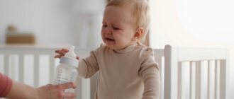
The first point in the medical care plan should be to call for specialized help. Next, it is necessary to calm the victim, without using medications for this, since they can distort the clinical picture during subsequent diagnosis. To slightly reduce pain, the person is placed on a flat surface, turning on his right side.
Doctors conduct an initial examination by interviewing the patient or the people around him. This is necessary for drawing up a subsequent detailed treatment program. To reduce discomfort, experts use antispasmodic drugs. To increase effectiveness, injectable medications are used as a basis, which are administered intramuscularly.
You should not try to inject a patient yourself, as there remains a high risk of developing an allergic reaction to the components of the medicine. The ambulance team has medications aimed at neutralizing the onset of anaphylactic shock in case of urgent need.
After providing assistance on the spot, doctors will hospitalize the victim if the severity of the disease is confirmed. In a hospital setting, specialists will conduct detailed diagnostics using ultrasound and other modern instrumental techniques.