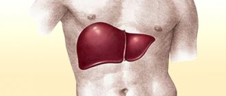
Liver cancer is a malignant tumor that quickly grows in size and metastasizes. It may be primary or represent metastases from other organs. The disease is asymptomatic for a long time or manifests itself with nonspecific symptoms.
Yusupovskaya's oncologists diagnose liver cancer using the latest research methods. To treat the disease, the most effective antitumor drugs are used, which have a minimal range of side effects. Surgeons use innovative surgical techniques. Medical staff provides professional patient care.
Causes
Primary liver cancer occurs on average 30 times less frequently than secondary liver cancer. As a rule, a malignant liver tumor occurs against the background of the presence of another oncological process in the body and its metastasis (for example, cancer of the uterus or stomach with metastases to the liver). Liver cancer occurs as often at 30 years old as at 50 and 60 years old - this disease has no age categories. The liver is susceptible to active metastasis due to intense blood circulation in this organ. There are a number of risk factors that influence the formation of malignant neoplasms in the liver:
- chronic inflammation of the liver. Cirrhosis, hepatitis C and B significantly increase the risk of developing cancer in the liver;
- alcoholism. Uncontrolled alcohol consumption destroys liver cells, providing favorable conditions for the formation of malignant neoplasms;
- poor nutrition and, as a result, excess weight. Excessive fat accumulation and slow metabolism increase the risk of cancer. The liver ceases to cope with the volumes of consumed hydrogenated fats and gradually loses its functions;
- hemochromatosis. This diagnosis involves a disorder of iron metabolism in the body, as a result of which its accumulation occurs in the gastrointestinal tract;
- heart failure;
- parasite infection;
- diabetes;
- syphilis;
- cholelithiasis;
- exposure to radiation and oncogenic viruses;
- disruption of the endocrine, lymphatic and circulatory systems;
- disturbances in the functioning of the biliary tract;
- accumulation of the contrast agent Thorotrast in the body. It can persist in the body for many years and eventually cause liver tumors;
- taking medications that have a toxic effect on the liver;
- smoking. Smoking negatively affects human health, and especially the functioning of the liver;
- elderly age. With age, organs stop working as well as in youth - the metabolic rate decreases and the likelihood of developing cancer becomes higher;
- burdened heredity.
Typically, malignant liver tumors develop under the influence of several causes and provoking factors.
Make an appointment
Patient reviews
| More information about liver cancer treatment at Euroonko: | |
| Liver cancer treatment | |
| Oncologist-gastroenterologist | RUB 5,100 |
| Emergency oncology care | from 12,100 rub. |
Bibliography:
- The prognostic value of tumor cells blood circulation after liver surgery for cancer lesions - Patiutko IuI, Tupitsyn NN, Sagaĭdak IV, Podluzhnyĭ DV, Pylev AL, Zabezhinskiĭ DA. — Khirurgiia (Mosk). 2011. [1]
- Surgical and combined treatment of multiple and bilobar metastatic affection of the liver. — Patiutko IuI, Sagaĭdak IV, Pylev AL, Podluzhnyĭ DV. — Khirurgiia (Mosk). 2005. [2]
- Modern approaches to the treatment of colorectal cancer metastases in the liver - A. L. Pylev, I. V. Sagaidak, A. G. Kotelnikov, D. V. Podluzhny, A. N. Polyakov, Patyutko Yu.I.,.. - Vestnik surgical gastroenterology - Number: 4, 2008 Pages: 14–28.
- Ten-year survival of patients with malignant liver tumors after surgical treatment - Yu. I. Patyutko, A. L. Pylev, I. V. Sagaidak, A. G. Kotelnikov, D. V. Podluzhny, M. G. Agafonova, - Annals of Surgical Hepatology 2010.
- Surgical and combined treatment of patients with metastatic liver and lymph nodes invasion by colorectal cancer — Patiutko IuI, Pylev AL, Sagaĭdak IV, Poliakov AN, Chuchuev ES, Abgarian MG, Shishkina NA. — Khirurgiia (Mosk). 2010. [3]
Book a consultation 24 hours a day
+7+7+78
Symptoms
In the first stages, liver cancer is practically asymptomatic. The patient may be concerned about weakness and fatigue, but this, as a rule, does not raise suspicions of liver cancer. Basically, the tumor begins to appear after spreading beyond the liver or metastasizing to other organs. The main clinical manifestations of liver cancer are:
- pain in the area of the right hypochondrium;
- increased body temperature;
- a feeling of heaviness that worsens after eating;
- causeless weight loss;
- decreased appetite;
- yellowness of the skin and sclera;
- nausea, vomiting, belching of air;
- tendency to diarrhea or constipation;
- palpable hard nodule in the liver area;
- an increase in abdominal volume due to the accumulation of pathological fluid;
- light colored stool;
- dark color of urine;
- anemia;
- skin itching;
- nosebleeds or gastrointestinal bleeding.
In order to establish a diagnosis as early as possible, you should contact a gastroenterologist when the first signs of liver dysfunction appear. After the examination, the patient will be consulted by an oncologist. Early diagnosis of malignant liver tumors allows for adequate therapy that increases life expectancy.
Rare symptoms
Some liver tumors secrete hormones, which cause specific symptoms. An excess of these hormones leads to the following changes:
- Increased levels of calcium in the blood (hypercalcemia), which is accompanied by nausea, confusion, constipation, muscle weakness;
- A decrease in blood sugar levels (hypoglycemia), which leads to rapid heartbeat, anxiety, irritability, and fainting;
- Enlarged breasts (gynecomastia) and/or testicular atrophy in men;
- An increase in the number of red blood cells in the peripheral blood (erythrocytosis), possible headaches that take the form of migraines, a reddish-bluish tint of the skin;
- Increased cholesterol levels.
Symptoms in women
Symptoms of liver cancer in women are no different from general symptoms. Doctors say that the danger for females lies in the later manifestations of the disease. Characteristic changes for women are:
- the appearance of male pattern hair;
- deepening of the voice;
- decreased libido;
- lumps in the chest and armpits.
Chemotherapy
There are now technologies that help deliver chemotherapy directly to the tumor in the liver. The main advantage of such procedures is that only a small amount of the drug enters the general bloodstream, so side effects are minimal. This allows the use of much higher dosages than with systemic chemotherapy. The problem of destruction of chemotherapy by liver cells becomes irrelevant.
- With intra-arterial chemotherapy, a puncture is made in the groin area, a catheter is inserted into the femoral artery, passed into the vessel feeding the tumor, and the chemotherapy drug is injected.
- Some patients undergo chemoembolization. A catheter is placed into the vessel feeding the tumor, and an embolic drug is injected through it along with the chemotherapy drug. The latter consists of special microscopic particles - microspheres, which block the lumen of the vessel. Tumor cells stop receiving oxygen and nutrients and die. Modern embolic drugs allow the procedure to be carried out as efficiently and safely as possible. Normal liver tissue is not affected because it receives its nutrition from another vessel.
Classification
Primary liver cancer mainly develops from hepatocytes - liver cells, or from bile duct cells. Experts distinguish several types of liver tumors:
- carcinosarcoma;
- sarcoma;
- liver lymphoma. Occurs due to the proliferation of atypical lymphocytes. This neoplasm is distinguished by its rapid growth and spread of metastases to distant organs;
- Hepatocellular carcinoma is an extremely rare form of tumor in the liver. The main cause of this type of cancer is cirrhosis or hepatitis B and C;
- cystadenocarcinoma. The structure of this neoplasm is similar to the structure of a cyst. The main manifestations of the disease are severe, sharp abdominal pain and dizziness. Cystadenocarcinoma tends to grow quickly, as a result of which it often causes compression of neighboring internal organs;
- hepatoblastoma is a tumor characteristic of childhood. Characterized by weight loss with a disproportionate increase in the abdomen;
- cholangiocellular liver cancer (portal liver cancer). A rather rare form of cancer associated with a mutation in the cells of the bile ducts. It is usually detected in the later stages, when treatment no longer brings results;
- angiosarcoma. Liver cancer is the most difficult and practically untreatable cancer. Metastases from angiosarcoma spread very quickly, preventing doctors from stopping or slowing down their growth. The reason for the appearance of this tumor is prolonged contact with poisonous and toxic substances in production;
- liver melanoma. One of the most severe liver tumors. Mainly occurs due to metastasis of another tumor or due to the development of melanoblastoma;
- fibrolamellar carcinoma. This liver tumor manifests itself as severe pain in the epigastrium and upper abdomen if the tumor has grown to a size of 20 centimeters or more. Treatable in early stages;
- undifferentiated sarcoma. In this case, the liver tumor grows and develops rapidly, spreading metastases to neighboring organs. It is often discovered in childhood and has virtually no treatment.
Timely diagnosis ensures a quick diagnosis, which is extremely important for cancer. You should not put off going to the doctor, because this is what can save your life.
Make an appointment
Other malignant neoplasms of the liver
Fibrolamellar cancer (FLC) is usually detected in young men with complete well-being, in a cirrhotic liver, without concomitant hepatitis B or C. Computed tomography for fibrolamellar cancer is performed to assess the resectability of the formation and determine the TNM stage. In a native study, LPR looks like an intrahepatic lesion, large in size - 5 cm or more, and with clear edges. This lesion has a density lower than the density of normal hepatic parenchyma, a lobular structure and - a peculiarity - a central area of fibrosis of a “star-shaped” shape. During the arterial phase, the tumor slightly intensifies in the peripheral parts, while the central part does not change its density.
Cholangiocarcinoma (CAC) is a malignant neoplasm of the bile duct epithelium. With CT, you can see uneven thickening of the wall of the duct against the background of its significant expansion. The formation accumulates contrast and remains hyperdense for a long time - this is a hallmark of cholangiocarcinoma.
Example of cholangiocarcinoma on computed tomography. Arrows highlight areas of the tumor that accumulate contrast agent in the delayed phase.
Hepatoblastoma is most often detected in childhood (3-5 years). On CT examination, it appears as a large hypodense lesion, occupying most of the section area. Approximately 1/5 of all hepatoblastomas are characterized by the presence of multiple foci. The structure of hepatoblastoma is heterogeneous - it may include areas of necrosis, calcifications, and connective tissue. Differential diagnosis is carried out with HCC, LPR, metastases.
Angiosarcoma of the liver is a rare tumor of the walls of the hepatic vessels. Its potential for malignancy is extremely low. It occurs more often in young women under 40 years of age; on CT it appears in the form of multiple cystic foci with clear boundaries, prone to fusion and the formation of polymorphic pseudocysts. It manifests itself as a “target” symptom due to uneven accumulation of contrast in the presence of a zone devoid of blood vessels along the periphery.
Liver lymphoma is extremely rare as a primary tumor; it is usually detected in systemic diseases, for example, lymphogranulomatosis. The CT picture is generally nonspecific - hypodense or isodense nodes of various sizes can be identified, and enlargement of the nearest lymph nodes can also be determined.
Undifferentiated embryonic cell sarcoma of the liver is a malignant neoplasm of sarcomatous cells. It looks like a large cyst, in some cases containing septa. However, despite its low density, it is actually solid, soft tissue. It sharply increases with contrast (in the periphery), differential diagnosis with the cystic variant of HCC is extremely difficult.
Angiosarcoma of the liver
An extremely aggressive malignant neoplasm that occurs upon contact with toxic chemicals (arsenic, copper, vinyl chloride) and upon exposure to radioactive radiation - liver angiosarcoma. Often, tumor growth begins against the background of accumulation in the body of Thorotrast, which was previously taken as a contrast agent when performing x-ray studies.
The symptoms of angiosarcoma are non-specific - the patient may experience abdominal pain, bloating, fever, loss of weight and appetite. During palpation, a compaction may be felt; during auscultation, the doctor may hear characteristic noises. It is difficult to establish a diagnosis at an early stage of development of the pathological process, since the onset of the disease is not accompanied by clinical signs of a malignant tumor.
Angiosarcoma is detected using the same methods as other types of liver malignancies. X-rays show thorotrast in the liver and spleen. During diagnostic laparoscopy, surgeons perform a biopsy and send areas of pathologically altered tissue for histological examination. Since angiosarcoma is detected at a late stage, patients are given palliative care in a hospice setting.
Factors for the development of HCC
Most often, the onset of cancer is preceded by cirrhosis of the liver (up to 90% of all cases). What is the difference between these two concepts? Cirrhosis is the replacement of normal liver parenchyma with atypical connective tissue, which in itself is not an oncopathology, but is considered a significant predisposing factor in its development. Cirrhosis can be either alcoholic or caused by hepatitis B or C. Exposure to carcinogenic factors and chemicals is important: sicasin (a toxic substance contained in some types of palm oil), aflatoxin (a toxin produced by fungi - aspergillus), thorotrast (a contrast agent, used at the dawn of x-ray diagnostics). Patients with hereditary pathology (Wilson-Konovalov disease, hemochromatosis, tyrosinosis) are also at risk for the incidence of HCC. It is also possible for liver tumors to occur in patients exposed to radiation (including after the accident at the Chernobyl nuclear power plant and other facilities).
Liver hemangioendothelioma
The clinical picture of liver hemangioendothelioma does not differ from other types of tumors. At first, the disease is asymptomatic, but later characteristic signs of liver cancer appear - yellowing of the skin, fever, weakness, fatigue, sudden and rapid weight loss. Often this disease entails particularly extensive internal bleeding. Scientists attribute this to damage to neighboring blood vessels by neoplasms and deterioration of blood clotting.
Effective methods of treating hemangioendothelioma are:
- radiation therapy. This technique is applied to primary pathology; a repeated course of radiation therapy in case of relapse is ineffective;
- surgical intervention. It is the most effective method of treating liver tumors, as it allows you to eliminate the source of the disease. However, surgery is not a guarantee of complete recovery, since metastases cannot be removed surgically;
- chemotherapy. This method involves the use of drugs that destroy malignant cells. Chemotherapy is fraught with a deterioration in the patient’s general well-being and is mainly used in cases of inoperable recurrent tumor.
Doctors at the Yusupov Hospital Oncology Clinic use only the most modern treatment methods in their practice, which can improve the patient’s quality of life.
What types of tumors are there?
If cancer first appeared in the liver, this is a primary process.
By type of source cells
tumors are divided into:
- the primary tumor arising from hepatocytes is hepatocellular liver cancer;
- primary tumor from the cells of the bile ducts - cholangiocellular liver cancer;
- mixed cancer (from the above two cell types);
- undifferentiated (cytologists cannot determine the type of tumor cells under a microscope due to their low differentiation);
- more rare - hemangiosarcoma, teratoma, carcinosarcoma, hepatoblastoma, embryonal sarcoma.
If the malignant cells are metastases, it is a secondary cancer. Such tumors are differentiated according to the place of appearance, number and nature of growth (single or multiple delimited nodes, diffuse proliferation of tumor tissue).
By disease stage
. Without going into complex letter designations (TNM), we can indicate that stage 1 cancer is a single node, without germination into any tissues or organs. Stage 2 cancer is several small tumors, or one large one with growth into the walls of blood vessels. Stage 3 liver cancer is a large tumor, but within an organ, or of any size, growing into the peritoneum, adjacent organs and nearby lymph nodes. Stage 4 liver cancer – there is a node in the organ, several groups of lymph nodes are affected and there is distant metastasis (in the spleen, for example).
Metastases
Metastases are the main danger of any cancer, since they are almost impossible to stop or prevent their spread.
Metastases of primary liver cancer can spread to the stomach, brain, lungs, esophagus, veins and arteries, heart muscle, blood vessels, bones and spine. Metastases from the liver enter the body in the following ways:
- through lymph flow;
- through the circulatory system;
- through growth beyond the liver and damage to neighboring tissues and organs.
Unfortunately, metastases occur in 40% of liver cancer cases and often affect vital organs, which are subsequently extremely difficult to treat.
Risk factors
Regardless of whether liver cancer has metastasized or not, it is still necessary to resort to urgent treatment. It is important to diagnose a malignant tumor on time. Therefore, a routine medical examination never hurts. There is a group of people who are more predisposed to developing this cancer. The most common form of liver cancer is a consequence of cirrhosis. Other risk factors include:
- excess weight.
- diabetes mellitus (type 2).
- use of anabolic steroids.
- exposure to toxic substances (aflatoxin, vinyl chloride, arsenic).
- smoking addiction.
- certain rare diseases (tyrosinemia, alpha-1-antitrypsin deficiency, porphyria cutanea tarda, etc.)
Complications
The uncontrolled course of liver cancer is dangerous due to the occurrence of complications. Their appearance depends on many factors. Possible complications include:
- bleeding from the tumor;
- suppuration of the tumor focus;
- impaired outflow of bile due to compression of the bile ducts;
- circulatory disorders due to compression of the abdominal organs by large tumor sizes;
- ascites.
The listed symptoms require immediate diagnosis and surgical intervention. Without this, death is possible.
Make an appointment
Why does the disease appear?
The liver has many functions in the body, the most important of which is detoxification. The liver, being a “living” chemical laboratory in the body, processes many aggressive substances every minute, transforming them into relatively safe compounds and promoting their elimination. The intensity of chemical reactions sometimes reaches millions per minute. The liver has an excellent ability to regenerate and restore.
Nature has programmed this ability due to the extreme load on the organ. Even 15% of the 100,000 existing liver cells - hepatocytes - are able to fully cope with normal loads. This is the “margin of safety” of this amazing organ. Unfortunately, modern features of life (unfavorable environmental situation; consumption of large amounts of fatty and salty foods; metabolic disorders in the body; sedentary lifestyle; use of foods containing many artificial additives - dyes, thickeners, sweeteners, etc.; drug abuse means, often unjustified use of antibiotics, immunomodulators, steroid hormones, and other pharmaceuticals) lead to the failure of reparative processes in liver cells (when uncontrolled proliferation of hepatocytes begins). The increase in the total number of oncological diseases leads to the frequent introduction of metastatic cells from other affected organs through the blood. The most common causes of primary cancers also include a high incidence of chronic hepatitis (B, C) and cholelithiasis among the population. All these factors can lead to liver cancer.
When cirrhosis of the liver develops against the background of alcoholism and especially viral hepatitis, this increases the risk of cancer by up to 20%. Alcoholism, as a predisposing cause of liver cancer, is dangerous not only by the development of liver cirrhosis. With excessive consumption of ethanol-containing drinks, it is the liver that processes (oxidizes) almost all the ethanol. And a toxic substance is formed - acetaldehyde, the processing products of which (oxidants) directly damage hepatocytes. Therefore, there is a high probability of developing malignant liver tumors without concomitant cirrhosis.
It has been noted that in regions where raw fish is traditionally consumed (Asian countries, India, South Africa, Eastern Siberia), the incidence of primary liver cancer is increased. This is associated with frequent parasitic lesions of the organ (opisthorchiasis, amoebiasis). And, as a consequence, the development of primary types of cancer occurs due to the oncogenic effect of helminths and protozoa. Harmful production factors predispose to the occurrence of tumors, including in the liver. The most dangerous substances that significantly increase the risk of cancer are fluoride compounds, phthalates, aromatic hydrocarbons, carbon tetrachloride, chlorine-containing pesticides, arsenic, and nitrosamines.
Diagnostics
Oncologists at the Yusupov Hospital conduct a comprehensive examination that allows you to quickly establish a diagnosis of liver cancer and verify the type of tumor. It includes the following studies:
- Complete blood count – the number of leukocytes and erythrocyte sedimentation rate increases;
- Biochemical blood test - the level of bilirubin, ALT, AST increases;
- Determination of the level of specific tumor antigen (tumor marker) ACE.
Using ultrasound, which is performed using expert-class equipment with high resolution, the location and size of the tumor are determined. Ultrasound is used both for primary diagnosis and for the purpose of dynamic monitoring of the condition of the tumor.
Magnetic resonance imaging and computed tomography are performed to clarify the size of the formation, the degree of invasion into surrounding tissues and organs, and distant metastases.
Laparoscopy is performed to clarify the spread of the tumor process. During the examination, the surgeon performs a biopsy - sampling tissue sections for histological examination.
Ablation
Metastases in the liver can be destroyed using radiofrequency ablation (RFA), a procedure during which a needle-electrode is inserted into the node and a high-frequency current is applied to it. The tumor tissue becomes very hot and dies. In order for RFA to be performed, certain conditions must be met:
- There are no more than 5 nodes in the liver. It is desirable that the diameter of each of them is no more than 4 cm.
- The lesions are clearly visible during ultrasound and computed tomography.
- The lesions are “conveniently” located: a needle can be inserted into them through the skin without damaging large blood vessels.
Stages
Determining the stage of the disease using clinical, instrumental and laboratory tests allows doctors at the oncology clinic to develop optimal tactics for patient management.
- In the first stage of liver cancer, the size of the tumor does not exceed two centimeters. There are no clinical symptoms. In some cases, patients experience increased fatigue and discomfort in the right hypochondrium.
- At the second stage of the disease, the size of the tumor reaches five centimeters. Invasion of atypical cells into blood vessels is observed. Patients are bothered by heaviness and dull aching pain in the right hypochondrium. As the pathological process progresses, the pain intensifies.
- The third stage of liver cancer is accompanied by the spread of the tumor to other organs and tissues. Severe pain syndrome forces the patient to see a doctor.
- Stage four liver cancer is characterized by multiple metastases to other organs. Due to increased pressure in the portal vein, the veins on the anterior wall of the abdomen and chest dilate. Patients are concerned about severe pain and constipation. Their abdominal volume increases due to the accumulation of fluid in the abdominal cavity, body weight decreases, and emotional lability occurs.
Diagnosis of liver cancer
As part of the diagnosis of liver cancer, studies are carried out aimed at detecting a tumor, determining the stage of the spread of the tumor process, as well as assessing the functional reserves of the liver.
The following tests are carried out as part of cancer detection.
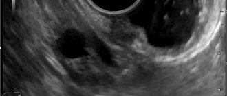
Ultrasound of the liver . The method is simple, safe, accessible and has sufficient sensitivity and specificity. Ultrasound also makes it possible to determine the invasion of cancer into the tissues adjacent to the liver, involvement in the process of the hilum of the liver, regional metastases and the presence of free fluid in the abdominal cavity. Also, expert-class devices allow you to assess the degree of fibrosis of the liver tissue.
Contrast-enhanced MRI has greater sensitivity and specificity than ultrasound and can detect tumors smaller than 1 cm against the background of cirrhosis.
To verify the diagnosis, a tumor biopsy under ultrasound guidance.
Liver biopsy
To search for metastases of liver cancer, an abdominal ultrasound, a plain chest X-ray or a CT scan of the chest organs is performed. A CT or MRI of the brain and bone scintigraphy are also prescribed.
To determine the functional reserve of the liver in the presence of cirrhosis, a series of tests are performed that take into account the presence of ascites, encephalopathy, the level of albumin and bilirubin in the blood serum, as well as the INR and prothrombin index.
Treatment
Doctors at the Yusupov Hospital Oncology Clinic take an individual approach to the choice of treatment methods for each patient diagnosed with liver cancer. The complex of treatment measures depends on the following factors:
- location of the pathological focus;
- stage of tumor development;
- presence or absence of metastases;
- patient's condition;
- accompanying illnesses.
The decision on the need and expediency of a particular method of therapy is made after a thorough diagnosis. Treatment for liver cancer includes:
- Medications. The active ingredients of medications have a toxic effect on cancer cells. In this case, healthy tissues are not damaged. Chemotherapy for liver cancer has no curative effect.
- Radiation therapy. Local impact on the tumor leads to a decrease in its size. As a result, the pain syndrome decreases. Radiation therapy is used at all stages of liver cancer treatment.
- Ablation. The essence of the method is the introduction of ethanol into the tumor formation. Then the pathological focus is exposed to microwave radiation. Ablation is effective for liver cancer that is less than 3 cm in diameter.
- Vascular embolization. Blood circulation in the tumor area is disrupted due to the introduction of special drugs into the vessels. As a result, the formation decreases in size. Vascular embolization is effective for tumors up to 5 cm in diameter. This method is often used in conjunction with other types of liver cancer treatment.
- Surgical intervention. In the presence of operable cancer, tumor removal or liver transplantation is indicated.
Briefly about the disease
According to statistics provided by the World Health Organization, the disease is widespread in the world (more than 700 thousand people are diagnosed annually) and is one of the leaders in the number of deaths from cancer (about 600 thousand people).
In liver cancer, metastasis processes are detected 30 times more often than primary cancer. It is difficult to treat at mature stages of development.
It affects men 3 times more often than women.
The average age of patients is 40-60 years.
Nutrition
The patient's condition improves with a special diet. The cooks at the Yusupov Hospital prepare dishes from high-quality products that do not contain nitrates, GMOs or carcinogenic food additives. In case of uncontrolled weight loss, increase daily caloric intake at the expense of protein products. When preparing dishes, preference is given to gentle culinary technologies: boiling, stewing, steaming and grilling, baking in the oven.
The following foods are excluded from the patient's diet:
- store-bought sauces;
- smoked meats and pickles;
- rich broths;
- margarine and other heavy fats;
- fresh baked goods;
- alcoholic drinks;
- strong tea and coffee.
Forecast
Since liver cancer is often diagnosed at the final stage of the tumor process, the prognosis for recovery is pessimistic in most cases. Palliative therapy is aimed at reducing the severity of symptoms of the disease and alleviating the general condition. In the presence of concomitant pathologies, death occurs 6 months or 1 year after diagnosis. The average life expectancy after the onset of this disease is about 2 years with properly selected effective treatment.
Primary and secondary cancer
In diagnosing liver cancer, in order to select a treatment method, it is important to identify its primary and secondary types.
Primary cancer is a pathology formed in the cellular structures of the gland. We are talking about stages 1 and 2 of the disease.
Primary cancer is:
- Hepatocellular carcinoma is the most common;
- Hemangiosarcoma - forms and develops in the blood vessels supplying the liver;
- Angiosarcoma - occurs infrequently, but is characterized by formation in people under 30 years of age;
- Cholangiocarcinoma - occurs in the bile duct;
- Hepatoblastoma – formed during fetal development;
- Hepatocellular carcinoma – more often found in men, appears in the liver epithelium.
A more severe form is secondary cancer. If it is present, metastasis processes are detected, i.e. These are stages 3 and 4 of the disease. As already noted, at this stage the disease is difficult to treat.
Unfortunately, the doctor has to deal with secondary cancer much more often than with primary cancer (95% to 5%), which means that the process of treating a patient causes many problems.
Prevention
You can reduce your risk of developing liver cancer by following the following medical recommendations:
- exclude alcoholic drinks;
- control weight;
- observe safety precautions when working with toxic substances;
- avoid unprotected sexual intercourse;
- do not take drugs;
- apply tattoos or make-up only in trusted salons;
- Vaccine against hepatitis B on time;
- undergo an annual preventive examination with a doctor.
Treatment of liver cancer at the Yusupov Hospital is prescribed by qualified oncologists depending on the nature of the tumor, its type and stage. All therapeutic procedures are carried out under the strict supervision of medical personnel, ready to provide assistance at any moment. In our clinic you can undergo any types of examinations using professional modern equipment.
Make an appointment
Popular questions
How can you prevent liver cancer?
First of all, care must be taken to avoid chronic liver diseases, which can lead to cirrhosis. If it is already present, then it is necessary to take tests for alpha-fetoprotein and perform an ultrasound of the abdominal cavity every six months.
What are the first symptoms of liver cancer?
The first symptoms can develop already at advanced stages, and they can also masquerade as signs of cirrhosis. Therefore, instrumental examination is necessary for early diagnosis. Symptoms most often include jaundice, loss of appetite, pain and heaviness in the right hypochondrium.
Does cirrhosis affect liver cancer treatment?
Yes, and cirrhosis itself can be a more serious problem than cancer, since with severe liver failure, the metabolism of various compounds and the synthesis of many proteins is disrupted, which in itself leads to the development of severe conditions. In addition, cirrhosis may influence the choice of treatment tactics for cancer patients.


