Stomach lymphoma accounts for 1/95-99 of all malignant processes in the organ, with cancer in the leading position. Externally and clinically, gastric lymphoma is similar to adenocarcinoma or a large ulcer. Previously, such lymphomas were treated surgically; modern therapy is focused on conservative methods.
- General information
- Causes of stomach lymphoma
- Classification of gastric lymphomas
- Symptoms of stomach lymphoma
- Diagnosis of gastric lymphoma
- Treatment of gastric lymphoma
- Diet for gastric lymphoma
- Recovery after treatment
- Complications
- Relapses
- Forecast
- Prevention of gastric lymphoma
General information
A rare tumor, gastric lymphoma refers to lymphoproliferative processes that develop not in the lymph node, but in the tissues of the stomach. In the group of non-Hodgkin lymphomas, which are very diverse in cellular structure, the proportion of gastric localization is less than 8%.
There are no incidence statistics, but out of 100 thousand adults, gastric lymphoma develops in hardly 5-6 people, mainly in mature and elderly people. It has been noticed that men get sick more often.
Lymphomas are treated by oncohematologists, and the process in the gastrointestinal tract is diagnosed by oncologists, which is explained by the standards for routing a patient with a suspected tumor.
Material and methods
The prospective study included 73 patients with GC who underwent radical surgery (R0) at the Orenburg Regional Clinical Dispensary from January 2009 to July 2010. The average age of the patients was 61.2±9.3 years (from 34 to 78 years, median - 61 years old). There were 43 men (58.9%), 30 women (41.1%). The tumor was localized in the upper third in 14 (19.2%) patients, in the middle third in 18 (24.7%) and in the lower third in 41 (56.1%). The intestinal type of gastric cancer was noted in 41 (56.2%) patients and diffuse - in 32 (43.8%). Well-differentiated adenocarcinoma (G1) was observed in 27 (36.9%) patients, moderately differentiated (G2) - in 14 (19.3%), poorly differentiated adenocarcinoma and undifferentiated GC - in 9 (12.3%) and signet ring cell carcinoma ( RCC) - in 23 (31.5%). The distribution of patients by disease stage was as follows: T1-2N0M0 - in 34 (46.6%), T3N0M0 - in 9 (12.3%), T3-4N1M0 - in 9 (12.3%) and T3-4N2M0 - in 21 (28.6%) patients.
Subtotal proximal resection was performed in 10 (13.7%) patients, subtotal distal resection in 56 (76.7%) and gastrectomy in 7 (9.5%). Lymphadenectomy in the D2 volume was performed in all patients, with D3 elements in 38 (52.0%). In 7 (12.3%) patients, combined operations were performed: splenectomy was performed in 3 cases, transverse colon resection was performed in 2 cases, and cholecystectomy was performed in one case. The study did not include patients with decompensation of chronic diseases, acute infectious pathology, severe allergic processes, or those receiving corticosteroids, antihistamines, non-steroidal anti-inflammatory drugs, or neoadjuvant chemotherapy.
After removal of the drug, the stomach was opened along the greater curvature. A biopsy of the tumor and macroscopically unchanged coolant was performed, 3-5 cm from the proximal edge of the tumor. The tissue samples taken were placed in neutral formalin. The material was carried out according to standard methods and embedded in paraffin. Sections 4–5 μm thick were stained with hematoxylin and eosin and immunohistochemically using anti-CD20 antibodies (Epitope Specific Rabbit Antibody, Thermo Fisher Scientific) at a dilution of 1:400. The material for study by immunohistochemical (IHC) methods was fixed with 10% neutral formalin for 24 hours, embedded in paraffin, sections 4 μm thick were prepared, which were applied to highly adhesive glasses and dried at a temperature of 37 °C for 18 hours. Deparaffinization and unmasking of antigens were carried out in the RT module at a temperature of 121 °C for 20 minutes, followed by cooling for 60 minutes. The imaging system used was Ultra Vision LP Detection System HRP Polymer & DAB Plus Chromogen. As a negative control, sections were similarly incubated with antibody dilution solution (UltrAb Diluent). In the peritumoral area, the presence of focal lymphoid B-cell infiltrates and the number of lymphoid follicles (LF) in the coolant (none, single - no more than 2 in the field of view, multiple - more than 2 follicles in the field of view) were assessed using a semi-quantitative method. The presence of normal and atypical light centers in them was also assessed. The density of diffuse B lymphocytes was counted using a magnification of 400, over an area of 1200×1800 pixels, taken as a conventional unit of area (ARU), with a resolution of 10 pixels/cm (0.42×0.28 mm2). The metric grid density was 216 points.
Statistical processing of the study results was carried out using the Statistica 6.0 application package. B cell density was expressed as mean (M) ± standard deviation (σ). Comparison of indicators between groups was carried out using parametric and nonparametric methods (LSD and Mann-Whitney tests). The relationship between indicators was determined using nonparametric methods (Spearman rank correlation and gamma). The reliability of differences in the frequencies of signs in the studied groups was assessed using the χ2 test. Analysis of overall and disease-free 3-year survival was performed using the Kaplan-Meier method. Comparison of survival rates between groups of patients was carried out using the long-rank test. Differences between indicators were considered significant at p
<0,05.
Causes of stomach lymphoma
The causes of the development of gastric lymphomas are unknown, and gene mutations leading to the degeneration of normal cells have not been identified. The family history of those suffering from gastric lymphomas often includes relatives with malignant diseases.
Immunity disorders are assumed to be involved in tumor initiation:
- excessive protection against allergies and autoimmune diseases;
- immunodeficiency due to HIV and hereditary syndromes;
- tolerance violations during antitumor therapy and chronic inflammation;
- depression due to increased radiation exposure.
A certain etiological role belongs to Helicobacter pylori infection; in most patients, Helicobacter pylori (H. pylori) is found in gastric secretions. Accumulations of B-lymphocytes in the gastric wall, from which a tumor develops, occur during chronic inflammation caused and maintained by bacteria. Detection of Helicobacter pylori infection and its treatment is a fundamental approach for the MALT (mucosal-associated) tumor variant.
Folk remedies and prevention
Hospital treatment is absolutely necessary
It is not possible to cure lymphoma with folk remedies. They only serve as supportive and strengthening therapy. Before taking any medication, be sure to consult your doctor. Uncontrolled use of herbs and infusions can significantly worsen your condition. Sea buckthorn juice has a healing effect. It soothes and restores the gastric mucosa. Sometimes it is recommended to mix vodka and vegetable oil and take it orally. A decoction of birch buds can also help the digestive system. Drops from Djungarian aconite can be consumed in limited quantities. It is used both internally and externally.
Preventive measures are the same as for stomach cancer. It is impossible to prevent the occurrence of lymphoma 100%, since it is impossible to accurately determine the causes of its occurrence. You can only reduce the risk factors that can lead to this disease. For example, it is recommended to avoid negative environmental influences such as radiation and polluted air. If you live in an ecologically unfavorable area and it is not possible to move, you need to periodically give your body a rest by going out into nature or to a village with a more favorable environment.
According to research, agricultural workers are at increased risk of developing lymphoma because they are exposed to pesticides, which is very dangerous for the body. With reduced immunity, the risk also increases. This is especially true for AIDS patients and people who have undergone organ transplants. During transplantation, therapy with immunosuppressants is carried out. Poor nutrition, consumption of carcinogens, and vegetables with pesticides also increase the chances of developing stomach lymphoma. The higher the quality of food, the less likely it is that the stomach will suffer from certain diseases.
Classification of gastric lymphomas
Lymphomas are heterogeneous in their cellular composition:
- The vast majority of 80% are tumors developing from B-lymphocytes of the marginal zone, or MALT lymphoma ; in some patients, H. pylori infection is found, which is in favor of a more favorable course.
- The remaining 20% are aggressive B-lymphocyte lymphomas or B-lymphomas , requiring difficult treatment with unenviable prospects for long life. Some researchers believe that this type of tumor results from transformation of a long-standing MALT lymphoma, as some patients have both types of tumor at the same time.
- The rarest type of benign tumor with possible transformation into aggressive B-lymphoma is pseudolymphoma , the process is localized only in the stomach, does not extend beyond its boundaries, and has a good prognosis for cure.
Staging of lymphomas is carried out not according to the usual oncological classification TNM or hematological Ann-Arbor, but according to Lugano, where the stage is:
- IE—small damage to the organ wall only;
- IIE - local tumor of the organ and nearby lymph nodes;
- IIIE - involvement of lymph nodes outside the abdominal cavity;
- IV - dissemination in the body.
Types of disease and its causes
Proper nutrition is the key to a healthy stomach
The increase in incidence can be associated with the deterioration of the environmental situation and the habit of eating unhealthy food. Most of the food consumed contains a significant amount of chemical additives that negatively affect the digestive system. Immunity also plays an important role. Some types of lymphoma are caused by infection. Lymphoid tissue contains lymphocytes, which are responsible for producing antibodies that attack harmful foreign cells. If a large amount of harmful substances enters the body, the immune system may malfunction, as a result of which antibodies will be produced in large quantities and attack the body or, conversely, will be produced insufficiently. Risk factors include old age, chronic infections such as hepatitis and AIDS, autoimmune diseases, etc.
Lymphoma may initially be cancerous or become cancerous as it grows. Stomach lymphoma is believed to be more treatable than cancer.
There are several types of lymphomas:
- Primary. Primary lymphoma is especially easy to confuse with stomach cancer. The symptoms are very similar. The gastric mucosa and submucous membranes are affected. The bone marrow is not affected. Often occurs against the background of other ulcerative and inflammatory diseases (gastritis, ulcers). It is somewhat easier to treat than cancer. The chances of recovery are relatively high.
- Secondary. Secondary lymphoma is characterized by a larger affected area.
- Lymphogranulomatosis. This disease is accompanied by an inflammatory process of lymphoid tissue and the transfer of this process to the gastric mucosa.
- Non-Hodgkin's lymphomas. Lymphomas of this type are clearly differentiated. They are caused by a certain type of infection caused by the bacterium Helicobacter. The degree of malignancy can vary greatly.
- Pseudolymphoma. Pseudolymphoma begins as a benign formation that does not have metastases. However, over time it can develop into malignant if not properly treated.
Symptoms of stomach lymphoma
Clinical manifestations of gastric lymphoma in the early stages are nonspecific and indistinguishable from the symptoms of carcinoma and ulcers. In various combinations, the following are possible: pain in the pit of the stomach, discomfort, heartburn, belching, feeling of heaviness after eating, loss of appetite, weight loss.
With a widespread process, the lymph nodes become enlarged, the lungs, intestines, liver, and spleen are affected.
Regardless of the size of the tumor, with the aggressive version of lymphoma, high fever with night sweats, weakness and rapid weight loss are possible - the so-called “B-symptoms”, indicating an unfavorable prognosis of lymphoma.
Diagnosis of gastric lymphoma
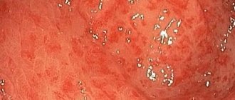
Diagnosis of lymphoma at the first stage does not differ from the detection of other malignant processes of the stomach: endoscopy with biopsy, ultrasound, CT of the abdominal cavity, CT of the chest.
As with all malignant processes of the blood and lymphatic tissue, a bone marrow trephine biopsy is necessarily performed.
After morphological verification, diagnostics clarifying the therapy are carried out:
- tests for the detection of Helicobacter Pylori in gastric contents;
- the sensitivity of Helicobacter to antibiotics is determined;
- cytogenetic analysis to detect the transfer of the t(11;18) region from one chromosome to another; when a translocation is detected, a clarifying FISH analysis is performed.
Clinical picture and diagnosis
Symptoms manifest in different ways, making diagnosis much more difficult. Some patients have no signs of the disease at all. It is generally accepted that primary lymphoma is characterized by pain in the epigastric region and periodic vomiting. Unreasonable weight loss and anemia are also possible. It is extremely important to differentiate between primary and secondary tumors. According to studies, in the first case, a larger (more than 5 cm) tumor focus and unique growth in the body or astral region are detected. In the second, in turn, the pathological process is localized in the bottom.
This disease can be asymptomatic for many years. Single clinical signs include:
- Pain and discomfort in the epigastric region (associated and unrelated to eating).
- Feeling of a full stomach, rapid satiety from food, belching of air, nausea and decreased appetite.
- Anemia (develops due to bleeding from a neoplasm into the lumen of the organ).
- General symptoms: increased body temperature, night sweats, causeless weight loss.
At the time of confirmation of the diagnosis, signs of generalization of the pathology are often detected - involvement of the bone marrow, abdominal lymph nodes and other organs.
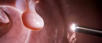
Lymphoma is most accurately diagnosed using a biopsy (a set of biomaterial for further microscopic examination). Also, if an oncological process is suspected, it is recommended to conduct a urease test with multiple biopsies from several parts of the organ. If endoscopic testing is negative, a serological test is performed. The stages of the disease are determined based on endoscopic ultrasound and are as follows:
- T1a - superficial layer of mucosa: does not leave the 1st layer.
- T1b - deep layers: within the 2nd layer.
- T2 - submucosal layer: within the 3rd layer.
- T3 - extends beyond the boundaries of the submucosal layer: the 4th and 5th are involved.
Histology is the most accurate and reliable way to identify pathology. However, MRI and CT cannot be ruled out.
Treatment of gastric lymphoma
Treatment for MALT lymphoma and B-cell lymphomas differs significantly, with the latter requiring aggressive chemotherapy.
The treatment option for early-stage MALT lymphoma is determined by Helicobacter pylori infection; if it is present, then at the first stage antibacterial treatment is carried out, similar to the eradication of H.pilory in peptic ulcer disease.
The second stage depends on the results of the initial treatment:
- If H.pilory is resistant to antibiotics, the treatment regimen is changed; if the prognosis is unfavorable with t(11;18), targeted therapy with rituximab or radiation is added in parallel.
- When the gastric secretion is cleared of Helicobacter pylori infection and the lymphoma is completely regressed, the patient is observed with regular examinations according to the schedule.
- If there is a proven absence of infection and residual tumor, radiation to the stomach or monotherapy with rituximab is performed.
MALT tumors that have developed without the participation of Helicobacter and are limited to the gastric walls, especially in the presence of a chromosomal abnormality, are irradiated at the first stage; if radiation therapy is not possible, a course of targeted drug is given. Surgery to remove the stomach is performed only for lymphomas complicated by bleeding ulcers.
For advanced MALT lymphoma, preference is given to drug therapy.
Complications
Any tumor in the stomach is characterized by complications:
- pain syndrome,
- ulceration with acute or chronic bleeding,
- perforation of the organ wall with the development of peritonitis.
In addition, with lymphoma, there may be a decrease in the specific gamma globulin IgG produced by normal immune cells, which contributes to frequent infections and inflammatory processes. The deficiency is compensated by intravenous administration of immunoglobulin.
In all cases, before starting chemotherapy, prevention of tumor lysis syndrome, which occurs when malignant cells are highly sensitive to drugs, is carried out.
Relapses
MALT lymphoma of the stomach has a slow course, however, as the stage increases, the likelihood of relapse increases. Starting from stage IIE, tumor recurrence is possible in every second patient, but the average period of relapse has not been determined due to the rare occurrence of the disease. The ability of some recurrent nodes to shrink without treatment—self-limitation—has been noted.
With aggressive lymphoma, relapses occur more often and remissions are shorter. In all situations, in case of relapse, chemotherapy is resorted to.