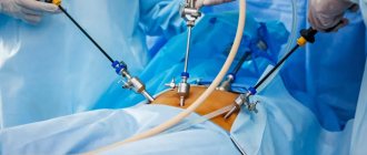
Intestinal operations are considered one of the most complex and require special professionalism of the surgeon. It is important not only to restore the damaged integrity of the organ, but also to do this so that the intestines continue to function normally and do not lose their contractile function.
Intestinal anastomosis is a complex operation that is performed only in cases of extreme necessity and in 4-20% of cases leads to various complications.
results
The creation of colorectal anatomoses after anterior and low anterior resections of the rectum is often accompanied by the formation of a “temporary” protective stoma in order to reduce the consequences of possible anastomotic leakage (AL). However, in some patients these “temporary” stomas remain unclosed. By the time of discharge from the hospital, 111 (52%) patients had a colostomy on the anterior abdominal wall: in 103 of them, a preventive double-barreled transversostomy was formed during the primary surgical intervention, the remaining 8 patients required its removal in the postoperative period due to the development of AN. The average follow-up period was 32.3 months (12-70 months). Of the 111 patients, 96 (86.5%) underwent reconstructive surgery. Multivariate analysis revealed the following factors that were predictors of “non-closure” of the stoma after anterior resection: death from disease progression (p=0.24) and chemotherapy (p=0.22).
Conclusions. The risk that a “temporary” stoma will remain permanent, even in a specialized center, is 13.5%. When performing sphincter-sparing operations for all stages of rectal cancer, the most significant risk factors for “non-closure” of the stoma are progression of the disease and the associated need for chemotherapy, as well as the death of the patient.
If intestinal obstruction develops
The occurrence of obstruction can cause swelling of the anastomosis area and cicatricial narrowing. In case of acute symptoms, a repeat laparotomy is performed (an incision in the abdomen and opening of the abdominal cavity) to eliminate the pathology.
In case of chronic obstruction in the long-term postoperative period, intensive antibacterial therapy and removal of intoxication are prescribed. The patient is examined to decide whether surgical intervention is necessary.
Any complications require treatment
Results
Creation of colorectal anastomoses after anterior and low anterior resections is quite often accompanied by the formation of “temporary” protective stoma to reduce consequences of possible anastomotic leak (AL). However, in some patients these “temporary” stomas are never reversed.. By the moment of discharge from hospital 111 (52%) patients had colostomies: in 103 of them were preventive double-barreled transverse colostomies were created at primary surgical intervention, the the remaining 8 patients underwent colostomy formation in the postoperative period because of development of AL. Average follow-up was 32.3 months (12 to 70 months). Among 111 patients the restorative operations were done in 96 (86.5%). At multivariant analysis the following factors were identified as predictors of non-closure of stoma after anterior resection: death due to disease progression (correlation coefficient = 0.24) and chemotherapy treatment (correlation coefficient = 0.22).
Conclusions
The risk of “temporary” stoma nonclosure even in specialized center is 13.5%. When sphincter-sparing operations are performed for all stages of rectal cancer the most essential risk factors of non-closure of stoma were progression of disease and chemotherapy treatment and patient death related to it, and patient's death.
The development and widespread introduction of staplers standardized the technique of anastomosis in the depths of the small pelvis and made it possible to form it at any distance from the edge of the anus, including the anal canal itself [18]. Due to this, about 80% of planned operations for rectal cancer today are sphincter-preserving in nature [2]. However, this does not mean that all of these patients will eventually empty their bowels naturally. The main reason is considered to be failure of the hardware colorectal anastomosis, for the prevention and treatment of which most medical institutions use the formation of a proximal stoma [13].
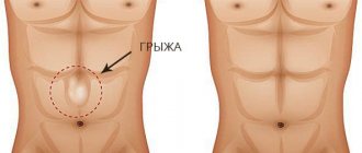
When the section of intestine carrying the anastomosis is temporarily excluded from the passage, a double-barreled stoma is formed, but if the anastomosis is disconnected, then the intestinal contents are drained by forming a single-barrel stoma [9]. Meanwhile, a clear understanding of the likelihood of preserving the natural course of the intestine if an anterior resection is planned will allow the multidisciplinary council to correctly plan possible treatment options, and the surgeon to build relationships with the patient and his relatives at the preoperative stage. The relevance of this kind of knowledge is explained by the fact that underestimation of the probability of formation and the impossibility of subsequent closure of the stoma during surgical treatment of the primary tumor, often in combination with the surgeon’s overestimation of his own strength, are the main components of the fact that anastomotic leakage (AF) after rectal resection often becomes a tragedy both for the patient and his relatives, and for the doctor who performed the operation.
The purpose of this study is to analyze the reasons and frequency of leaving colonic stomas created for prophylactic or therapeutic purposes after performing anterior and low anterior resections with the formation of a hardware colorectal anastomosis for rectal cancer.
Description and characteristics of the essence of anastomoses
The formation of an intestinal anastomosis is usually preceded by the removal of part of the intestine (resection). Next, it becomes necessary to connect the adducting and efferent ends.
End to end type
Used to sew together two identical sections of the large intestine or small intestine. Performed with a two- or three-row seam. It is considered the most advantageous in terms of compliance with anatomical features and functions. But technically difficult to implement.
The condition for connection is that there is no big difference in the diameter of the compared sections. The end that is smaller in clearance is cut to ensure full compliance. The method is used after resection of the sigmoid colon, in the treatment of intestinal obstruction.
First, the posterior wall of the anastomosis is formed, then the anterior one
End-to-side anastomosis
The method is used to connect sections of the small intestine, or the small intestine on one side and the large intestine on the other. Usually the small intestine is sutured to the side of the wall of the large intestine. Provides 2 stages:
- At the first stage, a dense stump is formed from the end of the efferent intestine. The other (open) end is applied to the intended site of anastomosis from the side and sutured along the back wall with a Lambert suture.
- Then an incision is made along the abductor colon along a length equal to the diameter of the adductor section and the anterior wall is sutured with a continuous suture.
It is used for various complex operations, for example, after complete removal (extirpation) of the esophagus with adjacent lymph nodes and fatty tissue.
Side to side type
It differs from previous options in the preliminary “blind” closure with a double-row suture and the formation of stumps from connected intestinal loops. The end above the stump is connected with the lateral surface to the underlying area by a Lambert suture, which is 2 times longer than the diameter of the lumen. It is believed that technically performing such an anastomosis is the easiest.
It can be used both between homogeneous sections of the intestine and to connect dissimilar areas. Main indications:
- the need for resection of a large area;
- danger of overstretching in the anastomosis area;
- small diameter of connected sections;
- formation of an anastomosis between the small intestine and the stomach.
The advantages of the method include:
- no need to suture the mesenteries of different areas;
- tight connection;
- guaranteed prevention of intestinal fistula formation.
With side-to-side anastomosis, the preliminary creation of stumps is one of the disadvantages of the technique
Side-to-end type If this type of anastomosis is chosen, this means that the surgeon intends to sew the end of the organ or intestine after resection into the created hole on the side surface of the afferent intestinal loop. More often used after resection of the right half of the large intestine to connect the small and large intestines.
The connection can have a longitudinal or transverse (more preferable) direction with respect to the main axis. With a transverse anastomosis, fewer muscle fibers are crossed. This does not disrupt the peristalsis wave.
Material and research methods
The materials of a prospectively filled database were analyzed, as well as medical records of patients treated in the department of coloproctology with pelvic floor surgery of the Russian Scientific Center for Surgery named after. B.V. Petrovsky RAMS in the period from June 2006 to June 2011.
The inclusion criteria for the study were: primary histologically confirmed rectal cancer and sphincter-preserving resection in the amount of R0. The exclusion criteria were:
- resection of the rectum without the formation of a primary colorectal anastomosis (obstructive resection of the rectum, or Hartmann-type surgery);
- resection of the rectum with the formation of a primary colorectal anastomosis manually;
- previous surgical interventions on the colon or rectum, as well as the pelvic organs;
- primary multiple tumors of the colon;
- the patient's death within 30 days after surgery.
Thus, patients were selected who underwent surgery for rectal cancer with preservation of the anal sphincter and the formation of a hardware colorectal anastomosis using various circular staplers - DST (Covidien, USA), KYGW (Kangdi, China), CDH (Ethicon Endosurgery, USA). By the method of collecting material, the study is prospective-retrospective, and by the type of observation - cohort.
For the analysis, the following data from the electronic database and archived medical records were used: gender, age of the patient, body mass index (BMI), diabetes mellitus and diverticulosis, American Association of Anesthesiologists ASA index, information on chemotherapy, radiation or chemoradiation therapy, height location of the tumor, parameters of the tumor process and its complications (obstruction, anemia, hypoproteinemia), the nature of the surgical intervention (combined, combined), the volume of lymph node dissection, the formation of a preventive colostomy, the frequency and nature of the development of AN, the scope of therapeutic measures to eliminate it (conservative therapy, minimally invasive methods of treatment, repeated surgical intervention), the time of closure of the stoma (if it was formed), the reasons for its late liquidation or refusal to close it, as well as measures aimed at treating the cavity in the area of insolvency of the anastomotic sutures, the date of the last visit and the presence of signs of recurrence of the disease, if in the event of death - the date and cause of death, if a local recurrence of rectal cancer is detected - the date, location and extent of the disease.
To assess AN, the classification proposed in 2010 by the International Colorectal Cancer Study Group was used. According to the classification, three degrees of severity of this complication are distinguished: grade A - corresponds to failure without clinical manifestations, usually detected during control proctography; grade B - to stop the complication, antibacterial therapy and/or minimally invasive (conservative) measures are required; grade C—repeated surgery is required [17].
In case of suspected AN, proctography was performed in the early postoperative period using a water-soluble contrast agent. According to indications, computed tomography of the pelvic organs was performed to determine hidden leaks and abscesses. The state of the formed hardware colorectal anastomosis in late follow-up periods (more than 30 days after surgery) was determined using liquid barium suspension proctography.
Given that the work is based on a retrospective analysis of prospectively collected clinical material, the patient's signing of informed consent for participation in the study and approval by the ethics committee were not required.
When comparing intergroup parameters, quantitative characteristics were assessed using Student's i-test, and qualitative characteristics were assessed using Fisher's exact test. To determine the risk factors for refusal to perform reconstructive surgery, the multiple logistic regression method and the compilation of a correlation matrix were used, followed by multivariate analysis and calculation of the correlation coefficient (CC). Patient survival was calculated using the Kaplan-Meier method. Differences at p values <0.05 were considered statistically significant.
Examination before surgery
Before applying a biliodigestive anastomosis, doctors at the Yusupov Hospital conduct a comprehensive examination of the patient. Cancer patients undergo a general blood test, urine test, and coagulogram. They perform instrumental studies:
- radiography of the biliary tract;
- ultrasound endoscopic examination;
- computed tomography;
- endoscopic retrograde cholangiography.
Before surgery, patients are examined by an anesthesiologist. He determines the degree of surgical risk, selects the type of anesthesia that is planned to be used during surgery, and prescribes premedication.
Research results
Between June 2006 and June 2011, 293 sphincter-sparing rectal resections were performed. We excluded from the study 68 patients in whom the anastomosis was formed manually, 8 patients after obstructive resection of the rectum using the Hartmann procedure, and 2 more patients who died on the 16th and 22nd days from progressive mesenteric thrombosis of the superior mesenteric artery.
Thus, 215 patients (among them 117 men) who underwent anterior or low anterior resection of the rectum with the formation of a primary colorectal anastomosis using a circular stapler were analyzed. The average age of the patients was 61.2±11.3 years (age range 29-87 years). 42 (19.5%) patients had a BMI over 30, corresponding to obesity. In 90% of observations, the degree of local spread of the process corresponded to T3-T4. Only 12% of patients received preoperative chemotherapy and/or radiation therapy, which was associated with refusal of neoadjuvant treatment for stage I, II, and IV disease. In addition, of all patients with stage III in the absence of severe concomitant diseases and complications of the tumor process, only those who were diagnosed with a “high-risk” tumor were referred for preoperative combined treatment. Clinical and morphological characteristics of patients included in the study are presented in Table. 1.
In 103 (47.9%) patients, a preventive double-barreled transversostomy was formed during the primary surgical intervention. In 99 of them, a low anterior resection of the rectum was performed with the formation of a supranal colorectal anastomosis: the creation of a preventive colostomy in such cases is standard practice in our clinic. In another 4 cases, the reasons for the formation of a stoma were the entry of air through the staple suture area during an air test and/or unsatisfactory preoperative bowel preparation. Among 112 patients in whom a preventive colostomy was not formed during the primary operation, 8 (7.1%) required its removal in the postoperative period due to the development of failure of the hardware anastomosis - in 6 cases a double-barreled colostomy was formed and in 2 cases the anastomosis was disconnected with the removal of an end colostomy. Thus, of the 215 patients who underwent a hardware colorectal anastomosis during surgery, 111 (52%) had a colostomy on the anterior abdominal wall by the time of discharge from the hospital.
Table 1
Characteristics of patients included in the study
| Options | Number of patients (n=215) | |
| abs. number | % | |
| Accompanying illnesses: | ||
| anemia | 45 | 20,9 |
| hypoproteinemia | 38 | 17,7 |
| colon diverticulosis | 21 | 9,8 |
| diabetes | 18 | 8,4 |
| ASA status: | ||
| 1-2 | 143 | 66,5 |
| 3 | 70 | 32,6 |
| 4 | 2 | 0,9 |
| Depth of tumor invasion: | ||
| T1 | 3 | 1,4 |
| T2 | 19 | 8,8 |
| T3 | 133 | 61,9 |
| T4 | 60 | 27,9 |
| Presence of lymphogenous metastases: | ||
| rSh-2 | 98 | 45,6 |
| rSh | 117 | 54,4 |
| Presence of hematogenous metastases: | ||
| sM1 | 68 | 31,6 |
| cm0 | 147 | 68,4 |
| Combination of lymphogenous and hematogenous metastasis | 43 | 20,0 |
| Neoadjuvant radiation and chemotherapy | 26 | 12,1 |
| Location of the tumor in the rectum: | ||
| superior ampulla | 89 | 41,4 |
| mid-ampullary section | 49 | 22,8 |
| inferior ampulla | 77 | 35,8 |
| Type of operation: | ||
| anterior resection | 116 | 54,0 |
| low anterior resection | 99 | 46,0 |
| Characteristics of the surgical intervention: | ||
| combined | 85 | 39,5 |
| combined | 28 | 13,0 |
| Formed by colostomy: | ||
| preventive | 103 | 47,9 |
| medicinal | 8 | 3,7 |
The average follow-up period was 32.3 months (9-70 months). Among 103 patients who had a colostomy for preventive purposes, in 57 (55.3%) its closure was performed within the standard time frame for our clinic - within 4- 8 weeks after surgery. In another 32 (31.1%) patients, the elimination of double-barreled colostomy was carried out at a later date (more than 8 weeks after surgery): in 17 patients this intervention was postponed due to the need for postoperative chemotherapy and in 15 due to long-term treatment measures, aimed at treating AN (conservative therapy, marsupilization, reresection of the anastomotic zone). In the remaining 14 patients with preventive colostomy, it was not possible to restore the natural course of the intestine. One of these patients refused re-intervention. As a result of AN, he developed a long-term non-healing fistula; for 10 months after the operation, conservative treatment was carried out, including marsupilization of the residual cavity, as a result of which the fistula healed, but the patient did not want to expose himself to additional risk during a repeat operation, citing the fact that that his quality of life with a stoma is quite satisfactory. The remaining 13 patients, whose colostomy was not eliminated, had distant metastases at the time of surgery, which rapidly progressed in the postoperative period: 7 of them died from metastatic foci, 5 continued chemotherapy, one patient was lost to follow-up, his fate is currently unknown .
Of the 8 patients who had a colostomy due to a leaky colorectal anastomosis that occurred in the early postoperative period, 7 underwent reconstructive surgery after eliminating the consequences of AN. In one patient with stage IV disease, restoration of the natural course of the intestine was not performed due to the progression of distant metastases: despite adjuvant chemotherapy, death occurred 7 months after surgery.
Consequently, the elimination of a formed preventive or therapeutic colostomy was carried out in 96 patients, in 91 (94.8%) of them during the first year after the main operation, while the average period between the primary intervention and closure of the colostomy was 3.4 ± 3.7 months. (interval 0.7–22.9 months).
Thus, out of 215 patients who underwent anterior resection of the rectum with the formation of a hardware colorectal anastomosis, it was possible to achieve preservation of the natural course of the intestine in 200 (93.0%) with an overall failure rate of 12.8% and a level of clinically significant NA corresponding to grade C, 5.6%. At the same time, out of 111 patients discharged from the hospital with a stoma after the main operation, reconstructive operations were performed in 96 (86.5%) cases.
In a univariate analysis of a large number of factors, it was not possible to establish a significant influence of any of them on the fact of performing a repeat operation aimed at restoring the natural course of the intestine (Table 2).
table 2
Univariate analysis of the influence of various factors on stoma closure
| Factor | R |
| Floor | 0,26 |
| Age | 0,71 |
| ASA index | 0,6 |
| BMI | 0,2 |
| Anemia | 0,8 |
| Hypoproteinemia | 0,58 |
| Diverticulosis | 0,28 |
| Chemotherapy | 0,052 |
| Radiation therapy | 0,93 |
| T4 | 0,9 |
| N | 0,5 |
| M | 0,12 |
| Operation duration | 0,76 |
| Volume of blood loss | 0,66 |
| Scope of lymph node dissection | 0,71 |
| Formation of a preventive colostomy | 0,16 |
| Anastomotic leakage (grade C) | 0,24 |
| Disease progression | 0,11 |
| Death from disease progression | 0,09 |
| Presence of signs of local recurrence | 0,47 |
Multivariate analysis revealed the following factors that were predictors of “non-closure” of the stoma after anterior resection: death from disease progression (CC = 0.24) and chemotherapy (CC = 0.22). Despite obtaining a possible connection between stage M and the risk of “non-closure” of the stoma when compiling a correlation matrix, a multivariate analysis did not establish a significant correlation (CC = 0.17).
Advantages of the Clinical Hospital on Yauza
The clinic on Yauza successfully performs surgical interventions of any complexity using laparoscopic and open methods. Hospital treatment has many benefits. The main ones include:
- the latest diagnostic devices;
- modern rehabilitation wards;
- highly qualified personnel;
- individual approach to each patient;
- affordable prices.
You can book an appointment with a surgeon using the hotline number or on the clinic’s website.
Sign up for the procedure
Discussion of the research results
The presented study reflects the results of the current protocol for the formation of preventive and therapeutic stomas when performing anterior resections using circular staplers in a specialized coloproctology department.
From a historical point of view, anterior resection with the formation of a colorectal anastomosis began to be used about 80 years ago [10]. However, its heyday began in the late 70s and early 80s of the last century after the advent of circular staplers. And here it would not be out of place to mention that the idea and the first practical implementation of a circular stapler belonged to Soviet engineers and surgeons [4]. The era of total mesorectumectomy that followed radically changed the understanding of the oncological effectiveness of sphinter-sparing operations, which made anterior rectal resection the most commonly used intervention for malignant lesions [18]. Further development of hardware technologies has led to the possibility of forming an anastomosis at an arbitrarily low level, even intraanally [2].
Meanwhile, the euphoria of the first years of using staplers when performing anterior resection crashed against the “rock” of postoperative complications in the form of AN, especially after performing a total mesorectumectomy with the formation of a “low” colorectal anastomosis [13]. If on average the frequency of clinically significant failure of the colon-rectal anastomosis was at the level of 5-10%, then after “low” resections it was diagnosed in every tenth operated patient, and its frequency sometimes reached 15% [6]. This circumstance has given rise to the widespread use in practice of double-barreled stomas, which are formed for the purpose of temporarily diverting feces from the area of the colorectal anastomosis and for the prevention of life-threatening complications in the form of infection of the pelvis or abdominal cavity due to the failure of the colorectal anastomosis.
However, the question of the advisability of forming preventive stomas still remains the focus of attention of colorectal surgeons for several reasons. Only a few of the many studies conducted indicate that exclusion of the anastomosed section of the intestine from the passage helps to reduce the number of cases of colorectal anastomotic failure [14, 16]. Most studies demonstrate that the formation of a preventive stoma only neutralizes the formidable consequences of incompetence, improving the immediate results of anterior resections of the rectum, and contributes to a more complete restoration of excretory function and control over the release of intestinal contents, reducing the likelihood of developing local relapse, in comparison with the group of patients in whom no stoma was created [5, 12]. In addition, opponents of the use of “preventive” ostomies point to the possibility of complications associated with both its formation and elimination [11, 15].
The lack of a sufficient number of studies on the use of preventive stomas in rectal cancer surgery and their retrospective nature are the main reasons that almost all national recommendations do not have clear instructions in this regard. However, the frequency of their creation when performing total mesorectumectomy in individual centers, according to Dutch researchers, is 70% [7]. It should be noted that the standard elimination of a stoma formed for prophylactic purposes is carried out within 6 to 12 weeks after surgery, but only if the colorectal anastomosis is completely solved. In some cases, it is necessary to resort to creating a stoma on the anterior abdominal wall during treatment due to developed incompetence.
What happens to those patients for whom a stoma was formed for preventive or therapeutic purposes and what is their subsequent fate? The answer to this question is by no means rhetorical, because the correct attitude towards the operation of both the patient and the surgeon depends on it. Traditionally, after completing the examination and making a decision to perform an anterior resection of the rectum using hardware technologies, the surgeon tells the patient the news he desires about the reality of preserving the natural course of the intestine, adding that it may be necessary to resort to the formation of a preventive stoma or a therapeutic one if preventive stoma is not available formed, and a complication will develop. At the same time, as a rule, confidence is expressed in the final restoration of the natural course of the intestine at the end of treatment. In this kind of conversations, issues related to the possible healing time of the anastomosis in case of its failure and the likelihood that the formed “temporary” stoma may remain for the rest of life are not addressed. This is due not only to the fact that the surgeon, trying to protect the patient’s psyche, avoids “unpleasant” topics before starting treatment, disguising them under the definition prescribed in the patient’s consent to the operation as “possible unforeseen complications,” but also because today There is very little information on this issue.
In the 80-90s of the last century, a preventive stoma was formed in less than half of the patients who underwent total mesorectumectomy with low colorectal anastomosis, while the frequency of clinically significant AN exceeded 10%, which was accompanied by the need for repeated surgical interventions and 7% mortality [13]. More recent data, based on the results of the Dutch trial, demonstrate that when a low colorectal anastomosis is formed, the frequency of preventive stoma formation exceeds 50%, reaching 70% in some centers [9]. In addition, among patients who did not have a preventive stoma formed during the primary operation, about 20% were reoperated due to complications in the anastomotic area. In our sample of patients, after the formation of a hardware colorectal anastomosis, only 8 (7%) people underwent repeated surgical interventions due to AN. A more than twofold reduction in the number of reoperations with a comparable level of NA in the analyzed study is the result of correctly selected indications for the formation of a preventive stoma. In addition, this is influenced by the significantly lower frequency of preoperative radiotherapy, which is reflected in better regeneration conditions in most patients.
As in the Dutch study (97%), in our series the vast majority of patients (94.8%) had their stoma closed within the first year after surgery, with an average period of about 12 weeks. The factors that had the most significant negative impact on the decision to close the stoma were the progression of the underlying disease and the associated death of patients, as well as chemotherapy carried out to aggravate the process. It should be noted that not all of these factors appear in previous studies and are presented in our work for the first time. The explanation for this is that currently in world practice there has not yet been a clear understanding of the site of resection of the primary tumor in the treatment of stage IV rectal cancer, much less the possibility of using sphincter-preserving operations for this category of patients [3].
On the other hand, the presented material did not reveal the influence of such factors as age and associated comorbidity, as well as the gender of patients on stoma closure, as indicated in a number of previously conducted studies[8]. There may be several reasons for this. First of all, there is an extremely small number (n=2) of single-barrel colostomies, which were ultimately imposed during the formation of a hardware colorectal anastomosis. This is probably due to the protocol for the mandatory use of preventive disconnection of a section of the intestine with a low-lying anastomosis when performing a total mesorectumectomy. In addition, the use of a multidisciplinary approach to the management of elderly patients [1] and with severe concomitant diseases in a multidisciplinary highly specialized hospital led to the fact that in our series these factors did not cause any refusal of restorative intervention in any observation.
What is intestinal anastomosis, and in what cases is it prescribed?
Fistulas are a cause of colon cancer.
Anastomosis is the joining of two hollow organs and their suturing. In this case, we are talking about stitching together two parts of the intestine.
There are two types of intestinal operations that require subsequent anastomosis - enteroctomy and resection.
In the first case, the intestine is cut to remove the foreign body from it.
During resection, you cannot do without an anastomosis; in this case, the intestine is not just cut, but part of it is also removed, after which only two parts of the intestine are stitched together in one way or another (varieties of anastomosis).
Bowel anastomosis is a major surgical procedure. It is performed under general anesthesia, and after it the patient requires long-term rehabilitation, and complications are possible. Bowel resection with anastomosis may be prescribed in the following cases:
- Colon cancer. Colon cancer occupies a leading place among cancer diseases found in developed countries. The cause of its occurrence may be fistulas, polyps, ulcerative colitis, and heredity. Resection of the affected area followed by anastomosis is prescribed in the initial stages of the disease, but can also be carried out in the presence of metastases, since leaving the tumor in the intestine is dangerous due to possible bleeding and intestinal obstruction due to tumor growth.
- Intestinal obstruction. Obstruction may occur due to a foreign body, tumor, or severe constipation. In the latter case, you can rinse the intestines, but for the rest, you will most likely have to undergo surgery. If the intestinal tissue has already begun to die due to compressed vessels, part of the intestine is removed and an anastomosis is performed.
- Intestinal infarction. With this disease, the flow of blood to the intestines is disrupted or completely stops. This is a dangerous condition that leads to tissue necrosis. It is more common in older people with heart disease.
- Crohn's disease. This is a whole complex of different conditions and symptoms that lead to intestinal dysfunction. This disease cannot be treated surgically, but patients have to undergo surgery, since life-threatening complications can arise during the course of the disease.
This video will tell you about colon cancer:
conclusions
The use of hardware technologies to restore the natural course of the intestine in the treatment of rectal cancer is accompanied by the need to form a preventive stoma in 52% of patients and a therapeutic stoma in 7%. At the same time, the risk that the stoma will remain permanent even in a specialized center is 13.5%.
When performing sphincter-sparing operations for all stages of rectal cancer, the most significant risk factors for “non-closure” of the stoma are progression of the disease and the associated need for chemotherapy, as well as the death of the patient.
Tsarkov Petr Vladimirovich - Doctor of Medical Sciences, Professor, Head of the Department of Coloproctology with Pelvic Floor Surgery of the Russian Scientific Center for Surgery named after. B.V. Petrovsky RAMS
Tsarkov Petr V. – MD, PhD, professor, head of department of coloproctology with pelvic floor surgery, “Russian Research Center of Surgery named after acad. BV Petrovsky Russian Academy of Medical Science"
Kravchenko Alexander Yurievich - senior researcher at the Department of Coloproctology with Pelvic Floor Surgery of the Russian Scientific Center for Surgery named after. B.V. Petrovsky RAMS. Contact information: [email protected] ; 119991, Moscow, Abrikosovsky lane, 2
Kravchenko Aleksander Yu. — senior research associate of department of coloproctology with pelvic floor surgery, “Russian Research Center of Surgery named after acad. BV Petrovsky Russian Academy of Medical Science"
Technical reasons
Sometimes complications are associated with inept or insufficiently qualified surgery. This is caused by excessive tension of the suture material and unnecessary application of multi-row sutures. Fibrin falls out at the junction and mechanical obstruction forms.
Intestinal anastomoses require compliance with the surgical technique, careful consideration of the condition of the tissues, and the skill of the surgeon. They are applied as a result of surgery only in the absence of conservative methods of treating the underlying disease.