Atrophy of the gastric mucosa is a pathological process that develops as a result of inflammation. With atrophy, there is a gradual death of functioning cells and their replacement with scar tissue, and then its thinning.
Foci of atrophy can be detected in any gastritis, but in the classification of stomach diseases, a special form is distinguished - atrophic gastritis, for which such changes are most characteristic. It is important that this disease is a precancerous pathology. Therefore, all patients require treatment and medical supervision.
In the International Classification, chronic atrophic gastritis is taken into account under code K 29.4.
Characteristics of the atrophy process
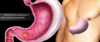
The most common location for atrophy of the gastric mucosa is the lower third of the body or the antrum. One of the main damaging factors is considered to be Helicobacter, which lives closer to the pyloric zone.
At the initial stage, glandular (goblet) cells produce hydrochloric acid even in excess. Perhaps this process is associated with the stimulating effect of the bacterial enzyme system.
Then the synthesis of gastric juice is replaced by mucus, and the acidity gradually decreases.
By this time, the protective role of the mucous membrane is lost. Any food chemicals can harm the cells lining the inside of the stomach. Toxic products and remnants of destroyed cells become foreign to the body.
The autoimmune mechanism is involved in the destruction process. Antibodies are produced against damaged cells, which continue to fight their own epithelium. Blocking recovery processes plays an important role.
In a healthy stomach, the epithelial layer is completely renewed every 6 days. Here, old dysfunctional cells remain in place or they are replaced with connective tissue.
On histology, instead of clear outlines of the epithelium (look along the upper edge), destroyed cells are visible, there are no pyriform glands
In any case, the atrophied mucosa cannot replace gastric juice with mucus. There is a gradual thinning of the stomach wall. In practice, the organ is excluded from digestion, and gastrin production increases. The bolus of food enters the small intestine unprepared, which leads to failure of other successive stages.
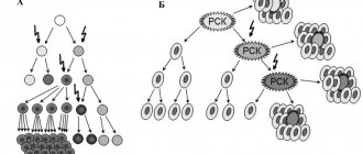
The process doesn't end there. The most dangerous period of atrophic changes begins: the epithelium begins to produce similar, but not true, cells. Most often they can be classified as intestinal. They are not able to produce gastric secretions. This process is called metaplasia and dysplasia (transformation), and precedes cancerous degeneration.
Atrophied areas on the mucous membrane cannot be completely restored, but with the help of treatment there is still a chance to support the remaining functioning cells, compensate for the lack of gastric juice and prevent disruption of the overall digestive process.
What is vaginal atrophy?
Vaginal atrophy (also called atrophic vaginitis or colpitis) is a condition in which the vaginal lining becomes drier and thinner. Most often occurs during menopause, a “lifestyle change” due to a decrease in the level of the hormone estrogen. Hormones are produced, stored and secreted by the endocrine system, a network of glands and organs. Women need estrogens for good health, especially during childbearing years. When menopause occurs around age 50, the ovaries produce fewer hormones and the woman stops having periods. But the condition of vaginal atrophy also occurs in young women when their estrogen levels are disturbed.
The mechanism of development of atrophic changes in the walls of the vagina
The vaginal mucosa is a stratified squamous epithelium, which before menopause is moist and thick with wrinkles. During menopause, when estrogen levels decrease, this epithelium becomes thinner. Fewer epithelial cells result in less cell shedding in the vagina (see diagram). When epithelial cells slough off and die, they release glycogen, which is hydrolyzed to glucose. Glucose, in turn, is broken down into lactic acid by lactobacilli, normal inhabitants of a healthy vagina. Without this cascade, vaginal pH rises, leading to loss of lactobacilli and overgrowth of other bacteria, including group B streptococci, staphylococci, coliforms, and diphtheroids. These bacteria can cause symptomatic vaginal infections and inflammation. This results in itching, burning and pain during sex, among other symptoms, and includes urinary tract infections (UTIs) and frequent urination. Recently, the term “vaginal atrophy” has been proposed to be replaced by the new term “genitourinary menopausal syndrome.” It involves not only vaginal but also urinary symptoms, which can be caused by low estrogen.
Who is at risk for vulvovaginal atrophy?
Women aged 50 years and older during menopause are most susceptible to vaginal atrophy. Other reasons that increase your likelihood of developing it include:
- Lack of sexual intercourse.
- Decreased ovarian function due to chemotherapy or radiation.
- Immune disorders.
- Medicines with antiestrogenic properties.
- Ovariectomy (removal of the ovaries).
- Postpartum estrogen loss.
- Breast-feeding.
Causes of atrophy
During menopause, your body produces less estrogen. Without estrogen, the lining of your intimate area may become thinner and less elastic. The vaginal canal may also narrow and shorten. Low amounts of estrogen reduce the amount of normal vaginal fluids. It also changes vulvovaginal acid balance.
Women who have just given birth and are breastfeeding also experience a decrease in estrogen levels. These symptoms also occur in patients who have had their ovaries removed or who are taking certain medications (such as aromatase inhibitors for breast cancer). And the first sign of mucosal atrophy is usually insufficient vaginal moisture.
Risk factors
There are two main factors that can increase your chances of vaginal atrophy:
- Smoking. It restricts blood flow, including in the vaginal area. Also reduces the natural amount of estrogen in your body.
- No vaginal birth. Those who have not had a vaginal birth are more likely to have these problems than those who have.
Clinical manifestations
Symptoms of vaginal wall atrophy may include:
- Vaginal dryness.
- Burning and/or itching in the vagina.
- Dyspareunia (pain during sex).
- Vaginal discharge is usually yellow in color.
- Spotting and spotting
- Bleeding from the vagina after sex.
- Itching of the vulva (desire to scratch between the legs).
- Feeling of pressure in the perineum.
Signs of atrophic changes in the urinary tract:
- Desire to wash yourself more often.
- Pain when going to the toilet.
- Urinary tract infections (UTIs).
- More acts of urination.
- Stress urinary incontinence.
- Painful urination (dysuria).
- Blood in the urine (hematuria).
- Burning when urinating.
The first symptom is usually a lack of lubrication during intercourse. Over time, persistent vaginal dryness may occur. Thinning of the epithelial lining can also cause itching, tenderness, and stinging pain in the vaginal and vulvar areas, which in turn can further contribute to dyspareunia. Spotting may also occur due to small tears in the vaginal epithelium. Women with genitourinary menopause syndrome may report a thin yellow or gray watery discharge without much itching or odor.
Photo 1. Microphotographs of superficial and intermediate cells (left) and atrophic cells (right). (Pap test, original magnification x20).
Photo of a normal smear and with atrophy of the vaginal mucosa
Diagnostic methods
A gynecologist can diagnose vaginal atrophy based on symptoms and a gynecological examination, which will show what the vagina looks like. This will help you know if you have reached menopause. Classic signs of vulvulvaginal atrophy during a pelvic examination include:
• Shortened or narrowed vagina. • Dryness, redness and swelling. • Loss of skin elasticity. • Whitish discoloration of the mucous membrane. • Rare pubic hair. • Bulge along the back wall of the vagina. • Skin diseases of the vulva (dermatoses). • Lesions of the vulva and/or redness of the vulva (erythema). • Bladder sagging in the vagina. • Changes in the external urethral meatus. • Minor cracks (tears) at the vaginal opening.
What tests need to be taken:
- Oncocytology of the cervix.
- General urine analysis.
- Ultrasound of the pelvic organs.
- Serum hormone test.
- Vaginal pH test.
- Microscopy for flora (see the entire catalog of analyzes).
Table 1. “Difference between vulvovaginal atrophy and other pathology of the intimate area.”
| Lichen planus | Painful red plaques or erosions, sometimes with white lacy edges or purple edges; may spread to the vagina |
| Lichen sclerosus of the vulva | Hypopigmented, wrinkled, wax-like tissue with confluent ivory and pink plaques, often in a butterfly or figure-eight pattern, enclosing the labia majora, minora and clitoral hood and extending around the anus; may lead to labial agglutination |
| Contact dermatitis | Redness, swelling and itching, sometimes with blistering and painful bright red swelling |
| Lichen simplex chronicus (hyperkeratosis) | Thick, lichen-like skin, often erythematous, caused by prolonged rubbing or scratching |
| Intraepithelial neoplasm of the vulva | Red, white or dark raised or eroded lesions, multifocal |
| Vulvar cancer | More often it is an ulcer with a raised or thickened edge |
| Extramammary Paget's disease | Brick-red scaly eczematoid plaque with a sharply defined border and sometimes rough surface |
Causes
The most common causes of the disease are: exposure to Helicobacter and autoimmune factors. Researchers have proposed distinguishing external (exogenous) and internal (endogenous) damage factors that can cause atrophic changes in the mucosa. External ones include toxic substances entering the stomach and malnutrition.
Toxic to the stomach are:
- nicotine, a decomposition product of tobacco products;
- dust particles of coal, cotton, metals;
- arsenic, lead salts;
- alcohol-containing liquids;
- medications from the Aspirin group, sulfonamides, corticosteroids.
Food can turn into exogenous damage factors if:
- a person eats irregularly, periods of hunger alternate with overeating;
- mainly eat fast food, spicy and fatty dishes, “dry food”;
- cold or too hot food (ice cream, tea) enters the stomach;
- insufficiently chewed food in the mouth due to diseases of the teeth, gums, poor prosthetics, lack of teeth in old age.
This "workaholic's dream" saturates the body, but is not a healthy food
Internal reasons include:
- any disorders of the neuroendocrine regulation of secreting cells, leading to disruption of regeneration processes (stress, chronic diseases of the nervous system, myxedema, diabetes mellitus, dysfunction of the pituitary gland and adrenal glands);
- general human diseases that disrupt blood flow in the wall of the stomach and regional vessels (thrombosis, severe atherosclerosis), congestion in the veins against the background of increased pressure in the portal system;
- cardiac and respiratory failure, accompanied by tissue hypoxia (lack of oxygen);
- deficiency of vitamin B12 and iron in the body;
- hereditary predisposition - consists of a genetically determined lack of factors to restore the cellular composition of the mucosa.
Risk factors
The root cause - the decrease in estrogen production lies not only in the natural aging of the female body. Sometimes involutional processes are observed against the background of various hormonal abnormalities, for example, when the relationship between the pituitary gland, hypothalamus and ovaries is disrupted or when the genital organs are removed.
Vaginal atrophy can be temporary or permanent. The pathology occurs periodically, for example, during lactation (especially accompanied by amenorrhea) or with long-term use of hormonal drugs (for breast cancer, for example).
Risk factors for developing vaginal atrophy:
- Smoking;
- Immune disorders;
- Non-vaginal (not through the birth canal) birth;
- Lack of regular sex life.
Vaginal atrophy does not always develop. If a woman adheres to a healthy lifestyle and understands how important sex is for her body, unpleasant symptoms and complications can be avoided. Timely identified signs are of great importance in the treatment of the disease.
Signs of atrophy
Symptoms of atrophy of the gastric mucosa appear late, when acidity reaches zero. Young and middle-aged men are more often affected. Pain syndrome is absent or very mild, which is why they consult a doctor at an advanced stage of the process.
Signs of atrophy do not differ from the general symptoms of gastric disorders. Patients note a feeling of heaviness in the epigastrium immediately after eating, sometimes nausea, belching, bloating, loud rumbling, bad breath and unstable stool.
Attacks of nausea and dyspeptic disorders are symptoms of pathology
The presence of signs of impaired digestion is indicated by:
- weight loss;
- symptoms of vitamin deficiency (dry skin, hair loss, bleeding gums, mouth ulcers, headaches);
- hormonal problems, expressed in men as impotence, in women as disrupted menstrual cycles, infertility;
- increased irritability, tearfulness, insomnia.
Diagnostics
Atrophy of the gastric mucosa can only be diagnosed visually. It used to be determined by a pathologist or surgeon, but nowadays the widespread use of fibrogastroscopic technology makes it possible not only to record the picture in different parts of the stomach, but also to take material for histological examination, to divide the process into types and degrees of functional disorders.
Histologically, the infiltration of cells of the mucous layer by lymphocytes, destruction of the glandular epithelium, thinning of the wall, and impaired folding are revealed. Cracks and erosions may appear.
Symptoms and treatment of atrophic hyperplastic gastritis
Depending on the size of the affected area, the following are distinguished:
- focal atrophy - areas of atrophy with normal tissue alternate on the mucosa; this process is most favorable for treatment, because there are still cells capable of taking on a compensatory function;
- diffuse - a severe, widespread process that covers the entire antrum and rises to the cardia, almost all cells are affected, instead of a mucous layer, continuous fibrosis appears.
Based on the number of lost and remaining healthy cells, the degrees of atrophic changes are distinguished:
- mild - 10% of cells do not function, but 90% work correctly;
- medium - atrophy covers up to 20% of the area of the gastric mucosa;
- severe - more than 20% of the epithelium is replaced by scar tissue, transformed cells appear.
With subatrophy, shortening of the cells of the epithelial layer is observed
Depending on the severity of the atrophic process, histological changes are assessed as:
- mild changes or subatrophy - the size of the glandular cells decreases, their slight shortening is determined, additional glandulocytes appear inside the cells (formations where the secretion is synthesized), some are replaced by mucous (mucoid);
- moderate atrophy - more than half of the glandular cells are replaced by mucus-forming ones, foci of sclerosis are visible, the remaining part of the normal epithelium is surrounded by infiltrate;
- pronounced disorders - very few normal glandular cells, large areas of sclerosis are visible, infiltration of various types of inflammatory epithelium is observed, intestinal metaplasia is possible.
In diagnosing pathology, it is not enough to establish that the gastric mucosa is atrophic; in order to try to stop the process, the doctor needs to know the cause of the changes, the degree of dysfunction of the organ.
To do this, the patient undergoes the following studies: detection of antibodies to Helicobacter and to Castle factor (components of parietal cells) in the blood, determination of the ratio of pepsinogen I, pepsinogen II (protein components for the production of hydrochloric acid), the method is considered a marker of atrophy, since it allows one to judge the remainder of intact epithelial glands.
It is also necessary to study gastrin 17, a hormonal substance responsible for the endocrine regulation of the secretion of epithelial cells, their restoration and motility of the gastric muscle tissue, and daily pH measurements to identify the nature of acid formation.
To identify Helicobacter, all patients with atrophic gastritis are prescribed a urease breath test by the attending physician.
What types of gastritis develop based on epithelial atrophy?
Depending on the degree of development and localization of the inflammation process in the stomach with mucosal atrophy, it is customary to distinguish several types of gastritis.
Surface
The mildest form of the disease. The acidity of gastric juice is almost normal. There is an abundant secretion of mucus by the glands, so protection is maintained. Histology shows signs of degeneration.
Focal
Acidity is maintained by areas of healthy epithelium. The mucosa shows alternating areas of atrophy and sclerosis with healthy tissue. Symptoms include intolerance to milk and eggs. This suggests a role for immune dysfunction.
Diffuse
The surface of the stomach is covered with a proliferation of immature cells, pits and ridges, and the structure of the mucosal glands is disrupted.
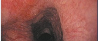
The atrophied mucosa has a grayish color, accumulations of blood vessels are visible
Erosive
In the atrophy zone, circulatory disturbance occurs, which gives a picture of spotty hemorrhages and accumulation of blood vessels. The course is severe with gastric bleeding. More often observed in alcoholics and people who have had a respiratory infection.
Antral
Named for the predominant localization of the lesion. It is characterized by cicatricial changes in the antral zone, narrowing of the pyloric region, and a tendency to develop into an ulcerative process.
Treatment
The problem of how to treat mucosal atrophy depends on the predominant aggressive action, the identified cause of the process, and the residual ability to recover (reparation). Given the absence of severe symptoms, patients are often treated on an outpatient basis. Mandatory recommendations include: regimen and diet.
It is not recommended to engage in strenuous sports; it is necessary to reduce physical activity to moderate. It is required to stop smoking and drinking alcoholic beverages, including beer. It is prohibited to take any medications without permission, including those for headaches and flu.
Diet Requirements
The patient's diet includes choosing foods that do not damage or irritate the gastric mucosa. Therefore, it is strictly prohibited:
- fried, smoked, salted and pickled dishes;
- strong tea, coffee, sparkling water;
- ice cream, whole milk;
- confectionery, fresh baked goods;
- spices, sauces, canned food;
- legumes
The patient is advised to maintain meals in small, frequent meals. Use stewed, boiled, steamed, baked dishes. In case of pain, it is advised to switch to semi-liquid pureed food (meatballs, low-fat broths, oatmeal with water, jelly) for several days.
If pain does not play a serious role in the clinic, then the diet should be varied, taking into account the given restrictions. Allowed:
- fermented milk products (low-fat sour cream, kefir, cottage cheese);
- egg omelet;
- vegetable stew;
- The most popular cereals are rice, buckwheat, and oatmeal;
- Fruit juices are best diluted with water.
The patient should consult a doctor regarding mineral water, since the choice depends on the acidity of the gastric juice, and it can vary during the process of atrophy.
Drug therapy
To restore the gastric mucosa, it is necessary to get rid of the harmful effects of Helicobacter, if present, and block a possible autoimmune process. To combat bacterial infection, a course of eradication is used.
A combination of tetracycline and penicillin antibiotics with Metronidazole (Trichopol) is prescribed. The course and dosage are chosen by the doctor individually.
Good results are accompanied by treatment with De-Nol (based on bismuth citrate)
To confirm the effectiveness, control studies are carried out on Helicobacter. In the initial stage of atrophy, when acidity may be increased, proton pump inhibitor drugs are recommended. They suppress the mechanism of hydrochloric acid production.
The group includes:
- Omeprazole,
- Esomeprazole,
- Rabeprazole,
- Ranitidine.
When hypo- and anacid states occur, these drugs are contraindicated. Acidin-pepsin and gastric juice are prescribed to replace one's own secretion. Stimulates the regeneration process Solcoseryl, Aloe in injections. Domperidone and prokinetics can support and improve gastric motor function.
Preparations based on bismuth and aluminum (Vicalin, Kaolin, bismuth nitrate) protect the mucous membrane from chemicals and bacteria from food products. If during the diagnostic process it becomes obvious that the body is in an autoimmune state, the patient is prescribed corticosteroid hormones to suppress an excessive immune response.
In severe cases of atrophy, the pathology is supplemented by a disruption in the production of enzymes by all organs involved in digestion. Therefore, enzymatic agents may be required: Panzinorm, Festal, Creon.
In the case of B12-deficiency anemia, courses of vitamin B12 and folic acid are prescribed.
So far, the fibrogastroscopic method is the only most accessible way for patients to confirm the diagnosis of atrophy
Folk and herbal remedies
The traditional method of treatment should be approached with caution, taking into account acidity. With normal secretory function, you can take decoctions of chamomile and calendula.
If it is reduced, rosehip decoction and diluted juices of tomatoes, lemon, and potatoes are recommended to stimulate acid formation. At the pharmacy you can buy herbal teas with plantain, thyme, wormwood, and St. John's wort. It is convenient to use the herbal medicine Plantaglucid. It consists of granulated plantain extract, diluted in warm water before taking.
The most significant problem of modern medicine is identifying patients and preventing cancer degeneration. It is difficult to organize fibrogastroscopic examinations of patients if they have little concern. Members of a family in which more than one case of atrophic gastritis has been identified and there have been deaths from stomach cancer are much more attentive to prevention.
Such patients should undergo fibrogastroscopy once a year, follow a diet, stop smoking and drinking alcohol. No one can be sure what difficulties these people will have to overcome in life, and how their stomach will tolerate genetic predisposition.
Treatment of vaginal atrophy
When should I see a doctor about vaginal atrophy? Even if you are not going through menopause, be sure to report any symptoms of dryness, pain, burning/itching, trouble urinating, UTIs, unusual spots, bleeding or discharge to your doctor. If weeks go by and the over-the-counter medications you take for dryness don't work, you should see your doctor. If you see unusual discharge or bleeding, this is also a reason to contact a specialist. Also, always consult a good gynecologist for any symptoms that are negatively affecting your daily life.
How to treat vaginal atrophy
The patient and her physician will work closely to develop a treatment plan for vaginal atrophy that is most effective based on the symptoms and their severity. Some methods are designed to treat symptoms of atrophy. Others aim to compensate for the loss of estrogen. According to reviews, estrogen therapy is considered the most effective addition to intimate rejuvenation procedures.
1. Lubricants and moisturizing creams to moisturize and relax the vagina can treat dryness. This improves comfort during sex, but will not completely restore the health of the intimate area. Vaseline is NOT recommended for use in the vagina as it can cause a yeast infection. Although many women use olive or coconut oil as a moisturizer and lubricant, it can sometimes cause allergic irritation in the area. Vitamin E and mineral oils should be avoided.
2. Dilators are devices that widen (widen) the vagina so you can get back to having sex. Women often start with a narrow dilator and move up to larger sizes over time. This is done until the vagina is wide enough for the penis to enter without pain for sexual activity. The best results are achieved when dilators are used in combination with topical hormonal therapy.
3. Hormone replacement therapy not only effectively improves the symptoms of vaginal atrophy, but also restores health to the skin by restoring the normal acid balance of the vagina, thickening the mucous membrane of the walls (to its original state), maintaining natural moisture and improving the balance of bacteria.
For women who only have symptoms of vaginal wall atrophy, there are several options that deliver estrogen only to the vagina. These options may help avoid high hormone levels in the rest of the body. Women who experience many other menopausal symptoms, such as hot flashes and trouble sleeping, may choose higher doses of hormone therapy to treat all of their symptoms (called systemic hormone therapy). Topical hormone administration will not treat any symptoms of menopause other than vaginal ones.
Next, look at the most important auxiliary therapeutic techniques for influencing the intimate area, which effectively complement the main course or are an important part of it.
Additional Methods
The specialists of our clinic offer a wide range of effective means of relieving and even relieving the symptoms of vaginal atrophy, proven by many years of experience.
For example, this could be the injection of autoplasma into intimate areas, injections of hyaluronic acid to relieve dryness of the mucous membrane of the vagina and vulva, treatment of stress urinary incontinence, etc. The sooner you start treatment, the less likely it is that vaginal atrophy will worsen. For example, the longer you go without estrogen, the drier your vagina becomes. Yes, without treatment, vaginal atrophy can get worse. Sometimes it can become so severe that it can significantly narrow the vaginal opening. This can make treatment difficult if it is started too late.
assignment_ind
Intimate contouring
Atrophy and sex life
You should not avoid sexual activity if your gynecologist has diagnosed you with vaginal atrophy. Lack of sexual activity actually makes the condition worse. Sex stimulates blood flow in the mucous membrane and promotes fluid production, so sex actually keeps the vagina healthy. If you experience dryness and discomfort during sex, vaginal moisturizers or water-based lubricants may help. Use them every few days and just before sex. What else contributes to his health can be found out in detail on this page.
Where to go in Moscow
Vaginal atrophy is serious for women during menopause. It affects your quality of life with discomfort, frequent trips to the toilet, frequent UTIs, burning, pain during sex and much more. Fortunately, there are many treatments available, and your doctor can help you find the best option to treat your symptoms. Seek treatment. Don't be afraid to talk to your doctor and your partner. Always follow the instructions of your gynecologist and do everything possible to prevent vulvar and vaginal atrophy from getting worse! We invite you to visit specialists from the gynecology department of our Moscow clinic.
Helpful information:
✍ Make an appointment with a gynecologist ☎ +7(495)790-0779 ☎ +7(495)797-7825

