- home
- general surgery
- Benign tumors and cysts of the liver - clinical picture, diagnosis, modern methods of surgical treatment
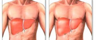
Most benign tumors (BTUs) are clinically oligosymptomatic or asymptomatic liver tumors, originating either from epithelial tissue (hepatocellular adenoma, etc.) or from stromal and vascular elements.
Spread of the disease.
Puchkov K.V., Bakov V.S., Ivanov V.V. Simultaneous laparoscopic surgical interventions in surgery and gynecology: Monograph. - M.: ID MEDPRACTIKA, 2005. - 168 p. Puchkov K.V., Ivanov V.V. and others. Technology of dosed ligating electrothermal effects at the stages of laparoscopic operations: monograph. - M.: ID MEDPRACTIKA, 2005. - 176 p.
Data on the epidemiology of DOP are very scarce. Sufficiently clear information is available only regarding the most common benign liver tumor - hemangioma. These tumors occur in 1-3% of the population, more often in women. Non-parasitic liver cysts occur in approximately 1% of the population. Other types of benign liver tumors are found much less frequently.
Classification of benign liver tumors
Benign liver tumors include hemangiomas, lymphangiomas, fibromas, lipomas and mixed tumors - hamartomas (teratomas). It is logical to classify non-parasitic cysts as benign liver tumors. Among them are true cysts (dermoid, retention cystadenomas) and polycystic liver (in more than half of the patients it is combined with cystic changes in other organs - kidneys, pancreas, ovaries). False cysts (traumatic, inflammatory) are also often observed. True cysts are usually solitary; false ones can be either single or multiple. The volume of multiple cysts is usually several milliliters, while the volume of solitary (true and false) cysts can reach 1000 ml or more.
Diagnosis of benign liver tumors
Two important signs are common to DOP: 1) the absence of an increase in the concentrations of alpha-fetoprotein, carcinoembryonic antigen CA - 199 in blood serum; 2) the absence of a clear increase in the activity of aspartic and alanine aminotransferases (AST and ALT), alkaline phosphatase (ALP), gamma-glutamyltransferase (GGTP) and lactate dehydrogenase (LDH).
These signs are reliable only in the absence of chronic or acute diffuse liver diseases, which themselves can cause changes in the above tests. Significant assistance is provided by the use of ultrasound and CT (or NMR) with bolus contrast, which have high resolution.
Liver cyst
Differential diagnosis of DOP usually begins with the exclusion of cysts. Non-parasitic cysts are more common. The possibility of polycystic disease, as well as solitary and multiple true and false liver cysts is taken into account.
Most cysts are small (1-5 cm in diameter) and are more common in women. A significant part of them are asymptomatic. A number of patients experience pain in the right hypochondrium, some have constant pain, others have periodic pain. Significant assistance is provided by the use of ultrasound and CT (or NMR), which have high resolution. The possibility of polycystic liver disease must be considered.
Differential diagnosis of simple cysts is also carried out with parasitic liver cysts (echinococcosis). The latter are supported by positive reactions with echinococcal antigen and Katsoni, as well as the detection of calcifications in the area of tumor-like formation, although hemengiomas can occasionally become calcified.
For a free written consultation, in order to determine the type of liver cyst, its localization to the main structures of the organ and indications for surgery, as well as choosing the correct surgical treatment tactics, you must send me a complete description of the abdominal ultrasound, MSCT data of the liver with contrast, and analysis to my personal email address blood for echinococcus, indicate age and main complaints. Then I will be able to give a more accurate answer to your situation.
Liver cyst treatment
Some non-parasitic liver cysts are also subject to surgical treatment, due to the real possibility of their rupture, infection and hemorrhage into the lumen of the cyst. In addition, rapidly growing large cysts lead to impaired liver function due to atrophy and replacement of the liver parenchyma with a cystic formation. Among the operations, liver resection, pericystectomy and cyst enucleation are the most commonly used.
In recent years, transparietal punctures of cysts under ultrasound or CT control have become widespread. After aspiration of the contents, a 96*ethyl alcohol solution is injected into the lumen of the cyst to harden the inner lining of the cyst. This operation is effective for cysts up to 5 cm in size. If there is no effect from these treatment methods or the cyst is larger, an operation is indicated - laparoscopic excision of the cyst area, followed by de-epithelialization of the inner lining of the cyst with argon-enhanced plasma or a defocused laser beam. Similar tactics are used for polycystic liver disease. In case of complicated polycystic liver disease (suppuration, bleeding, malignancy, compression of the bile ducts, portal or vena cava by large cysts), surgical treatment is indicated. Typically, fenestration is performed (opening cysts protruding above the surface of the liver), followed by deepithelialization of the inner lining of the cyst.
Watch a video of operations for liver cysts performed by Professor K.V. Puchkov. You can visit the website “Video of operations of the best surgeons in the world.”
Hepatocellular adenoma
Clinically, this is an asymptomatic benign liver tumor, which has signs of an adenoma developing from hepatocytes and is often delimited by a capsule. It most often affects women, usually due to long-term use of estrogen-progestational contraceptives. Occurs less frequently with long-term use of anabolic steroids. Adenoma develops quite rarely: in 3-4 people per 100,000 who use contraceptives for a long time.
As a rule (90%), it is single. It is found more often in the right lobe, subcapsularly. If located in the anteroinferior regions, it is palpated as a smooth, loose formation. Adenomas that developed while taking anabolic steroids have a more “aggressive” course. Complications such as intraperitoneal bleeding are rarely observed. Very rarely, an adenoma degenerates into a malignant tumor.
Focal nodular hyperplasia
Clinically, this is an oligosymptomatic benign tumor that does not have a capsule. The central part of the tumor is represented by scar connective tissue, and the peripheral part is represented by nodularly transformed hepatocellular tissue. Most often located subcapsularly. Foci of necrosis and hemorrhage are often observed in the tumor. As a rule, it does not develop in a cirrhotic liver, so it is sometimes called “focal cirrhosis.” Usually happens solitary. This is a rare benign liver tumor observed primarily in women taking oral contraceptives.
Nodular regenerative hyperplasia
This tumor resembles focal nodular hyperplasia of the liver, and is sometimes combined with it. In contrast to the latter, elements of connective tissue are significantly less represented. May be considered a prestage of hepatocellular carcinoma. Sometimes, as the cellular elements of this tumor grow, compression of large bile ducts or large branches of the portal vein occurs. As a rule, it is not detected in a cirrhotic liver. Sometimes it develops against the background of malignant diseases of extrahepatic localization (myeloproliferative processes, sarcomas, etc.).
All these types of DOP are asymptomatic diseases; in most cases, their detection can be attributed to accidental findings. The liver in most of these patients is not enlarged.
Radionuclide scintigraphy usually reveals a focal process measuring 3-5 cm. If the tumor is located in the marginal zones of the liver, then formations of smaller sizes may be detected.
Similar data are obtained with ultrasound and CT, as well as with the help of selective angiography and nuclear magnetic resonance (NMR). Therefore, a significant portion of small tumors are visible. Only morphological methods can clarify the nature of these three types of tumors. The material for these studies is usually obtained using a puncture biopsy with Shiba needles under ultrasound or CT control.
Patients with hepatocillary adenoma, focal nodular hyperplasia and nodular regenerative hyperplasia of the liver do not require drug treatment. Surgical treatment is used infrequently. Indications for it are either compression of the bile ducts or blood vessels, or the appearance of pain. The operation is performed if any complication develops and the tumor grows rapidly.
Secondary prevention methods and surveillance systems are as follows. It is prohibited to take oral contraceptives, estrogens, and anabolic steroids. Work related to the production of vinyl chloride is not recommended. Taking phenobarbital and zyxorine is undesirable. Abstinence from alcohol is recommended.
When a tumor is first detected, examinations are carried out 3-6-9-12 months and then once a year. In addition to the usual examination with determination of liver size according to Kurlov, studies of bilirubin content, aminotransferase activity, alkaline phosphatase, GGTP, alpha-fetoprotein, carcinoembryonic antigen and CA 19-9 antigen are performed. An ultrasound of the liver is also performed.
Hemangioma
Clinically, it is an asymptomatic benign tumor originating from the vascular, mainly venous, elements of the liver. Refers to the most common type of DOP.
It is represented by two options: cavernoma, which is like dilated blood vessels, and true hemangioma, developing from vascular embryonic tissue. It is most often located subcapsularly, in the right lobe, and often has a pedicle. Often covered by a fibrous capsule that may become calcified.
Spontaneous ruptures are very rare, but life-threatening. Clear clinical manifestations are observed in only 5-10% of tumors. As a rule, in these cases the tumor diameter exceeds 5 cm.
In many cases, the detection of hemangioma, like other DOPs, is an incidental finding. With large sizes and corresponding localization, symptoms of compression of the biliary tract or, less commonly, symptoms of portal hypertension sometimes appear. Sometimes a patient consults a doctor due to pain in the upper abdomen.
Instrumental studies provide important information. Radionuclide scintigraphy of the liver is performed, as usual, if a space-occupying process in the liver is suspected in two projections. Thanks to this method, as a rule, it is possible to detect a tumor with a diameter of 4-5 cm. For hemangiomas with a diameter of 4-5 cm or more, the tumor is detected in 70-80% of those examined. Ultrasound in the presence of hemangioma reveals a hyperechoic, well-defined formation. NMR provides similar information. Often, especially in the less massive left lobe, the vascular pedicle is clearly visible. Hemangiomas with a diameter of 3-5 cm or more are detected by ultrasound in 70-80% of those examined. Sometimes areas of calcification are observed in hemangiomas.
CT allows you to obtain data close to the results of ultrasound, although it often brings significant additional diagnostic information. This additional information primarily concerns the condition of surrounding tissues and organs. Celiacography allows you to obtain the most accurate data when recognizing hemangiomas. Usually, hypervascularized areas with clear boundaries are clearly visible, making it possible to detect a hemangioma with a diameter of 2-3 cm or more in 80-85% of those examined.
Indirect radionuclide angiography, performed using a gamma camera, brings similar, but less accurate results compared to celiacography. NMR often provides significant information.
When making a diagnosis of hemangioma, malignant liver tumors are excluded. In recent years, a kind of focal fatty degeneration of the liver has increasingly become the object of differential diagnosis, especially in cases where rounded areas of the intact liver are found against the background of focal fatty degeneration. These areas have a different density from fatty degeneration, and this difference is quite clearly recorded with ultrasound and CT. These pseudotumor formations are usually not visible on radionuclide scintigraphy of the liver. However, this differential diagnostic sign is not very reliable. A targeted liver biopsy plays a decisive role in identifying focal fatty degenerations.
Treatment of liver hemangiomas. With small hemangiomas without a tendency to grow, patients, as a rule, do not need medical and surgical treatment. For large tumors that compress the bile ducts or blood vessels, there are indications for resection of the corresponding liver segments. More often this rule applies to hemangiomas with a diameter of more than 5 cm.
Liver lymphangiomas are extremely rare; their clinical picture makes them difficult to distinguish from hemangiomas. Suspicion of lymphangioma arises only if there is an extrahepatic location of the tumor in the mediastinum and neck.
Fibromas, myxomas, lipomas, liver neuromas, which have the features of benign tumors: slow development, clear boundaries, normal ESR, are extremely rare. Absence of tumor markers and increased activity of serum enzymes such as AST, ALT, ALP, GGTP, LDH.
The treatment strategy is similar to that for hemangiomas.
Secondary prevention methods and surveillance systems are essentially the same as for the benign tumors described above. For all types of DOP, medications such as oral contraceptives and anabolic steroids are prohibited. It is undesirable to take drugs such as phenobarbital and zyxorin. Work related to the production of vinyl chloride is not recommended.
All patients with DOP require constant medical supervision. When a tumor is detected for the first time, examinations are carried out after 3-6-9-12 months and then once a year. In addition to the usual examination with determination of the size of the liver according to Kurlov, studies of bilirubin content are performed, determining the activity of ALT, AST, ALP, GGTP, GDH and LDH, alpha-fetoprotein and carcinoembryonic antigen.
Hepatocellular carcinoma (HCC)
HCC is a malignant tumor that develops from hepatocytes. Refers to primary liver carcinomas. In 60-80% of patients, it is associated with the persistence of hepatitis B and C viruses; in 70-85% of patients in developed countries, HCC develops against the background of liver cirrhosis. Around the world, approximately 750,000 people die from HCC each year.
Basically, morphological classifications of HCC have been proposed. The most common division of HCC is into nodular, massive and diffuse forms. The TNM system is also used. We have developed a classification (1988), which includes the main clinical variants of the disease: hepatomegalic (covers about 50% of patients), cystic (3-5%), cirrhosis-like (about 25%), hepatonecrotic, or abscess-like (6-10%), ictero-obstructive ( 6-10%), masked(6-10%).
Some researchers value ultrasound data more highly. A. Maringhini et al. (1988) when examining 124 patients with HCC, found hyperechoic areas in 47 of them, hypoechoic areas in 30, and mixed areas in 47. The sensitivity of ultrasound, according to the authors, was 90%, specificity - 93.3%.
As reported by JC Ellis (1988), tumors with a diameter of less than 2 cm are difficult to distinguish from hemangiomas, solitary regenerative nodules and adenomas. Diagnosis of tumors located directly under the diaphragm, in the superolateral part of the right lobe, is especially difficult.
CT gives approximately the same results as ultrasound, sometimes slightly higher. However, identifying small tumors (2-4 cm in diameter), especially against the background of cirrhosis, is very difficult. JM Henderson et al. (1988) during a CT examination of 15 out of 100 patients with liver cirrhosis revealed focal anomalies suspicious for HCC.
Treatment of hepatocellular carcinoma.
In all cases where possible, surgical treatment of tumors is performed. Most often, resection is feasible for tumors of the left lobe. Long-term results of surgical treatment are not encouraging. In this regard, control examinations of patients after resections are recommended every 3 months.
A relatively small proportion of patients undergo liver transplantation. It is performed in persons under 60 years of age, in the absence of metastases and severe extrahepatic diseases. Long-term results are unfavorable.
If surgical treatment is not possible, some patients undergo chemotherapy.
Metastatic liver carcinoma (MLC)
The primary focus of MCP is located outside the liver - in the lung, stomach, colon and other organs. Refers to secondary liver tumors.
The frequency of metastasis of tumors of different primary localizations to the liver is different.
Tumors of the gallbladder metastasize to the liver in 75% of cases, pancreas - in 70%, colon, breast, ovaries, and melanoblastoma - in 50%, stomach and lungs - in 40%. However, primary tumors themselves occur with varying frequencies. Therefore, the doctor most often observes metastases in the liver coming from the colon, stomach and lungs, and in women - also from the mammary gland and ovaries.
To confirm or exclude the metastatic nature of a malignant liver tumor, a thorough examination of a number of organs is carried out. For some localizations this is especially important.
The examination plan includes:
- blood serum test (ACE, carcinoembryonic antigen, CA antigen - 199, acid phosphatase);
- chest x-ray;
- gastroscopy;
- colonoscopy or sigmoidoscopy in combination with irrigoscopy;
- Ultrasound of the pancreas, kidneys, ovaries, prostate;
- breast examination and mammography in women;
- consultation with a gynecologist and urologist.
Particular attention is paid to the possibility of primary localization of the tumor in the colon, prostate gland (in men) and ovaries (in women), since metastases of these localizations seem to be relatively curable in some patients.
Features of ultrasound visualization
Using ultrasound, the following focal changes in the liver can be detected:
- non-parasitic cystic formations;
- bacterial, parasitic foci;
- benign neoplasms (adenoma, vascular anomalies, hyperplasia);
- malignant;
- postoperative, post-traumatic changes.
Every year the number of patients with liver pathology is growing steadily. This is due to low-quality products, uncontrolled use of medications, alcohol abuse, as well as late diagnosis of diseases.
Focal liver lesions can be visualized using ultrasound, computed tomography, and magnetic resonance imaging. In this case, one can suspect a benign or malignant course of the disease based on the structure of the formation.
Due to its high information content and harmlessness, ultrasound diagnostics can be used as a preventive method for the initial detection of pathology, as well as for assessing the dynamics (rate of disease progression).
Of course, such a study will not verify the diagnosis, but it is quite possible to detect a pathological focus using ultrasound.
To confirm the diagnosis, tomography and biopsy of the gland are prescribed.
Ultrasound examination can reveal changes in the structure of the liver tissue, visualize additional formation, evaluate its contents, size, density, and also analyze the outline of the liver itself, its volume, vascular blood flow and the condition of surrounding organs.
Let us briefly describe in the table the visualization features of common liver tumors.
| Education | Ultrasound picture | Course of the pathology |
| Adenoma |
| Benign |
| Hemangioma |
| Benign |
| Lipoma |
| Benign |
| Hyperplasia |
| Benign |
| Cystadenoma |
| Benign |
| Cysts (parasitic, non-parasitic, post-traumatic, postoperative) |
| Benign |
| Hepatocellular carcinoma, angiosarcoma, hepatoblastoma |
| Malignant |
Note that even a benign course of the disease under certain conditions can take on a malignant form.
Liver cancer symptoms
The first signs of liver cancer are not specific; they are also possible in many other pathologies of the organ (acute and chronic hepatitis, alveococcosis, cirrhosis, damage to the bile ducts or liver metastases). In the early stages, the pathology may not manifest itself in any way, since the organ has a high reserve. Therefore, liver cancer is often first detected by ultrasound, which is performed for various pathologies of internal organs located near the liver. If the tumor is significant in size, patients may experience the following complaints and manifestations:
- decreased or complete lack of appetite (anorexia);
- decrease in the amount of hemoglobin and red blood cells;
- discomfort, pain in the hypochondrium, closer to the right side or in the upper abdomen;
- nausea, vomiting with bile or food;
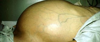
- an increase in the size of the abdomen due to the growth of a tumor formation that can be felt through the abdominal wall;
- ascites (growth of the abdomen due to the accumulation of fluid in it);
- weight loss, sometimes very significant (up to several kilograms in a couple of months);
- yellowing of the skin and whites of the eyes (development of jaundice);
- severe skin itching;
- nose bleed;
- stool disorders - alternating constipation or diarrhea;
- change in color of urine (dark) and stool (discoloration);
- expansion of small capillaries under the skin (formation of telangiectasia).
As the tumor grows and disintegrates, general malaise, fever, and weakness may occur.
Benign formations
In most cases, such lesions do not manifest significant symptoms. Their structure can be represented by epithelial tissues, as in adenoma, stromal tissues - in nodular hyperplasia, or vascular elements, which is typical for hemangioma.
Benign tumors practically do not show symptoms, so their detection by ultrasound is usually accidental.
Only with a significant increase in the formation can heaviness in the right hypochondrium be bothersome. Therapeutic tactics depend on the size of the tumor and the course of the disease. The prognosis is often favorable.
Now in more detail about each benign neoplasm.
Adenoma
Adenoma is not so common in the parenchyma of the gland. It may consist of cells that resemble hepatocytes (liver cells) - hepatocellular adenoma. This type of pathology is in most cases diagnosed in the female population of childbearing age.
The lesions are located one at a time or in groups of nodules, limited from the normal parenchyma by a capsule. Considering the risk of rapid growth of the adenoma (up to 20 cm in diameter), surgical intervention is indicated for therapeutic purposes. It is necessary to prevent tumor rupture, vascular damage and the development of massive bleeding.
In addition, the adenoma may consist of small bile ducts with cysts and mucous accumulations. This type of pathology is more typical for the male half of the population.
Hemangioma and lipoma
Changes in the liver in the form of hemangioma are the most common type of benign lesion of the gland. The structure of the formation is represented by venous elements. It is characterized by slow growth, lack of metastasis and damage to healthy liver tissue.
Despite this course, it is still recommended to regularly perform preventive ultrasound due to the risk of complications:
- compression of the bile ducts with difficulty in the outflow of bile;
- compression of blood vessels, which disrupts the blood supply to the organ;
- rupture of blood vessels with bleeding;
- malignant tissue degeneration.
As for lipoma, it is formed from adipose tissue. Its diameter often does not exceed 5 cm.
Diagnosis of lipoma begins with ultrasound, but often requires additional examination, for example, MRI. In most cases, the wen is localized in the right lobe; it can be single or located in groups. Over time, such formations merge into conglomerates and are surrounded by a connective tissue capsule.
Among the complications, it is worth highlighting the risk of developing liposarcoma - a malignant liver lesion.
Hyperplasia and cystadenoma
With hyperplasia, no cell changes are observed, but the lobulation of the gland is disrupted. In most cases, the pathology is of genetic origin and is diagnosed mainly in the right lobe of the organ in the female population.
Given the similarity to malignant lesions, additional diagnostics are required. Ultrasound reveals a heterogeneous structure, as well as varying echogenicity (increased or decreased).
With nodular hyperplasia, the examination reveals many nodules up to 4 cm, and the size of the gland remains within normal limits, and changes in the parenchyma are minimal.
The difference between a benign process is:
- slow growth;
- lack of germination into surrounding organs;
- good response to therapy;
- no metastasis.
Cystadenomas are benign in origin, but in 10% of cases tissue malignancy is observed. On ultrasound they appear as cystic single-chamber structures with a fibrous capsule. Inside the cyst there may be septa, papillary growths and mucus. Such formations can be located both intrahepatically and in the biliary tract or bladder.
Causes and risk factors
The exact causes of liver cancer have not yet been established. Most often, a malignant lesion occurs against the background of a long-term inflammatory process in the liver tissue. Most often, the first signs of liver cancer are detected against the background of cirrhosis. In regions where viral hepatitis is widespread, cases of liver cancer are detected much more often. Thus, approximately 25% of all patients with signs of liver cancer are diagnosed with viral hepatitis C, and approximately 40% are chronic carriers of the hepatitis B virus.
Other risk factors that increase the risk of developing liver cancer include:
- poor nutrition leading to obesity;
- drinking large amounts of alcohol;
- long-term use of drugs that have hepatotoxicity (including steroids, oral contraceptives);
- the influence of external factors - toxins that damage liver cells (vinyl chloride, aflatoxins);
- long history of diabetes mellitus;
- Budd-Chiari syndrome (these are episodes of thrombosis in the hepatic veins);
- hereditary pathologies - Wilson's disease, hemochromatosis, tyrosinemia, glycogenosis, alpha-trypsin deficiency, intestinal polyposis.
In some cases, the causes of liver cancer cannot be determined, and the cases are considered idiopathic.
Cystic formations
These neoplasms differ in origin, structure and size. They can be inflammatory, parasitic or congenital, and have a capsule and liquid content. Usually they are filled with a clear or yellowish liquid, but a brown or green tint may appear, which indicates an admixture of blood or bile.
Cysts can be located superficially or inside the gland, and can also reach 25 cm. If during diagnosis a cyst is found in each segment, it is customary to talk about polycystic disease.
Non-parasitic cysts
They are liquid formations with a capsule that are formed from the bile ducts. They are registered in 5% of the population, mainly in women. They can be single or multiple, affecting no more than 30% of the liver tissue.
In most cases, cysts are located in one lobe. With polycystic disease, more than 50% of the glandular tissue is affected, and the cysts are localized in both lobes without preserving the normal glandular tissue between them.
If we consider false cysts, they form in the post-traumatic period. The wall of the neoplasm is represented by fibrous tissue. In addition, such cysts can form after treatment of ulcers or excision of an hydatid cyst. Their contents are a light liquid, which can sometimes contain an admixture of bile.
Clinically, non-parasitic formations do not appear; only occasionally, with a significant increase in size, heaviness or pain in the right hypochondrium is observed. Discomfort can be associated with both stretching of the gland capsule and compression of surrounding organs.
Parasitic cysts
Thanks to modern ultrasound machines, a diagnostician can accurately determine the location of the tumor and the nature of its contents. Immunological methods are also used in diagnosis, for example, RIFA.
Alveococcosis develops as a result of infection with cestodes of echinococcus, which differs from the causative agent of echinococcosis in morphological and biological characteristics.
First, let's take a closer look at echinococcosis. It is considered a rather serious disease and develops as a result of infection of the body with echinococcus. The main problem of diagnosis is the long asymptomatic course, which is why a person consults a doctor at a late stage of the pathology. The volume of cyst contents can reach 5 liters.
Postoperative and post-traumatic cysts
Considering the liver abscess, it should be said about the infectious origin of the pathology. Pathogenic microorganisms penetrate the gland tissue with bile, lymph or blood flow. Often the lesions are localized in the right lobe, have a round shape and are accompanied by discomfort and pain in the right hypochondrium.
An infected cavity in the gland can form in the presence of intra-abdominal infection, after wounds, traumatic damage to the organ, or surgical interventions.
In addition to pain, the disease is manifested by fever, pronounced sweetness and profuse sweating. In terms of frequency of occurrence, the leading cause of abscess is infection of the bile ducts (cholecystitis, cholangitis). Inflammation can also occur after endoscopic manipulation or parasitic infection of the biliary tract.
In second place among the causes of abscesses is an intra-abdominal infection, which penetrates the liver through the portal vein. This is observed with diverticulitis (inflammation of the intestinal processes), a violation of the integrity of the intestine or its ulcerative lesion.
Hematomas form after injury or surgery, when blood accumulates in the cavity, which has entered the parenchyma from a damaged blood vessel.
During an ultrasound examination, the following may be detected:
- formation filled with liquid with clots, which indicates the initial stage of cyst formation;
- lesion with dense masses, partitions of varying thickness and dense walls (stage of progression);
- at the last stage, a false cyst with liquid contents or fibrous areas may be detected, which indicate resorption of the cyst.
Types of primary liver cancer
There are several classification options for primary liver cancer. There are various types of tumors, which differ in course and prognosis. Experts identify several of the most typical types of primary liver cancer (the tumor is formed from the cells of the organ).
Hepatocellular cancer
is the most common tumor (carcinoma) that occurs in the liver area.
As the name suggests, these tumors form in hepatocytes, which form the basis of liver tissue. The course of hepatocellular cancer is possible in two clinical forms - diffuse
(when a large part of the liver is affected) or
nodular
(one focus is formed - a node or several areas).
A more favorable variant of this cancer is possible - fibrolamellar carcinoma
, with it there are possibilities for more active treatment.
Cholangiocarcinoma
is a malignant tumor that originates in the area of the bile ducts.
Cholangiohepatoma
is a mixed type of tumor in which cancer cells originate both in the area of hepatocytes and in the area of epithelial cells in the bile ducts.
Variants of tumors such as angiosarcomas, mesodermal formations
are lesions that grow from lymphatic capillaries, blood vessels or connective tissue cells.
Possible hepatoblastoma
cystadenocarcinoma
is also identified . It occurs as a result of the degeneration of a benign tumor.
Malignant tumors
During laparoscopic examination of the abdominal cavity, material is collected, which is subsequently sent for histology. In addition, laparoscopy makes it possible to examine surrounding organs, which is necessary to determine the extent of the malignant process.
The morphological structure of the tumor can only be determined using histological analysis.
It is not always possible to use the puncture technique under ultrasound guidance, since the material can be collected from the unaffected part of the organ. In most cases, the pathology is diagnosed at a late stage, when the tumor is considered inoperable and metastasis is observed.
It is not always possible to suspect a malignant lesion using ultrasound examination, since it may have the same echogenicity as normal gland tissues. Only computed tomography and magnetic resonance imaging make it possible to more accurately establish the localization of the lesion, assess its size, density, and relationship with surrounding tissues.
When using elastography, as well as elastometry, the information content of ultrasound increases significantly. An important part of the diagnosis is the assessment of blood flow in the tumor.
A malignant lesion can be of primary or secondary origin. In the first case, malignant transformation of cells occurs directly in the liver. As for the secondary process, the gland is affected by metastases from the main tumor, which may be located in another organ. Often the liver is affected secondarily.
Among the types of cancer it is worth highlighting:
- hepatocellular carcinoma, which is characterized by rapid progression and high mortality. The risk group is the male part of the population after 50 years of age;
- angiosarcoma, which is also characterized by high aggressiveness;
- hepatoblastoma - manifested by nodes without a capsule, yellowish tint. Pathology is diagnosed in infants.
Symptomatically, the malignant process manifests itself:
- severe malaise;
- icteric syndrome (yellowing of the skin, mucous membranes, darkening of urine and discoloration of feces);
- rapid loss of body weight;
- pain syndrome in the area of the right hypochondrium;
- dyspeptic disorders (nausea, vomiting and flatulence);
- lack of appetite.
On palpation, the gland is felt as a dense, lumpy, painful formation. Therapeutic tactics depend on the stage of the oncological process and the morphology of the tumor. If the formation is considered operable, it is removed.
Treatment of liver tumors is based on:
- type of disease;
- stages of the pathological process;
- functional state of the gland;
- general condition of the patient (presence of allergic reactions and concomitant pathologies);
- the risk of complications (this applies to cases where the formation affects large vessels, intestines and diaphragm).
A feature of the malignant process is the rapid growth of the tumor, metastasis, germination into surrounding organs, inhibition of organ functions and often an unfavorable outcome due to late diagnosis and aggressiveness of the tumor.
Prevention of liver cancer
Among the main preventive measures are avoidance of alcohol consumption, vaccination against viral hepatitis B, as well as protection against infection with other types of infection. In addition, timely recognition and treatment of liver pathologies, treatment of fatty hepatosis and cirrhosis are important.
The information in this article is provided for reference purposes and does not replace advice from a qualified professional. Don't self-medicate! At the first signs of illness, you should consult a doctor.
Diffuse lesion
If an ultrasound examination reveals diffusely altered tissue, hepatosis or cirrhosis should be suspected. Depending on the type of provoking factor, hepatosis can be:
- lipid, when fat is deposited in hepatocytes. Ultrasound reveals an increase in signal throughout the gland, as well as its compaction. There are three degrees of progression of fatty hepatosis. At the first stage, the content of fatty inclusions in the organ exceeds the upper limit of normal. On the second, steatohepatitis is diagnosed when the tissue is diffusely affected. As for the third degree, it is characterized by fibrotic changes localized around the vessels. At the same time, the gland becomes dense;
- alcoholic type;
- hepatosis of pregnant women;
- non-alcoholic form;
- hepatosis in diabetes.
Cirrhotic changes involve the replacement of normal gland tissue with connective tissue. Ultrasound diagnostics reveals tissue compaction. Without treatment, the risk of liver malignancy increases.
Ultrasound examination is a safe diagnostic technique that is widely used for preventive examination, assessment of the rate of disease development, and analysis of dynamics during treatment. Ultrasound is prescribed for children and pregnant women, which confirms its harmlessness.
If an ultrasound examination reveals a suspicious lesion in the liver, it is recommended to continue the diagnosis to clarify its origin and the nature of the pathology. This will help diagnose the disease at the initial stage, start therapy on time and avoid serious complications.
Primary liver cancer: diagnosis
All people who are at high risk for developing liver cancer are regularly screened. First of all, doctors look for ultrasound signs
Liver cancer in people suffering from chronic inflammatory processes of the organ have different degrees of cirrhosis. They are advised to perform an ultrasound examination every six months, even in the absence of complaints.
In addition, they are advised to perform laboratory blood tests to determine the tumor marker of primary liver cancer - AFP
(stands for alpha-fetoprotein). With this type of observation, it is possible to identify tumor lesions at an early stage (there are no symptoms or complaints yet), when the cancer can be radically cured. Changes in tests for liver cancer with an increase in AFP typically occur in up to 50-90% of patients. Levels greater than 400 ng/mL may occur in individuals with large tumors or rapidly growing tumors. But a temporary increase in the indicator is possible against the background of cirrhosis or inflammation of hepatocytes. However, high AFP in combination with ultrasound data and MRI of the liver confirms the diagnosis in almost 100% of patients.
Performing a CT
or
MRI with contrast
helps to identify a tumor and clarify its size and location, which is important for drawing up a treatment plan for liver oncology.
If all this data is not enough to make a diagnosis, a percutaneous biopsy
(puncture or aspiration) under ultrasound control. The risk of complications with such diagnostic methods is minimal.
Complications
The uncontrolled course of liver cancer is dangerous due to the occurrence of complications. Their appearance depends on many factors. Possible complications include:
- bleeding from the tumor;
- suppuration of the tumor focus;
- impaired outflow of bile due to compression of the bile ducts;
- circulatory disorders due to compression of the abdominal organs by large tumor sizes;
- ascites.
The listed symptoms require immediate diagnosis and surgical intervention. Without this, death is possible.
Make an appointment
Nutrition
The patient's condition improves with a special diet. The cooks at the Yusupov Hospital prepare dishes from high-quality products that do not contain nitrates, GMOs or carcinogenic food additives. In case of uncontrolled weight loss, increase daily caloric intake at the expense of protein products. When preparing dishes, preference is given to gentle culinary technologies: boiling, stewing, steaming and grilling, baking in the oven.
The following foods are excluded from the patient's diet:
- store-bought sauces;
- smoked meats and pickles;
- rich broths;
- margarine and other heavy fats;
- fresh baked goods;
- alcoholic drinks;
- strong tea and coffee.
Forecast
Since liver cancer is often diagnosed at the final stage of the tumor process, the prognosis for recovery is pessimistic in most cases. Palliative therapy is aimed at reducing the severity of symptoms of the disease and alleviating the general condition. In the presence of concomitant pathologies, death occurs 6 months or 1 year after diagnosis. The average life expectancy after the onset of this disease is about 2 years with properly selected effective treatment.
Metastases
Metastases are the main danger of any cancer, since they are almost impossible to stop or prevent their spread.
Metastases of primary liver cancer can spread to the stomach, brain, lungs, esophagus, veins and arteries, heart muscle, blood vessels, bones and spine. Metastases from the liver enter the body in the following ways:
- through lymph flow;
- through the circulatory system;
- through growth beyond the liver and damage to neighboring tissues and organs.
Unfortunately, metastases occur in 40% of liver cancer cases and often affect vital organs, which are subsequently extremely difficult to treat.