Reasons for appearance
Normally, bile is synthesized by the liver, flows into the gallbladder, where it reaches the required concentration, and then moves through the bile ducts to the duodenum to take part in the breakdown of fats. They can block her path:
- malignant and benign tumors both inside the duct (papillomas) and external, compressing the canal from the outside (including lymphomas and metastases);
- stones in the common bile duct (choledocholithiasis);
- scars and strictures resulting from past operations;
- inflammatory processes;
- congenital anomalies;
- cysts;
- helminthic infestations.
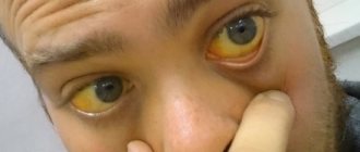
Bile is forced to accumulate in the ducts and bladder. The pigment bilirubin, contained in bile, sweats along with its liquid part through the walls, spreads throughout the body with the flow of lymph and blood and stains the skin and sclera.
Cholestasis
With this pathological syndrome, manifestations and pathological changes are caused by an excess amount of bile in hepatocytes and tubules. The severity of symptoms depends on the cause that caused cholestasis, the severity of toxic damage to liver cells and tubules caused by impaired bile transport.
Any form of cholestasis is characterized by a number of general symptoms: an increase in the size of the liver, pain and discomfort in the area of the right hypochondrium, itching, acholic (discolored) feces, dark urine, disturbances in the digestive processes. A characteristic feature of itching is its intensification in the evening and after contact with warm water. This symptom affects the psychological comfort of patients, causing irritability and insomnia. As the severity of the pathological process and the level of obstruction increase, the stool loses color until it becomes completely discolored. At the same time, the stool becomes more frequent, becoming liquid and foul-smelling.
Due to a deficiency in the intestines of bile acids, which are used for the absorption of fat-soluble vitamins (A, E, K, D), the level of fatty acids and neutral fat in the feces increases. Due to impaired absorption of vitamin K during a protracted course of the disease, blood clotting time increases in patients, which is manifested by increased bleeding. A lack of vitamin D provokes a decrease in bone density, as a result of which patients suffer from pain in the limbs, spine, and spontaneous fractures. With prolonged insufficient absorption of vitamin A, visual acuity decreases and hemeralopia occurs, manifested by deterioration of eye adaptation in the dark.
In the chronic course of the process, a disturbance in the metabolism of copper occurs, which accumulates in the bile. This can provoke the formation of fibrous tissue in organs, including the liver. Due to an increase in lipid levels, the formation of xanthomas and xanthelasmas begins, caused by the deposition of cholesterol under the skin. Xanthomas have a characteristic location on the skin of the eyelids, under the mammary glands, in the neck and back, and on the palmar surface of the hands. These formations occur with a persistent increase in cholesterol levels for three or more months; when its level normalizes, they may disappear on their own.
In some cases, the symptoms are mild, which complicates the diagnosis of cholestasis syndrome and contributes to the long course of the pathological condition - from several months to several years. A certain proportion of patients turn to a dermatologist for treatment of itchy skin, ignoring other symptoms.
Symptoms of obstructive jaundice
Even before yellowing appears, the patient begins to experience pain in the right hypochondrium. Depending on the cause, the sensations may vary in intensity and frequency. Normally, bile is released from the bladder into the ducts when the gallbladder contracts when the food mass reaches the duodenum. Therefore, at the beginning of the disease, pain is felt mainly after eating, and may disappear completely or subside before a new attack. And when bile accumulates, pressure on the walls of the ducts and bladder increases, and constant severe debilitating pain appears. Most often encircling or radiating to the area of the shoulder blades.
Feces become colorless and greasy, since without bile fats are not digested and there is no coloring pigment.
The urine is dark as a result of pigment being excreted by the kidneys.
If there is inflammation, the temperature rises, chills and weakness appear.
Bile components that enter the bloodstream poison the body and lead to itchy skin, blood clotting disorders, and decreased blood pressure and heart rate (bradycardia). The patient is anxious, panics, sleeps poorly.
When the duct is not completely blocked, the symptoms are less pronounced.
Reasons for the development of postcholecystectomy syndrome
The causes of postcholicystectomy syndrome and its development are often associated with disruption of the normal functioning of the sphincter of Oddi (orbicularis muscle). The sphincter of Oddi is a smooth muscle that is located in the lower part of the duodenum and is responsible for regulating the supply of bile and pancreatic juice to the duodenum3.
If we consider that PCES most often affects patients who have undergone surgery to remove the gallbladder4, then the mechanism of occurrence of this syndrome can be explained as follows:
- after the operation, the sphincter, which normally opens when the gallbladder is full, does not receive a signal about filling, as a result of which it is almost constantly under tension;
- due to the absence of a bladder, bile enters the duodenum in a diluted state, which increases the pressure inside the intestinal walls. In addition, bile itself has a bactericidal effect and a change in its composition can lead to intestinal infection5.
But sphincter of Oddi dysfunction may not always be caused by removal of the gallbladder. Sometimes the cause of the syndrome is advanced gastrointestinal diseases* (chronic colitis or gastritis, peptic ulcer) and chronic pancreatitis) - a disease of the pancreas, as well as errors in preoperative examination.
Diagnostics
Since obstructive jaundice is not an independent disease, but a syndrome, the task is not to confirm, but to find and differentiate the cause. First of all, an examination is carried out and data about the patient is collected: whether there have been operations for cholelithiasis, the presence of chronic diseases, dietary habits, and so on.
Clinical and biochemical laboratory tests of blood, feces and urine will show the general condition, degree of poisoning, the presence of inflammation, and the state of the coagulation system. Obstructive jaundice is characterized by a strong increase in direct bilirubin, alkaline phosphatase, and an increase in ALT and AST.
Instrumental methods:
- Ultrasound examination (ultrasound) of the abdominal organs. With its help, you can see the enlargement of the gallbladder and ducts, the presence of stones, and tumors. The diagnostic value of ultrasound is approximately 75%. Used as a screening procedure - painless for the patient and not requiring the creation of special conditions for implementation;
- Computed tomography (CT) with contrast can detect tumors of the duodenum, pancreas, common bile duct;
- Magnetic resonance imaging (MRI) detects stones and tumors and shows the structure of the liver and pancreas;
- ERCP - diagnosis using X-ray contrast agent. Read more about it in the treatment section.
The main goal of the examination is to distinguish hepatic jaundice from mechanical jaundice and to identify the specific cause and location of blockage of the bile flow.
Causes of bile stagnation
Cholestasis can develop for many reasons, the most common of which are:
- obstruction of the gallbladder or bile ducts due to the presence of stones in them, as well as when they are compressed by a cyst or tumors;
- abnormalities in the structure of the gallbladder or ducts, when they are bent;
- inflammation of the gallbladder or ducts;
- dysfunction of the valves responsible for the free movement of bile;
- endocrine and enzymatic disorders, in which thickening of bile and changes in its chemical properties are observed.
Diagnosis
Stagnation of bile is characterized by changes in the color of the sclera and skin (yellowing), as well as complaints of itching, unstable stools (alternating constipation and diarrhea), discolored feces and dark urine. If there are even several of these symptoms, the doctor has reason to prescribe a biochemical blood test (the level of bilirubin, bile acids, liver enzymes, etc. is examined), a urine test for urobilin, ultrasound of the liver and gallbladder, fibrogastroduodenoscopy, etc. When diagnosing cholestasis, it is important to distinguish between these condition from viral, parasitic and other liver diseases that have a similar clinical picture.
Treatment
The task of doctors in the treatment of obstructive jaundice is to eliminate the cause of obstruction (blockage), restore the outflow of bile and prescribe symptomatic treatment.
For the treatment of choledocholithiasis, the most commonly used method is ERCP (endoscopic retrograde cholangiopancreatography). An endoscopic examination is performed using special equipment in an X-ray room. Then a contrast agent is injected through the major duodenal papilla and the condition of the bile and pancreatic ducts is analyzed using a series of x-rays. If small stones are found, they are removed. Large ones are crushed and also removed. ERCP makes it possible to make an incision in the sphincter and remove papillomas that obscure the lumen. The procedure has a number of contraindications, but in some cases it is performed for health reasons.
Drainage of the bile ducts in obstructive jaundice is used to stop the pathological process and remove pressure from the bile ducts. A puncture is made through the skin and stagnant bile is drained through the drainage into a special container. In patients with neoplasms, the tube remains until the issue with the tumor is resolved. The operable area is removed surgically. For inoperable cases, stenting is performed - a special prosthetic expander is installed inside the ducts to normalize the flow of bile.
Surgery in the treatment of obstructive jaundice can be abdominal or laparoscopic. In case of cholelithiasis, the gallbladder filled with stones is removed, the ducts are freed from stones, and the patency of the bile ducts is restored.
For drug treatment the following is used:
- painkillers;
- antispasmodics;
- transfusion solutions for the rapid removal of toxins and restoration of electrolyte balance;
- drugs to regulate blood clotting;
- antibacterial and anti-inflammatory agents and so on.
The Yusupov Hospital is well equipped and has all the capabilities to correctly diagnose and eliminate the causes and consequences of bile duct blockage. Doctors will select the optimal treatment regimen and methods, taking into account the patient’s condition, individual characteristics of the disease and examination results.
Possible complications
If you seek medical help in a timely manner, the prognosis for recovery is favorable. However, with prolonged poisoning of the body with bilirubin, damage to the liver, kidneys, heart, blood vessels, central nervous system, and blood coagulation system is possible. If medical recommendations are not followed, complications can arise in any of the listed organ systems.
In the area of the outbreak itself, irreversible processes in the tissue of the liver and pancreas can occur. Gallstone disease tends to recur, so it is especially important to follow the diet prescribed by your doctor.
Why is it necessary to consult a doctor at the first symptoms?
Pain and other signs of illness in the liver area cannot be ignored, especially if there have already been operations in the anamnesis. By self-medicating, we only remove symptoms and waste time. So, in the case of tumors, at an early stage they can be removed and treated. Stones blocking the gap increase in size over time and become more difficult to remove. Timely detection of the cause is 50% of successful treatment.
If the skin and sclera turn yellow, emergency medical attention is needed. Failure to see a doctor promptly can lead to death.


